Physical Activity After Spinal Cord Injury

By | | No Comments

Author: Sharon Jang | Reviewer: Sonja de Groot | Published: 20 April 2022 | Updated: XX October 2024

Physical activity after spinal cord injury (SCI) can provide many health benefits, as in the able-bodied population. This page covers the benefits of exercising with an SCI, precautions, and adaptations to exercising with an SCI.

Key Points

  • Exercising after an SCI can improve muscle strength, type, and size, your abilities to do things on a day-to-day basis, your well-being, and decrease risks for secondary complications.
  • There are many ways to get physically active, including sports, being active in the community, and going to the gym.
  • Many exercises and sports can be adapted for those with SCI using adaptive equipment.
  • Although rare, some secondary complications such as autonomic dysreflexia (AD), orthostatic hypotension (OH), skin breakdown, and temperature regulation, may arise.

After SCI, there is deconditioning of muscles, bones, joints, and changes in the heart and blood vessels due to inactivity. This can lead to various secondary complications, such as heart disease, breathing complications, weakening of the bones (osteoporosis), pain, spasticity, and diabetes. Exercise has many positive changes for those with an SCI including muscle type and size, improved muscle strength, independence, well-being, and helping to prevent secondary health complications.

Muscle type, size, and strength

In the body, there are 2 main types of muscle fibers: slow twitch (type I) and fast twitch (type II). Slow twitch muscles are known as the endurance muscles, as they are able to hold a contraction for a long period of time before getting tired. For example, the muscles that are used to keep your head upright are mostly made up of slow twitch muscle fibers. Type II fibers are known for their short burst of speed or strength. They can generate more strength, but get tired really quickly. Over time with an SCI, the muscles with the endurance type (type I) tend to turn into the more fatigable type (type II). There is some moderate-weak evidence that shows that among those with limited movement in their legs, the use of functional electrical stimulation (FES) can help shift muscle fibers from being more fatigable to more endurance based.

After injury, muscles in the body slowly begin to become smaller (atrophy). However, there is moderate to weak evidence that indicates that moving your arms and legs, either passively or actively, can help build muscle up again. Two (weak evidence) studies found that amongst those with limited to no leg function, electrical stimulation (Neuromuscular electrical stimulation (NMES) or FES) can increase the size of the thigh muscles. In addition, there is weak evidence that the use of a body-weight support treadmill can also increase the size of the lower leg muscle, resulting in a partial reversal of muscle shrinking.

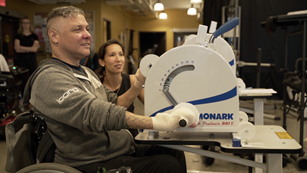
There is strong-moderate evidence that exercising can help individuals of any injury level improve their strength. Among those with paraplegia, there is strong evidence that strength training (i.e., doing weight training) can improve muscle strength in the arms. There is also strong evidence showing that body weight support training can improve overall muscle strength, and moderate evidence that arm cycling can help strengthen the arms and the front of the shoulder. Among those with tetraplegia, there is strong evidence that the use of FES on the arm and shoulder can improve muscle strengthening. Moreover, strong evidence suggests that neuromuscular stimulation (NMES) can improve strength among those with cervical level injuries. If you are unable to access specialized equipment, strength training with free weights or using an arm cycle can show similar benefits as well.

Activities of Daily Living

There is some moderate evidence that shows that exercising can enhance the ability to perform daily tasks by yourself. Exercising improves your fitness level (such as your strength and endurance), which can help you perform daily tasks. More specifically, tasks may become easier by reducing physical strain and a decrease in the amount of time required to do an activity.  One moderate evidence study found that doing physical therapy exercises in addition to neuromuscular stimulation enhanced participant’s ability to perform self-care (e.g., dressing, feeding, toileting) and mobility (e.g., transferring, wheelchair pushing). Other weak evidence supports these findings, as they found that exercise can help improve transferring and the ability to put on/take off clothing, wheeling and cleaning. Furthermore, increased fitness levels have also been associated with return to work.

Well-being

Some evidence suggests that exercise can help individuals improve perceptions of well-being. Well-being has been defined as how well an individual feels in their mind, their satisfaction with their health and functioning, and their overall satisfaction in life. Two aspects of well-being that are relatively well-researched are the impact of physical activity on depression and quality of life. There is weak evidence that found that all types of physical activity can help improve depressive symptoms and can improve quality of life. This relationship between physical activity and depressive symptoms and quality of life can be explained by a strong evidence study, which indicates that exercise can lead to decreased stress and pain. For example, strong evidence has shown that exercise can reduce shoulder pain, which can allow individuals to perform a greater variety of movements without consequences. The reduction in stress and pain, in turn, is thought to improve quality of life and depressive symptoms. However, many of these studies lack a control group. As a result, we are unable to determine if physical activity alone has an influence on subjective well-being.

Secondary complications

After sustaining an SCI, multiple secondary complications can occur. However, research suggests that exercise can help prevent or reduce the severity of secondary complications, including:

  • Conditions impacting the heart and blood vessels, by improving the strength of the heart and balancing out the sympathetic (fight or flight; stimulation) and parasympathetic  (relax and slowing) nervous systems,
  • Breathing complications, through strengthening the muscles required for breathing and through increasing the amounts of oxygen taken up by the body,
  • Weakened bones, by increasing bone mass density,
  • Type II diabetes, through improving the balance of blood sugar (glucose),
  • Pain, through strengthening, and
  • Spasticity, which can be reduced short term with exercising.

There are many ways for you to remain physically active, even after SCI! Strength training can be done at a local community center or private gym, most often with the equipment already there. Strength training can also be done at home with free weights and exercise bands. Some equipment that can be used for strength training include free weights, exercise bands, and pulleys. For aerobic exercise, some alternatives include using an arm ergometer (arm cycle), a rowing machine (if possible), and adaptive rowers, such as the Ski-Erg.

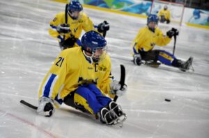
Adaptive sports is another way to get active. There are a variety of adaptive sports, including court sports (e.g., basketball, rugby, tennis), water sports (e.g., sailing, kayaking), race sports (e.g., cycling, track and field), and winter sports (e.g., Nordic and alpine skiing).

Refer to our article on Adapted Sports & Equipment for more information!

Alternatively, specialty equipment is available to help facilitate exercise after SCI. However, this equipment is more commonly used in rehabilitation settings, as they are very expensive and additional assistance is often required. A Functional Electrical Stimulation (FES) bike can be used to simulate the legs while cycling, and has been shown improve strength and endurance. Body-weight Support Treadmills are specialized treadmills with a sling attached. This type of treadmill allows an individual to move their legs on the treadmill, while having their bodyweight supported by a sling. Some models are available to allow users to control how much of their bodyweight they feel while in the treadmill, which can alter the challenge of walking.

If going to the gym or playing sports is not your thing, there are still other ways to get active! Performing daily tasks can be hard work as well. For example, activities such as heavy gardening, going grocery shopping and carrying home groceries, doing a lot of housework such as vacuuming and cleaning the house, going for a push with family/friends are all ways of being active. However, if these are your activity of choice, you want to make sure you are pushing yourself enough to get your heart rate up and keep it up for a while.

In 2020, exercise guidelines for the SCI population were released. Currently, the starting level guidelines for fitness benefits are:

  • at least 20 minutes of moderate to vigorous intensity endurance (aerobic) exercise, 2 times per week
  • 3 sets of strength exercises for each major muscle group at a moderate to vigorous intensity, 2 times a week.

The advance level provides guidelines for additional fitness and health benefits, such as reducing your risk for diabetes. It is recommended to get at least 30 minutes of moderate to vigorous intensity aerobic exercise at least 3 times a week, in addition to the 3 sets of strength exercises twice a week.

Refer to our article on Exercise Guidelines for Adults with SCI for more information!

Another way to gauge your effort is through a Rating of Perceived Exertion (RPE). The RPE is a subjective rating scale where the individual rates how hard they feel they are working, where 0 is not working at all, and 10 is working at your absolute maximum. If someone is just starting off with exercising, starting between a 5-7 on the RPE scale is a good idea.


Watch SCIRE’s YouTube Video explaining how to use RPE when exercising!

Another way to evaluate how hard you are working is through using the talk test. The talk test uses your ability to carry out a conversation while performing exercise to gauge exercise intensity. According to the talk test, a moderate intensity workout is achieved when one is able to talk to someone while working out, but not being able to sing. During a vigorous intensity workout, you would only be able to say a couple of words to someone, and speaking is difficult.

Watch SCIRE’s YouTube video explaining how to adapt exercises.

Going back into a gym after an SCI may be daunting given that much of the equipment may no longer be accessible. However, there are a number of ways to adapt gym equipment, including grip assistance,  transfer boards, chest straps, and using free weights and wedges. When exercising at a gym, you may require some additional assistance getting set up on pieces of equipment. If this is the case, consider going with a family member or a friend, and don’t be afraid to ask the gym attendant for help.

Adaptive grip aids include commercial gloves (left), tensor bandages (upper right) and weight lifting cuffs (bottom right).

After a high-level SCI, hand functioning may be impaired, resulting in a lack of ability to grip. To address this in a gym setting, some available options include using tensor bands, commercially available gloves, or weight-lifting cuffs. Tensor bandages can be used to wrap your hands around a handlebar. Benefits of using a tensor bandage include wide availability and low cost. Commercially available gloves, such as the Active Hands, are also available to assist with grip function on handles. These gloves provide a bit more support to the wrist and have a Velcro strap around the wrist. They also have a second Velcro that goes over the hand, which secures the hand to the handle. However, commercial gloves may not be as readily available and are usually expensive. Lastly, some individuals use weight-lifting cuffs, which are available at most gyms for use, to assist with grip function. These cuffs have a Velcro strap that goes around the wrist and a hook that can be connected to handlebars. Although commonly found in gyms, weightlifting cuffs only work for specific movements, such as pushing and pulling. In addition, they might not fit around handles of all sizes.

An abdominal binder (circled in red) being used to help keep an upright posture during rowing.

Abdominal (core) function is often impacted with an SCI, which may limit the types of activities you are able to do. One way to address this issue is through the use of a chest strap. A chest strap is a neoprene strap that comes in differing widths but is often wide enough to cover your abdominal area. The idea is to wrap the chest strap around the backrest of your wheelchair and around your torso, preventing you from falling forward if you are doing a pulling exercise. Chest straps are commonly used in various wheelchair sports as well, to provide additional support.

Refer to our article on Abdominal Binders for more information! 

When exercising in a wheelchair, you may find that the wheel lock still allows for some movement in the wheels, which may hinder an exercise. One way to address this situation is through adding additional support at the base of the wheel using wedges or free weights. Free weights can be placed behind the rear tire on both sides, or in front of the rear tire on both sides. In place, small wooden wedges (or door stoppers) can be placed under the tires on all four sides (in front and at the back) to help prevent rocking.

Watch SCIRE’s YouTube video explaining potential complications during exercise.

Exercise is relatively safe for individuals with SCI. However, there are some complications that, while rare, can arise.

Low blood pressure

When you first start exercising, it is common to possibly feel some nausea, or like you might pass out. This is a result of exercise-induced (exertional) hypotension, or a sudden drop in blood pressure due to exercise. One way to overcome this is to build up your exercise routine. When doing aerobic exercises, try a discontinuous approach: exercise for 2-3 minutes, then take a break. The idea is to slowly increase the length of exercising before you require a break, working your way up to 20-30 minutes of exercise. Once you are able to continuously exercise for 20-30 minutes, then you may consider increasing the resistance.

Autonomic Dysreflexia

Autonomic dysreflexia is a condition where blood pressure suddenly increases to dangerous levels. If this occurs, stop exercising. Sit up and try to lower your legs if possible, loosen any tight clothing, and move off of any high-pressure areas (e.g., sit bones, hands/wrists if you are using assistive grip). If symptoms do not go away, seek medical attention.

Refer to our article on Autonomic Dysreflexia for more information!

Temperature regulation

With a high level injury, temperature dysregulation, the body’s inability to control temperature, may be influenced. The ability to produce sweat can be compromised with higher levels of injury, resulting in an inability to cool down the body. In colder environments, it may be harder to warm up.

When exercising in hot or warmer environments, make sure you are drinking water consistently throughout your workout. Consider wearing looser clothing, and try to work out in an environment with ventilation, fans, or air conditioning. If you notice that you tend to overheat during exercise and are unable to sweat, you can also try carrying a spray bottle with you and spray your face down to cool off. When exercising in cooler environments, be mindful of your hands, arms, legs, and feet and make sure they aren’t getting too cold. Try dressing in layers so you can wear more if necessary, but also take layers off if you get warm.

Skin concerns

When exercising, it is important to be cautious of skin integrity, especially if you have no sensation. One area to be mindful of is the back when performing rocking or twisting motions. Rocking and twisting movements may cause the back to rub on the backrest of the wheelchair, creating a potential for skin breakdown. Another area to be mindful of is areas used with straps, such as the hands and sometimes the feet. For example, if using a grip aid for a longer duration of time to perform an activity, you may want to check for red spots that may have been caused by the straps. Ensure to check your skin after exercising for redness.

Refer to our article on Pressure Injuries for more information!

Overuse injuries

Overuse injuries occur when you exercise muscles that are already often used on a daily/frequent basis. An example of this is the shoulders, as it is used for pushing a wheelchair. To prevent overuse injury, make sure you have the correct posture when performing exercises. When working on the shoulder, try to consider alternatives to pushing your wheelchair as exercise, if possible. For example, the use of an arm bike could be an alternative to get around as they require less demand on your shoulders and arms. In addition, try to balance aerobic exercise and strength training in muscle groups prone to overuse injuries.

Benefits of physical activity as you age

Watch SCIRE’s video explaining to learn about ways to stay active as you age.20

It is normal as you age for the function of body systems and organs to gradually decline. People with SCI and other disabilities may find that this process starts earlier in life and has a bigger impact on daily function. Physical activity is important to keep your body and mind healthy. Physical activity can help someone aging with SCI prevent and slow down losses in function, independence, and quality of life. Small changes in muscle strength, heart health, breathing, or pain can have big impacts on the daily activities of someone with SCI. Physical activity can also be a good way to socialize if you are able to join a community group or program.

Physical activity can help keep your body healthy in many ways:

  • Improve or maintain muscle strength
  • Improve or maintain mobility
  • Improve respiratory fitness
  • Improve blood circulation
  • Reduce risk of heart health problems
  • Improve bone health and prevent osteoporosis
  • Improve your overall mood and wellbeing
  • Improve body composition (increase muscle, reduce unhealthy fat)
  • Boost the immune system
  • Improve chronic shoulder pain with strengthening and stretching
  • Reduce fatigue

Refer to the “What are the benefits of exercise after SCI?” section above for more information.

Risks and considerations

While physical activity is essential to maintaining health as you age, the impacts of aging on your body may also increase your risk for injury. These risks should be considered when doing physical activity, especially because problems like pain, fractures, pulled muscles, and strains can hinder participation.

Be aware of:

  • Pain that may be an indication of injury.
  • Increased risk of fractures due to bone loss (people with SCI are at high risk for osteoporosis).
  • Joint degeneration and injury in the shoulders and wrist (people with SCI develop overuse injury with age and have more arthritis and breakdown in the joints).
  • Skin breakdown and pressure injury from motions that cause friction during physical activity (skin becomes more thin and less elastic with age and heals more slowly from injury).

How to know when to stop

Many people with SCI live with a baseline level of pain. This can make it difficult to determine whether discomfort and pain while doing physical activity is acceptable or harmful. Expert advice includes starting small and taking note of any discomfort/pain before, during, and after physical activity.

  • If physical activity is not causing additional discomfort or pain that persists afterwards, you can maintain or make a small increase to the intensity and/or amount of physical activity.
  • If physical activity is causing additional pain that persists afterwards, consider changing the type, or reducing the intensity and/or amount of physical activity.

Adaptations

You may need to adjust your physical activity routines and methods as you age to keep it safe and accessible. Consult your physiotherapist for advice on how to safely do physical activity based on your unique abilities and barriers.

Some possible adaptations:

  • Changing the type of physical activity you do
    • Slower-paced sports
    • Less weight-bearing activities to accommodate injuries or arthritis
    • Hobbies that get you moving
  • People often get tired more quickly as they age. Start with shorter periods of less intense activity and increase gradually in small increments
  • Opportunities to do PA at home (online classes, walking the dog, gardening, etc.)
  • Opportunities in the community
    • Check with local organizations for online and in-person PA programs
  • Equipment and technology to facilitate PA
    • Lift or transfer technology to get onto workout equipment
  • Work with a trainer/coach who is knowledgeable about SCI

Can medication make you tired?

Some medications used to treat pain or spasticity may cause fatigue, reducing your motivation to be physically active. If you are concerned about the effects of your medication on energy levels, consult with your healthcare provider to see if there are opportunities to optimize your regimen.

Participating in physical activity after SCI can be intimidating, but it is beneficial. Being physically active can help improve your well-being and help reduce the impact of secondary complications after SCI. There are many ways to stay active after an injury, and many ways to adapt existing sports and equipment to help you get exercise. Although exercise is healthy, there are precautions to keep in mind when exercising. Overall, it is recommended that individuals with SCI stay active to promote a healthy lifestyle.

For a list of included studies, please see the Reference List. For a review of how we assess evidence at SCIRE Community and advice on making decisions, please see SCIRE Community Evidence.

Parts of this page has been adapted from SCIRE Professional “Physical Activity” Module:

Wolfe DL, McIntyre A, Ravenek K, Martin Ginis KA, Latimer AE, Eng JJ, Hicks AL, Hsieh JTC (2013). Physical Activity and SCI. In Eng JJ, Teasell RW, Miller WC, Wolfe DL, Townson AF, Hsieh JTC, Connolly SJ, Mehta S, Sakakibara BM, editors. Spinal Cord Injury Rehabilitation Evidence. Version 4.0.
Available from: https://scireproject.com/evidence/physical-activity/

Evidence for “What are the benefits of exercise after SCI?” is based on:

Alexeeva, N., Sames, C., Jacobs, P. L., Hobday, L., DiStasio M. M., Mitchell S.A., & Calancie B. (2011). Comparsion of training methods to improve walking in persons with chronic spinal cord injury: a randomized clinical trial. The Journal of Spinal Cord Medicine, 34, 362—379.Ref 2

Andersen, J. L., Mohr, T., Biering-Sorensen, F., Galbo, H., & Kjaer, M. (1996). Myosin heavy chain isoform transformation in single fibres from m. vastus lateralis in spinal cord injured individuals: effects of long-term functional electrical stimulation (FES). Pflugers Archiv – European Journal of Physiology, 431, 513-518.

Cameron T, Broton JG, Needham-Shropshire B, Klose KJ. An upper body exercise system incorporating resistive exercise and neuromuscular electrical stimulation (NMS). J Spinal Cord Med 1998;21(1):1-6.

Chen Y, Henson S, Jackson AB, Richards JS. Obesity intervention in persons with spnal cord injury. Spinal Cord 2006;44:82-91

Chilibeck PD, Jeon J, Weiss C, Bell G, Burnham R. Histochemical changes in muscle of individuals with spinal cord injury following functional electrical stimulated exercise training. Spinal Cord 1999;37(4):264-268.

Crameri RM, Weston A, Climstein M, Davis GM, Sutton JR. Effects of electrical stimulation- induced leg training on skeletal muscle adaptability in spinal cord injury. Scand J Med Sci Sports 2002;12(5):316-322.

Crameri RM, Cooper P, Sinclair PJ, Bryant G, Weston A. Effect of load during electrical stimulation training in spinal cord injury. Muscle Nerve 2004;29(1):104-111.

de Carvalho DC, Cliquet A, Jr. Energy expenditure during rest and treadmill gait training in quadriplegic subjects. Spinal Cord 2005;43(11):658-663.

De Groot PC, Hjeltnes N, Heijboer AC, Stal W, Birkeland K. Effect of training intensity on physical capacity, lipid profile and insulin sensitivity in early rehabilitation of spinal cord injured individuals. Spinal Cord 2003;41(12):673-679.

Ditor DS, Macdonald MJ, Kamath MV, Bugaresti J, Adams M, McCartney N et al. The effects of body-weight supported treadmill training on cardiovascular regulation in individuals with motor-complete SCI. Spinal Cord 2005;43(11):664-673.

Fukuoka Y, Nakanishi R, Ueoka H, Kitano A, Takeshita K, Itoh M. Effects of wheelchair training on VO2 kinetics in the participants with spinal-cord injury. Disability & Rehabilitation Assistive Technology 2006;1:167-74.

Glinsky, J., Harvey, L., Korten, M., Drury, C., Chee, S., & Gandevia, S. C. (2008). Short-term progressive resistance exercise may not be effective at increasing wrist strength in people with tetraplegia: a randomised controlled trial. Australian Journal of Physiotherapy, 54, 103- 108.

Grimby G, Broberg C, Krotkiewska I, Krotkiewski M. Muscle fibre composition in patients with traumatic cord lesion. Scand J Rehabil Med 1976;8(1):37-42.

Hetz SP, Latimer AE, Martin Ginis KA. Activities of daily living performed by individuals with SCI: Relationships with physical fitness and leisure time physical activity. Spinal Cord 2008;47(7):550-554.

Hicks AL, Adams MM, Martin GK, Giangregorio L, Latimer A, Phillips SM et al. Long-term body- weight-supported treadmill training and subsequent follow-up in persons with chronic SCI: effects on functional walking ability and measures of subjective well-being. Spinal Cord 2005;43(5):291-298.

Jacobs, P. L. (2009). Effects of resistance and endurance training in persons with paraplegia. Medicine & Science in Sports & Exercise, 41, 992-997.

Jeon JY, Weiss CB, Steadward RD, Ryan E, Burnham RS, Bell G et al. Improved glucose tolerance and insulin sensitivity after electrical stimulation-assisted cycling in people with spinal cord injury. Spinal Cord 2002;40(3):110-117.

Klose KJ, Schmidt DL, Needham BM, Brucker BS, Green BA, Ayyar DR. Rehabilitation therapy for patients with long-term spinal cord injuries. Archives of Physical Medicine & Rehabilitation 1990;71:659-62.

Le Foll-de Moro D, Tordi N, Lonsdorfer E, Lonsdorfer J. Ventilation efficiency and pulmonary function after a wheelchair interval-training program in subjects with recent spinal cord injury. Arch Phys Med Rehabil 2005;86(8):1582-1586.

Martin Ginis KA, Latimer AE, McKechnie K, Ditor DS, Hicks AL, Bugaresti J. Using exercise to enhance subjective well-being among people with spinal cord injury: The mediating influences of stress and pain. REHABIL PSYCHOL 2003;48(3):157-164.

Millar PJ, Rakobowchuk M, Adams MM, Hicks AL, McCartney N, MacDonald MJ. Effects of short-term training on heart rate dynamics in individuals with spinal cord injury. Auton Neurosci 2009; 150: 116-21.

Mohr T, Dela F, Handberg A, Biering-Sorensen F, Galbo H, Kjaer M. Insulin action and long- term electrically induced training in individuals with spinal cord injuries. Med Sci Sports Exerc 2001;33(8):1247-1252.

Mulroy, S. J., Thompson, L., Kemp, B., Hatchett, P. P., Newsam, C. J., Lupold, D. G., et al. (2011). Strengthening and Optimal Movements for Painful Shoulders (STOMPS) in chronic spinal cord injury: a randomized controlled trial. Physical Therapy, 91, 305—324.

Needham-Shropshire BM, Broton JG, Cameron TL, Klose KJ. Improved motor function in tetraplegics following neuromuscular stimulation-assisted arm ergometry. J Spinal Cord Med 1997;20(1):49-55.

Round JM, Barr FM, Moffat B, Jones DA. Fibre areas and histochemical fibre types in the quadriceps muscle of paraplegic subjects. J Neurol Sci 1993;116(2):207-211.

Sabatier, M. J., Stoner, L., Mahoney, E. T., Black, C., Elder, C., Dudley, G. A. et al. (2006). Electrically stimulated resistance training in SCI individuals increases muscle fatigue resistance but not femoral artery size or blood flow. Spinal Cord, 44, 227-233.

Silva AC, Neder JA, Chiurciu MV, Pasqualin DC, da Silva RC, Fernandez AC et al. Effect of aerobic training on ventilatory muscle endurance of spinal cord injured men. Spinal Cord 1998;36(4):240-245.

Sutbeyaz ST, Koseoglu BF, Gokkaya NK. The combined effects of controlled breathing techniques and ventilatory and upper extremity muscle exercise on cardiopulmonary responses in patients with spinal cord injury. Int J Rehabil Res 2005;28(3):273-276.

Stewart BG, Tarnopolsky MA, Hicks AL, McCartney N, Mahoney DJ, Staron RS et al. Treadmill training-induced adaptations in muscle phenotype in persons with incomplete spinal cord injury. Muscle Nerve 2004;30(1):61-68.

Evidence for “What are the exercise guidelines?” is based on:

Martin Ginis, K. A., van der Scheer, J. W., Latimer-Cheung, A. E., Barrow, A., Bourne, C., Carruthers, P., … Goosey-Tolfrey, V. L. (2018). Evidence-based scientific exercise guidelines for adults with spinal cord injury: an update and a new guideline. Spinal Cord, 56(4), 308–321.

SCIRE. (2020, March 6). Exercise after Spinal Cord Injury: How to Begin [Video file]. Retrieved from https://www.youtube.com/watch?v=P0EWCQawRbI&list=PLi2Dc1h0G7-vn6X1ROpMEMXJK6nmzinWu&index=2

Evidence for “How can I adapt exercises?” is based on:

SCIRE. (2020, March 6). Exercise after Spinal Cord Injury: How to Adapt Equipment [Video file]. Retrieved from https://www.youtube.com/watch?v=k7vTlHzYoug&list=PLi2Dc1h0G7-vn6X1ROpMEMXJK6nmzinWu&index=4

Evidence for “What should I be cautious of when exercising?” is based on:

SCIRE. (2020, March 6). Exercise after Spinal Cord Injury: Complications to Avoid [Video file]. Retrieved from: https://www.youtube.com/watch?v=HXVaLdhsBuk&list=PLi2Dc1h0G7-vn6X1ROpMEMXJK6nmzinWu&index=3

Evidence for ” How does aging affect physical activity after SCI?” is based on:

Cerrel Bazo, H. A., Demertzis, E., & Musumeci, A. (2018). The Aging Effects in Spinal Cord Injury Rehabilitation (pp. 379–402). https://doi.org/10.1007/978-3-319-57406-6_39

Cheung, L., Chiang, J., Kaiser, A., Patterson, K. K., & Musselman, K. E. (2023). Exploring the Experiences and Perceptions of Adults With Spinal Cord Injury or Disease Aged 50 Years or Older on Participation in Sports. Topics in Spinal Cord Injury Rehabilitation, 29(4), 37–50. https://doi.org/10.46292/sci23-00028

Galea, M. P. (2012). Spinal cord injury and physical activity: preservation of the body. Spinal Cord, 50(5), 344–351. https://doi.org/10.1038/sc.2011.149

Jiang, S.-D., Dai, L.-Y., & Jiang, L.-S. (2006). Osteoporosis after spinal cord injury. Osteoporosis International, 17(2), 180–192. https://doi.org/10.1007/s00198-005-2028-8

Krause, J. S., Vines, C. L., Farley, T. L., Sniezek, J., & Coker, J. (2001). An exploratory study of pressure ulcers after spinal cord injury: Relationship to protective behaviors and risk factors. Archives of Physical Medicine and Rehabilitation, 82(1), 107–113. https://doi.org/10.1053/apmr.2001.18050

Lee, A. K. Y., Miller, W. C., Townson, A. F., & Anton, H. A. (2010). Medication use is associated with fatigue in a sample of community-living individuals who have a spinal cord injury: a chart review. Spinal Cord, 48(5), 429–433. https://doi.org/10.1038/sc.2009.145

Lundström, U., Wahman, K., Seiger, Å., Gray, D. B., Isaksson, G., & Lilja, M. (2017). Participation in activities and secondary health complications among persons aging with traumatic spinal cord injury. Spinal Cord, 55(4), 367–372. https://doi.org/10.1038/sc.2016.153

Mulroy, S. J., Thompson, L., Kemp, B., Hatchett, P. P., Newsam, C. J., Lupold, D. G., Haubert, L. L., Eberly, V., Ge, T.-T., Azen, S. P., Winstein, C. J., & Gordon, J. (2011). Strengthening and Optimal Movements for Painful Shoulders (STOMPS) in Chronic Spinal Cord Injury: A Randomized Controlled Trial. Physical Therapy, 91(3), 305–324. https://doi.org/10.2522/ptj.20100182

Paterson, D. H., Jones, G. R., & Rice, C. L. (2007). Ageing and physical activity: evidence to develop exercise recommendations for older adults. Applied Physiology, Nutrition, and Metabolism, 32(S2E), S69–S108. https://doi.org/10.1139/H07-111

Tawashy, A. E., Eng, J. J., Lin, K. H., Tang, P. F., & Hung, C. (2009). Physical activity is related to lower levels of pain, fatigue and depression in individuals with spinal-cord injury: a correlational study. Spinal Cord, 47(4), 301–306. https://doi.org/10.1038/sc.2008.120

Tomasone, J. R., Wesch, N. N., Ginis, K. A. M., & Noreau, L. (2013). Spinal Cord Injury, Physical Activity, and Quality of Life: A Systematic Review. Kinesiology Review, 2(2), 113–129. https://doi.org/10.1123/krj.2.2.113

Waters, R., & Sie, I. (2001). Upper Extremity Changes with SCI Contrasted to Common Aging in the Musculoskeletal System. Topics in Spinal Cord Injury Rehabilitation, 6(3), 61–68. https://doi.org/10.1310/7TD2-Q5CX-5WH1-FDXH

Image credits

  1. Muscle ©Servier Medical Art, CC BY 3.0
  2. Woman on FES ©SCIRE, CC BY-NC 4.0
  3. Transferring ©SCIRE, CC BY-NC 4.0
  4. Wheelchair woman disability ©codipunnett, Pixabay License
  5. Modified from: Femur, Lungs, Heart ©Servier Medical Art, CC BY 3.0, and Lightning ©FLPLF, CC BY 3.0
  6. Arm cycling ©SCIRE, CC BY-NC 4.0
  7. Sledge Hockey: Italy/Sweden ©Mariska Richters, CC BY-NC-SA 2.0
  8. Bodyweight Support Treadmill ©SCIRE, CC BY-NC 4.0
  9. RPE Scale ©SCIRE, CC BY-NC 4.0
  10. RPE thumbnail ©SCIRE, CC BY-NC 4.0
  11. Adapted Exercise Thumbnail ©SCIRE, CC BY-NC 4.0
  12. Adaptive grip aids ©SCIRE, CC BY-NC 4.0
  13. Abdominal Binder ©SCIRE, CC BY-NC 4.0
  14. Dizzy ©Berkah Icorn, CC BY 3.0
  15. Exercise Video Series ©SCIRE, CC BY-NC 4.0
  16. High blood ©Eucalyp, CC BY 3.0
  17. Hot thermometer ©Abby, DE, CC BY 3.0
  18. Modified from: Man Resting on Long Chair ©Gan Khoon Lay, CC BY 3.0
  19. Shoulder injury ©ProSymbols, US, CC BY 3.0
  20. Aging Activity Thumbnail ©SCIRE, CC BY-NC 4.0


Disclaimer: This document does not provide medical advice. This information is provided for educational purposes only. Consult a qualified health professional for further information or specific medical advice. The SCIRE Project, its partners and collaborators disclaim any liability to any party for any loss or damage by errors or omissions in this publication.

Bowel Changes After Spinal Cord Injury

By | | No Comments

Authors: SCIRE Community Team | Reviewer: Bonnie Nybo | Published: 20 March 2018 | Updated: 23 October 2024

Changes to bowel function are common after spinal cord injury (SCI). This page provides an overview of bowel changes and basic bowel care after SCI.

Key Points

  • Most people experience changes to bowel function after SCI, which can affect their ability to sense bowel fullness, move stool through the bowel, and control emptying.
  • These changes can lead to constipation, inability to pass stools, and leaking or accidents (incontinence), which can have a major impact on health and well-being after SCI.
  • There are two main types of bowel changes after SCI:

Spastic bowel (reflex bowel) occurs with injuries at T12 and above and involves intact bowel reflexes and tight anal sphincters.

Flaccid bowel (non-reflex bowel) occurs with injuries below T12 and involves loss of bowel reflexes and relaxed anal sphincters.

  • Bowel care typically involves following a regular bowel routine of techniques like diet and lifestyle changes, hands-on emptying techniques, and medications to empty the bowel and prevent complications.
  • If a bowel routine alone is not effective, more invasive techniques like surgeries may be considered.

To better understand how bowel function changes after a spinal cord injury, it is helpful to first understand how the bowel works when the spinal cord is not injured.

The gastrointestinal tract

The gastrointestinal tract (GI tract or digestive tract) is a long tube that runs from the mouth to the anus. It is a part of the digestive system, which is responsible for extracting energy and nutrients from food and getting rid of waste.

When food is taken in through the mouth, it travels down a tube called the esophagus into the stomach, where mechanical forces and acidic stomach juices begin breaking it down. It then travels into the intestines (also called the bowel) where it is broken down further to form a stool.

The bowel

The bowel extends from the bottom of the stomach to the anus.1

The bowel (intestine or “gut”) is the segment of the gastrointestinal tract that runs from just below the stomach to the anus. It has two main parts, the small intestine and the large intestine. The bowel is where nutrients are extracted from food and indigestible waste products are formed into a stool (also called feces, pronounced “fee-sees”).

The stool is moved through the bowel by a wave-like movement of the intestinal walls called peristalsis. The last part of the bowel is called the rectum. When stool reaches the end of the bowel and stretches the walls of the rectum, signals are sent through the spinal cord to the brain, which is felt as an urge to empty.

Bowel emptying is controlled by two muscles called anal sphincters, which surround the opening of the anus:

• The internal anal sphincter is smooth muscle that is controlled automatically by a reflex.

• The external anal sphincter is skeletal muscle that is controlled consciously by the brain.

These muscles tighten to close the anus and “hold in” stool and relax to allow the bowel to empty.

The pelvic area also has pelvic floor muscles, which are a group of muscles that support the internal organs from below. These muscles also help to control bowel movements by contracting and relaxing in a coordinated fashion.

Bowel function

Part of bowel emptying occurs because of reflexes in the spinal cord. When a large amount of stool enters the rectum, it triggers emptying reflexes in the spinal cord. However, because the brain can consciously control tightening and relaxing of the external anal sphincter, the stool can be “held” until an appropriate time.

When ready to empty the bowel, these reflexes cause the internal anal sphincter to relax so that stool can leave through the anus and triggers movement in the walls of the rectum to push the stool out.

Neurogenic Bowel

After an SCI, the nerve signals that would normally allow the brain and bowel to communicate with one another cannot get through. This can contribute to a number of bowel changes that are known as neurogenic bowel dysfunction.

Loss of bowel control

Spinal cord injury can also interrupt nerve signals travelling from the brain to the anal sphincter muscles. This can lead to a loss of the ability to control when the anal sphincters tighten or relax, which cause difficulties emptying the bowel if they remain tightened all the time (stool retention) or leaking and accidents if they relax unexpectedly. The types of changes depend on whether you have spastic bowel or flaccid bowel (see below). SCI can also cause a loss of control over the pelvic floor and abdominal muscles, which also affects bowel control.

Charlie speaks about his initial fears of having a bowel accident while living in the community.

Slow movement of stool through the bowel

After an SCI, food continues to be digested and moved through the bowel. However, this movement happens at a much slower pace because signals from the brain that help coordinate this movement are blocked by the SCI. Slow movement through the bowel means that food takes much longer to digest, which can lead to dry, hard stools and constipation.

Reduced bowel sensation

Normally, nerve signals are sent to the brain when there is a sensation that the rectum is full. When the signal are blocked by an SCI, this can reduce the ability to feel when the bowel is full or recognize other sensations from the bowel, like discomfort.

Spastic Bowel

Spastic bowel can lead to constipation because of tightened sphincter muscles.3

Spastic bowel (also called reflex bowel or upper motor neuron bowel) usually occurs when the spinal cord is injured at T12 and above. With spastic bowel, the bowel’s natural reflexes are retained, but they no longer receive control from the brain. This causes increased muscle tension in the walls of the intestines, tightening of the anal sphincter muscles, and uncontrolled reflex emptying when something is present in the rectum.

Spastic bowel is typically experienced as constipation and an inability to pass a stool (stool retention). People with spastic bowel are usually able to empty the bowel by activating the bowel reflexes. This is usually done every other day through techniques like digital stimulation or the use of suppositories.

Flaccid Bowel

Flaccid bowel leads to a loss of muscle tension, and incontinence can be experienced.4

Flaccid bowel (also called non-reflex bowel or lower motor neuron bowel) usually occurs when the spinal cord is injured below T12. Flaccid bowel involves a loss of control from the brain and a loss of bowel reflex activity. This leads to a loss of muscle tension in the walls of the intestines and the anal sphincter muscles, which causes the bowel muscles to be floppy and loose.

Flaccid bowel is typically experienced as involuntary leaking of stool (incontinence) and constipation. With flaccid bowel, the stool usually needs to be removed by manual evacuation (also called disimpaction) once or twice per day to empty the bowel.

Leaking and accidents (Fecal incontinence)

Fecal incontinence is the inability to control bowel movements, leading to involuntary loss of stool. Incontinence can range from leaking a small amount of stool to a full bowel accident.

Bowel accidents are common after SCI. Most people will experience accidents early on before bowel routines have been established. It takes time and trial and error to develop a routine that allows for consistent and effective emptying that minimizes the risk of accidents.

Constipation and stool retention

Constipation is when stools are dry and hard and take a long time to pass. Constipation is common after SCI because movement of stool through the bowel is slow, which dries out the stool. The stool may also be difficult to pass if the anal sphincter muscles do not relax enough (called stool retention).

Constipation is usually treated by making lifestyle changes, such as increasing fibre and fluid in the diet, increasing physical activity and reviewing your medications. Constipation may also be treated with medications like stool softeners and occasionally other techniques like bowel irrigation.

Constipation can be treated by increasing fluid intake and physical activity.6

Understand the side effects of constipation through Josh’s experience.

Some medications and supplements may cause constipation

purple bottle on side with white tablets spilling out

Medications and supplements can sometimes cause constipation. For example, some antidepressants, antispasticity medications, pain medications and supplements may contribute to constipation. Speak to your health providers for more information about whether your medications could cause constipation.

Diarrhea

Diarrhea is a loose watery stool that happens when the stool moves too quickly through the intestines. Although most people with SCI experience slow movement through the intestines, diarrhea may happen in response to certain foods, illnesses, other bowel conditions (such as stool impaction) or as a side effect of medications. It is important to speak with your health providers to determine the cause of diarrhea if it happens.

Fecal impaction (stool impaction)

Fecal impaction (stool impaction) is when a solid bulk of stool builds up within the bowel over time and gets stuck. The stool cannot be emptied through regular emptying methods. Fecal impaction happens when a person has constipation for a long time.

Although impaction involves a stool that is stuck in the bowel, it can sometimes result in leakage of stool, which is sometimes mistaken for diarrhea. This happens because watery stool may be able to get around the impacted stool and leak from the anus.

Fecal impaction can sometimes have the same symptoms as a bowel obstruction, which is a serious condition where the bowel is blocked. It is important to speak to your health team right away if you have had constipation for a long time.

Hemorrhoids (piles)

Hemorrhoids (piles) are swollen or inflamed veins in the rectum and anus. They may be located inside or outside of the anus. Symptoms may include pain, blood or mucus in the stool, and swollen protrusions out of the anus. Hemorrhoids are usually treated with a combination of medicated creams, suppositories, and sometimes surgery.

Other related problems

Bowel changes after SCI can also lead to other problems such as loss of appetite, nausea, abdominal distension (a build-up of fluid or gas in the abdomen), ulcers, rectal fissures (cracks or breaks in the skin of the rectum), and rectal prolapse (a condition where the walls of the rectum slide out of place and can protrude out of the anus).

Bowel changes can also contribute to other medical conditions like skin breakdown, pressure wounds and autonomic dysreflexia.

The main way that bowel changes are diagnosed is through a bowel history. A bowel history typically involves several components:

  • Your health providers will ask you about your medical history, symptoms, and current management.
  • They will ask you about your bowel routine, such as how often you have bowel movements, how long it takes you to complete your bowel routine, and what the stool looks like.
  • A physical exam, including a neurological exam and a rectal exam, will also be done. This may involve an inspection of the abdomen and rectum, feeling the muscle tone in the anal sphincter muscles, and testing your bowel reflexes.

The Bristol Stool Scale

One method of communicating and keeping track of the qualities of a stool is through the use of the Bristol Stool Scale. This scale classifies stools into seven types and is a useful tool to quickly assess your stool. The goal is usually to maintain a Type 3 or Type 4 stool.

The Bristol Stool Scale classifies stools into seven different types.8


Other testing

Other testing may also be done if your health providers need further information.

  • Medical imaging, such as x-ray, computed tomography (CT), and magnetic resonance imaging (MRI) of the abdomen may be used for further investigation of bowel problems
  • A stool sample may be taken for analysis
  • Blood tests may be done to screen for infections or cancer
  • A colonoscopy (the insertion of a small camera or ‘scope’ into the colon) may be done to look for colon cancer, polyps, and hemorrhoids.

A bowel review should be done regularly

A cartoon professor with glasses
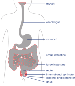
Your bowel function and symptoms may change over time. Most health providers recommend people with SCI to have a check-up once every two to three years or if there are changes to bowel function to keep track of changes to your bowel function and care.

Bowel care is an essential part of staying healthy after an SCI. For most people, a bowel routine is their main method of caring for their bowel health following SCI.

A bowel routine is a regular routine of bowel care techniques that is done every day or every other day to empty the bowel. There is a wide range of different components that may make up a bowel routine, such as hands-on emptying techniques, diet and lifestyle changes, and the use of suppositories, mini-enemas and medications.


Bowel routines are usually done at the same time each day, so the body gets used to the program. The goal of bowel routines is usually to be able to complete the routine within one hour on a regular basis to empty stool from the bowel and prevent accidents.

Every person’s bowel routine is different and often involves some trial and error to find the methods that best meet the person’s unique symptoms, physical abilities, preferences, and lifestyle. Although bowel routines can be complicated and time-consuming, they are a very important part of bowel care to allow for regular emptying and to prevent complications. Poorly managed bowel problems can lead to severe constipation, fecal impaction and other serious bowel complications.

Digital stimulation is a common spastic bowel emptying technique.11

Most people will need to use “hands-on” (manual) techniques to empty the bowel. Some people may need assistance from a care provider to use these techniques. They are usually done while sitting on a toilet or commode or while lying on your left side with the knees bent on an underpad.

Digital stimulation

People with spastic bowel typically use a technique called digital stimulationdigital rectal stimulation, or rectal touch to empty the bowel. Digital stimulation is a technique in which a gloved and lubricated finger (known in anatomy as a “digit”) is gently inserted into the anus and moved in circular motions along the walls of the rectum. This triggers a reflex that causes rectal contractions and loosening of the anal sphincters to allow emptying.

Digital stimulation is one of the most common techniques used for bowel emptying after SCI, however very little research has been done to determine if it is effective.

Watch for signs of autonomic dysreflexia

Cartoon lightning boltsPeople with injuries at T6 and above may trigger autonomic dysreflexia when performing digital stimulation and other bowel techniques. Keep an eye out for signs including sweating, headache, heart rate changes, goose bumps and increasing muscle spasms, and stop any techniques right away. Autonomic dysreflexia can also be triggered by bowel problems such as constipation or hemorrhoids.

People who experience autonomic dysreflexia during digital stimulation may use topical anesthetic medications like Lidocaine (Xylocaine) applied to their finger to numb the nerve endings during digital stimulation. There is moderate evidence that Lidocaine helps to reduce the symptoms of autonomic dysreflexia during digital stimulation.

Refer to our article on Autonomic Dysreflexia for more information! 

Manual removal of stool

People with flaccid bowel do not have bowel reflexes, so digital stimulation is not effective. Instead, the stool needs to be removed manually. This is called manual evacuation, digital disimpaction, or a rectal clear. This technique involves the use of a gloved and lubricated finger, which is inserted into the anus and used to remove stool. It may be done in conjunction with use of a lubricating suppository.

There is limited evidence that manual evacuation helps to reduce the number of bowel accidents, and it is unclear whether it helps to reduce time spent on bowel care.

Abdominal massageDigestive system with a clock for direction reference

Abdominal massage is a technique where a person massages the abdomen by stroking or kneading the lower torso in a clockwise motion to increase movement within the colon. However, research results on whether abdominal massage is effective for helping with bowel care after SCI have been conflicting. Use caution when using abdominal massage after a recent surgery or if you have an abdominal stoma.

Diet and fluid intake play a very important role in maintaining stools that can be moved easily through the bowel. Altering diet and fluid intake is often one of the first changes that are made to try to improve the consistency of stool and manage problems like constipation and diarrhea.

Fibre

The amount and type of fibre consumed can affect stool consistency.14

Fibre plays an important role in maintaining stool consistency so the stool can be moved easily through the bowel. Different types of fibre have different effects in the body. Soluble fibre, such as oatmeal and psyllium fibre, helps to trap waste and water along the digestive tract. It is important to increase water intake together with soluble fibre because it needs to absorb water to work. Soluble fibre may help improve stool consistency if stools are too loose or too hard. Insoluble fibre, such as whole grains, helps propel and move the contents through the colon more quickly.

There is weak evidence that diets high in fibre may actually slow down digestion after SCI. The optimal amount of fibre intake for people with SCI has not yet been studied and is highly variable between individuals, however, most health providers recommend moderate fibre intake to help with bowel care after SCI.

Refer to our article on Fibre to learn more about its role in your diet. 

Fluids

Water is important to bulk up the stool to allow it to move more easily. Health providers often recommend drinking at least 2 litres of healthy fluid per day, however, the optimal amount of fluids for improving bowel movement after SCI has not yet been studied.

It may take trial and error to find the right amount of fibre and fluidBalance scale with question marks on each opposing plate

The amount of fibre and fluids for optimal bowel function is different for everyone. It usually takes some trial and error to find the right amount of fibre for you. Keeping track of your fibre and fluid intake over a period of time can be helpful in figuring out how much you need to maintain your desired stool consistency. Fibre intake should be gradually adjusted to achieve the desired consistency of stool. Speak to your health providers for more information.

Foods contributing to other symptoms

Some people find that certain foods and drinks affect their bowel function in different ways. For example, some foods may cause loose stools or constipation. Every person is different, and it may take some trial and error to find the best diet for you.

Timing of food and drink

The gastrocolic reflex is when food or drink in the stomach triggers movement in the bowel. Some people eat food or drink warm fluids at least 30 minutes before their bowel routine to try to take advantage of this reflex, which is thought to be strongest in the morning. Many people use this technique as part of their bowel care; however the evidence is conflicting about whether the gastrocolic reflex is effective after SCI.

Find out how Charlie manages his fiber and fluid intake depending on the season.

Medications and suppositories may be added to your bowel routine if hands-on techniques and diet changes are not enough.

Stool softeners

Stool softeners are a type of laxative that increases the moisture in the stool to make it easier to pass. Stool softeners are usually taken once or twice per day or as needed. Some common stool softeners include Polyethylene glycol 3350 (PEG) and Docusate sodium (Colace).

Suppositories and Micro-enemas

A suppository is a solid (usually cone or cylinder-shaped) form of medication or other substance that is inserted into the rectum to absorb. Suppositories must make contact with the bowel walls to work, so care should be taken that they are not placed into a stool.

Alternatively, a micro-enema is a liquid form of a drug administered into the rectum before a planned emptying. These methods are used to allow for quick and direct action on the bowel walls.

Stimulant laxatives (including Stimulant suppositories)

Micro-enemas are administered into the rectum before a planned emptying.18

Stimulant laxatives increase the movements in the intestinal wall that help move the stool along. There are many different stimulant laxatives that may be taken by mouth or as a suppository or micro-enema. Some common stimulant laxatives include Bisacodyl (Dulcolax) suppositories and Sennosides (Senokot).

Stimulant suppositories contain medications (such as Dulcolax) which stimulate the bowel reflex. Suppositories are usually inserted 15 to 30 minutes before planned bowel emptying. However, the medication used and even the base that the medication is dissolved in can affect how quickly the medication is absorbed. For example, Dulcolax in a water-soluble polyethylene glycol base (such as a Magic Bullet) allows shorter times to emptying than Dulcolax in a vegetable oil base.

Lubricating suppositories

Lubricating suppositories contain non-medicated substances (such as glycerin) which holds water in the bowel to make the stool softer so it is easier to pass.

Prokinetic agents

Similar to stimulant laxatives, prokinetic agents are medications that increase digestive tract muscle activity to move the stool through digestion. However, prokinetic agents also speed up movement through the upper digestive tract as well as the bowel. Prokinetic agents are not routinely used and considered as a last resort for constipation or in preparation for a bowel procedure, such as a colonoscopy.

There is moderate evidence that the prokinetic agents fampridine, neostigmine, and metoclopramide help provide relief from constipation in people with SCI. There is also some evidence that prucalopride can help SCI individuals with impaired bowel emptying but may cause abdominal pain.

Standing and Exercise

Standing and exercise have long been thought to assist with bowel management after SCI. Standing and exercise are thought to help move abdominal contents toward the rectum. However, there is not enough evidence at this time to determine whether standing and exercise are effective to help with managing bowel changes after SCI.

Assistive devices

A number of devices also exist to help with bowel management. These devices are typically used together with regular bowel care techniques, and may include:

  • Commodes, raised or padded toilet seat, or automated toilet seat
  • Suppository inserter or digital stimulator
  • Mirrors
  • Anal plugs
  • Footstools
  • Bowel irrigation systems

However, limited research has been done on assistive devices for bowel care, so we do not yet know if these treatments are useful to help manage bowel changes after SCI.

Colostomy and ileostomy surgery

Most people are able to manage their bowel function effectively with a bowel routine made up of conservative techniques and medications. However, some people may require the use of additional treatments to achieve optimal bowel care.

Colostomy and ileostomy are surgical procedures that may be used if conservative techniques do not work well or as a way to streamline bowel care. Both of these procedures involve a surgically-created hole between the intestine and the outside of the abdomen. Once the opening is formed, a pouch (collection bag) is attached to allow stool to be collected outside of the body, bypassing the rectum and anus entirely.

A colostomy is when the hole (or stoma) is created between the colon (large intestine) and the outside of the body. An ileostomy is when the hole is created between the ileum (last part of the small intestine) and the outside of the body.

Image showing a person's abdomen with the lower gastrointestinal tract. The large intestine has a hole in the colon labelled as 'stoma' and a bag attached to the stoma labelled 'colostomy bag'.

A colostomy is a surgically created opening between the large intestine (colon) and an external collection device (colostomy bag).19

Although these types of surgeries are invasive, a number of different studies have shown that there are many benefits for bowel care. There is weak evidence that colostomy surgery:

  • Simplifies bowel routines and reduces the amount of time spent on bowel care
  • Reduces the need for use of laxatives and dietary changes in bowel care
  • Reduces the number of hospitalizations related to bowel problems
  • Improves independence and quality of life

These studies showed that, while there were some complications related to stoma surgeries, such as opening of the surgical wound, these were uncommon, and the majority of people were satisfied with the results of their surgery.

Bowel irrigation

Bowel irrigation is the use of water or other fluids (including medications) to stimulate the bowel muscles and flush stool from the bowel. This is done to help loosen stools and treat constipation. Bowel irrigation is sometimes referred to as an enema, although it typically uses more fluid than an enema. The fluid may be introduced into the bowel through the anus (called transanal irrigation) or through a surgically created stoma (called antegrade irrigation).

In MACE, the appendix is made into a stoma.20

Transanal irrigation is done through a catheter that is inserted into the rectum through the anus. The catheter may be surrounded by a balloon that can be inflated to hold the catheter in place and prevent leaking of water. The catheter is connected by a tube to a hand pump or electrical pump system to push water into the bowel. Pulsed water transanal irrigation involves quick pulses of water into the rectum to help loosen stools. There is weak evidence that transanal irrigation and pulsed water transanal irrigation helps improve bowel function after SCI.

When fluid is introduced through a stoma (antegrade irrigation), it is usually done through a small stoma into the beginning of the large intestine (the cecum) called a cecostomy or as part of a surgical technique called the Malone Antegrade Continence Enema (MACE) procedure, where the appendix is made into a stoma. Irrigation is not usually done through an ileostomy because this can cause dehydration. There are several studies which provide weak evidence that MACE is a safe and useful alternative for bowel irrigation after SCI.


Electrical and magnetic stimulation treatments (not in regular use)

Electrical and magnetic stimulation techniques are available to assist with bowel care. However, these treatments are not usually available in most healthcare settings.

Functional magnetic stimulation

Functional magnetic stimulation is a non-invasive procedure in which a magnet is placed over the spinal cord to administer magnetic waves which can stimulate nerve cells to fire. There is weak evidence that functional magnetic stimulation help improve bowel transit time after SCI.

Abdominal muscle stimulation

Abdominal muscle stimulation is another non-invasive procedure in which electrodes are placed on the abdominal muscles to stimulated muscle activity. It has been proposed that activating the abdominal muscles might help to increase pressure within the abdominal cavity, which may assist with bowel emptying for people who do not have abdominal control (injuries above T7).

Sacral anterior root stimulation

Sacral anterior root stimulation is an invasive procedure in which electrodes and a controlling device are implanted onto the sacral nerves during surgery. The device is controlled by an external transmitter and can stimulate the nerves which help empty the bowel.

There is moderate evidence that sacral anterior root stimulation may help improve constipation after SCI. Sacral root stimulation is an emerging area of research and further research in the future may provide more information about whether this treatment works.

Watch SCIRE’s video to learn more about bowel management as you age.21

All people age, and as they do they may notice changes in how they digest certain foods or that they need to go to the bathroom more frequently, or they may not go to the bathroom as successfully. As the body ages, nerve signals to the bowel may decrease and weaken bowel contractions and cause stool to move more slowly through the bowel. As a result, more water gets reabsorbed from the stool which often leads to more constipation. Some may experience more stool leaking.

Aging with SCI

For people aging with SCI, the changes in the bowel from typical aging may combine with the effects of neurogenic bowel dysfunction to worsen problems like constipation. Generally, as people age with SCI, symptoms of constipation increase, the frequency of bowel movements decrease, and the time required to complete a bowel movement increases. Medications and antibiotics taken to treat infections can also have significant effects on bowel function. They can increase constipation, increase leaks/accidents, or make the bowels less predictable.

Bowel changes that people aging with SCI may experience:

Other aging-related problems like pain, osteoarthritis, decreased strength and mobility, and skin issues, can affect the ability to carry out bowel routines. Bowel changes like constipation, hemorrhoids, and spending more time sitting on the toilet can trigger pain, spasticity, and autonomic dysreflexia. All these changes from aging could lead to a need to reassess bowel management strategies.

Refer to the “What bowel problems can happen after SCI?” section above for more information on bowel complications.

Managing bowel changes with age

To manage changes to bowel function from aging with SCI, consult regularly with a health care provider about your bowel management. Strategies to manage bowel changes may include:

  • Modifying your diet and fluid intake
  • Medications for bowel movements like suppositories and laxatives
  • Surgical procedures
  • Reviewing your current medications and supplements (some medications can cause constipation or diarrhea as a side effect)
  • Reviewing toilet and transfer equipment for injury to skin and joints
  • Additional caregiver help

Changing diet and water intake needs

Drinking water and the types of foods you eat both play an important part in how easily stool moves through the bowel. Needing to adjust what you eat and drink as you age is common since people often feel less appetite and thirst, and your body will not be as good at maintaining water and nutrient levels.

The optimal diet and daily water intake are different for each person, but finding the right balance can make a big difference for your bowels. You might also find that adjusting when in the day you eat and drink can affect your bowel movements.

Many people with SCI change their bowel management as they age. In one study, 63% of participants changed their bowel management methods over 20 years. People who used only medication and/or straining to manage their bowels were most likely to change their methods. Use of colostomy and bowel irrigation for management increased during this time.

For some people with long bowel routines (1-2 hours spent on bowel care per day), undergoing a colostomy was able to reduce time spent on bowel care to around 10-20 minutes per day. When considering surgical options for bowel management, people with SCI find it helpful to talk to stoma nurses, SCI doctors, and peers.

There is also research looking at the potential for nerve stimulation to improve bowel function and reduce time needed for bowel management. This may be available depending on location.

Refer to the “Diet and fluids”, “Medications and suppositories”, and “Other treatments and techniques” sections above for more information.

When do I need to review or change my bowel routine?

  1. Your current routine is not working anymore.
  2. The time and energy you spend on bowel management is limiting time spent with family/friends or doing things you love.

Questions to ask yourself to manage bowel changes

  • What has changed (issues, challenges, barriers)?
  • When did the change start?
  • What is the root cause of the change? (medical illness, injury, surgery, mobility, weakness, cognitive changes, social changes, financial changes)
  • What treatments or recommendations have been tried so far?
  • What has worked and what has not?
  • When was the most recent review of bowel with a family doctor or physiatrist?
  • Are current care team members actively involved to provide guidance and support?

Most people experience bowel changes after SCI. Even when well managed, bowel care can take up a lot of time and energy each day, which may interfere with maintaining a regular schedule. Bowel problems can also interfere with important activities like work, socializing and sexual intimacy.

Caring for bowel changes and establishing a bowel routine after SCI is one of the highest priorities for recovery after injury. Bowel care typically involves developing a conservative bowel management routine in combination with various manual techniques, diet changes, and medications. Several other procedures exist that are not as commonly used, such as cecostomy, colostomy and ileostomy surgeries and bowel irrigation.

For a review of how we assess evidence at SCIRE Community and advice on making decisions, please see SCIRE Community Evidence.

Parts of this page have been adapted from the SCIRE Professional “Bowel Dysfunction and Management” and “Autonomic Dysreflexia” Module:

Coggrave M, Mills P, Willms R, Eng JJ, (2014). Bowel Dysfunction and Management Following Spinal Cord Injury. In Eng JJ, Teasell RW, Miller WC, Wolfe DL, Townson AF, Hsieh JTC, Connolly SJ, Noonan VK, Loh E, McIntyre A, editors. Spinal Cord Injury Rehabilitation Evidence. Version 5.0. Vancouver: p 1- 48.
Available from: https://scireproject.com/evidence/bowel-dysfunction-and-management/

Krassioukov A, Blackmer J, Teasell RW, Eng JJ (2014). Autonomic Dysreflexia Following Spinal Cord Injury. In Eng JJ, Teasell RW, Miller WC, Wolfe DL, Townson AF, Hsieh JTC, Connolly SJ, Noonan VK, Loh E, McIntyre A, editors. Spinal Cord Injury Rehabilitation Evidence. Version 5.0. Vancouver: p 1- 35.
Available from: https://scireproject.com/evidence/autonomic-dysreflexia/

Mortenson WB, Sakakibara BM, Miller WC, Wilms R, Hitzig S, Eng JJ (2014). Aging Following Spinal Cord Injury. In Eng JJ, Teasell RW, Miller WC, Wolfe DL, Townson AF, Hsieh JTC, Connolly SJ, Noonan VK, Loh E, McIntyre A, editors. Spinal Cord Injury Rehabilitation Evidence. Version 5.0. Vancouver: p 1- 90.
Available from: scireproject.com/evidence/aging/

Evidence for “Hands-on bowel techniques” is based on the following studies:

[1] Sloots CE, Felt-Bersma RJ, Meuwissen SG, Kuipers EJ. Influence of gender, parity, and caloric load on gastrorectal response in healthy subjects: a barstat study. Dig Dis Sci 2003; 48: 516-521.

[2] Ford MJ, Camilleri MJ, Hanson RB, Wiste JA, Joyner MJ. Hyperventilation, central autonomic control, and colonic tone in humans. Gut 1995; 37: 499-504.

[3] Korsten M, Singal AK, Monga A, Chaparala G, Khan AM, Palmon R, Mendoza JRD, Lirio JP, Rosman AS, Spungen A, Bauman WA. Anorectal stimulation causes increased colonic motor activity in subjects with spinal cord injury. J Spinal Cord Med 2007; 30: 31-35.

[4] Furusawa K, Sugiyama H, Tokuhiro A, Takahashi M, Nakamura T, Tajima F. Topical anesthesia blunts the pressor response induced by bowel manipulation in subjects with cervical spinal cord injury. Spinal Cord 2009;47:144-148.

[5] Solomons J and Woodward S. Digital removal of faeces in the bowel management of patients with spinal cord injury: a review. British Journal of Neuroscience Nursing 2013; 9: 216-222.

[6] Menter R, Weitzenkamp D, Cooper D, Bingley J, Charlifue S, Whiteneck G. Bowel management outcomes in individuals with long-term spinal cord injuries. Spinal Cord 1997; 35: 608-612.

[7] Haas U, Geng V, Evers GC, Knecht H. Bowel management in patients with spinal cord injury — a multicentre study of the German speaking society of paraplegia (DMGP). Spinal Cord. 2005; 43: 724-30.

[8] Ayas S, Leblebici B, Sozay S, Bayramoglu M, Niron EA. The effect of abdominal massage on bowel function in patients with spinal cord injury. Am J Phys Med Rehabil 2006; 85: 951-955.

[9] Hu C, Ye M, Huang Q. Effects of manual therapy on bowel function of patients with spinal cord injury. J Phys Ther Sci 2013; 25: 687-688.

Evidence for “Diet and fluids” is based on the following studies:

[1] Cameron KJ, Nyulasi IB, Collier GR, Brown DJ. Assessment of the effect of increased dietary fibre intake on bowel function in patients with spinal cord injury. Spinal Cord 1996; 34: 277-283.

Evidence for “Medications and suppositories” is based on the following studies:

Stimulant laxatives

[1] House JG, Stiens SA. Pharmacologically initiated defecation for persons with spinal cord injury: effectiveness of three agents. Arch Phys Med Rehabil 1997; 78: 1062-1065.

Prokinetic agents

[1] Cardenas DD, Ditunno J, Graziani V, Jackson AB, Lammertse D, Potter P, Sipski M, Cohen R, Blight AR. Phase 2 trial of sustained-release fampridine in chronic spinal cord injury. Spinal Cord 2007; 45: 158-168.

[2] Korsten MA, Rosman AS, Ng A, Cavusoglu E, Spungen AM, Radulovic M, Wecht J, Bauman WA. Infusion of neostigmine-glycopyrrolate for bowel evacuation in persons with spinal cord injury. Am J Gastroenterol 2005; 100: 1560-1565.

[3] Segal JL, Milne N, Brunnemann SR, Lyons KP. Metoclopramide-induced normalization of impaired gastric emptying in spinal cord injury. Am J Gastroenterol 1987; 82: 1143-1148.

[4] Krogh K, Jensen MB, Gandrup P, Laurberg S, Nilsson J, Kerstens R, De Pauw M. Efficacy and tolerability of prucalopride in patients with constipation due to spinal cord injury. Scand J Gastroenterol 2002; 37: 431-436.

Evidence for “Other treatments and techniques” is based on the following studies:

Assistive Devices

[1] Hoenig H, Murphy T, Galbraith J, Zolkewitz M. Case study to evaluate a standing table for managing constipation. SCI Nursing 2001;18: 74-7.

[2] Uchikawa K, Takahashi H, Deguchi G, Liu M. A washing toilet seat with a CCD camera monitor to stimulate bowel movement in patients with spinal cord injury. Am J Phys Med Rehabil 2007; 86: 200-204.

Colostomy and Ileostomy surgery

[1] Frisbie JH, Tun CG, Nguyen CH. Effect of enterostomy on quality of life in spinal cord injury patients. J Am Paraplegia Soc 1986; 9: 3-5.

[2] Stone JM, Wolfe VA, Nino-Murcia M, Perkash I. Colostomy as treatment for complications of spinal cord injury. Arch Phys Med Rehabil1990b; 71: 514-518.

[3] Kelly SR, Shashidharan M, Borwell B, Tromans AM, Finnis D, Grundy DJ. The role of intestinal stoma in patients with spinal cord injury. Spinal Cord 1999; 37: 211-214.

[4] Rosito O, Nino-Murcia M, Wolfe VA, Kiratli BJ, Perkash I. The effects of colostomy on the quality of life in patients with spinal cord injury: a retrospective analysis. J Spinal Cord Med 2002; 25: 174-183.

[5] Branagan G, Tromans A, Finnis D. Effect of stoma formation on bowel care and quality of life in patients with spinal cord injury. Spinal Cord 2003; 41: 680-683.

[6] Munck J, Simoens Ch, Thill V, Smets D, Debergh N, Fievet F, Mendes da Costa P. Intestinal stoma in patients with spinal cord injury: a retrospective study of 23 patients. Hepatogastroenterology 2008; 55: 2125-2129.

[7] Coggrave MJ, Ingram RM, Gardner BP, Norton C. The impact of stoma for bowel management after spinal cord injury. Spinal Cord 2012; 50: 848-852.

Bowel irrigation

[1] Del Popolo G, Mosiello G, Pilati C, Lamartina M, Battaglino F, Buffa P, Redaelli T, Lamberti G, Menarini M, Di Benedetto P, De Gennaro M. Treatment of neurogenic bowel dysfunction using transanal irrigation: a multicenter Italian study. Spinal Cord 2008; 46: 517-522.

[2] Christensen P, Kvitzau B, Krogh K, Buntzen S, Laurberg S. Neurogenic colorectal dysfunction – use of new antegrade and retrograde wash-out methods. Spinal Cord 2000; 38: 255-261.

[3] Faaborg PM, Christensen P, Kvitsau B, Buntzen S, Laurberg S, Krogh K. Long-term outcome and safety of transanal colonic irrigation for neurogenic bowel dysfunction. Spinal Cord 2009; 47: 545-549.

[4] Kim HR, Lee BS, Lee JE, Shin HI. Application of transanal irrigation for patients with spinal cord injury in South Korea: a 6-month follow-up study. Spinal Cord 2013; 51: 389-394.

[5] Teichman JMH, Harris JM, Currie DM, Barber DB. Malone antegrade continence enema for adults with neurogenic bowel disease. Journal of Urology 1998; 160: 1278-1281.

[6] Christensen P, Kvitzau B, Krogh K, Buntzen S, Laurberg S. Neurogenic colorectal dysfunction – use of new antegrade and retrograde wash-out methods. Spinal Cord 2000; 38: 255-261.

[7] Teichman JMH, Zabihi N, Kraus SR, Harris JM, Barber DB. Long-term results for Malone antegrade continence enema for adults with neurogenic bowel disease. Urology 2003; 61: 502-506.

[8] Worsoe J, Christensen P, Krogh K, Buntzen S, Laurberg S. Long-term results of antegrade colonic enema in adult patients: assessment of functional results. Dis Colon Rectum 2008; 51: 1523-1528.

[9] Puet TA, Jackson H, Amy S. Use of pulsed irrigation evacuation in the management of the neuropathic bowel. Spinal Cord 1997; 35: 694-699.

Electrical and magnetic stimulation

[1] Korsten MA, Fajardo NR, Rosman AS, Creasey GH, Spungen AM, Bauman WA. Difficulty with evacuation after spinal cord injury: Colonic motility during sleep and effects of abdominal wall stimulation. JRRD 2004; 41: 95-99.

[2] Tsai PY. Wang CP, Chiu FY, Tsai YA, Chang YC, Chuang TY. Efficacy of functional magnetic stimulation in neurogenic bowel dysfunction after spinal cord injury. J Rehabil Med 2009; 41: 41-47.

[3] Lin VW, Kim KH, Hsiao I, Brown W. Functional magnetic stimulation facilitates gastric emptying. Arch Phys Med Rehabil 2002; 83: 806-810.

[4] Lin VW, Nino-Murcia M, Frost F, Wolfe V, Hsiao I, Perkash I. Functional magnetic stimulation of the colon in persons with spinal cord injury. Arch Phys Med Rehabil 2001; 82: 167-173.

[5] Binnie NR, Smith AN, Creasey GH, Edmond P. Constipation associated with chronic spinal cord injury: the effect of pelvic parasympathetic stimulation by the Brindley stimulator. Paraplegia 1991; 29: 463-469.

Evidence for “How does aging with SCI affect the bowel?” is based on:

Britton, E., & McLaughlin, J. T. (2013). Ageing and the gut. Proceedings of the Nutrition Society, 72(1), 173–177. https://doi.org/10.1017/S0029665112002807

Cameron, K. J., Nyulasi, I. B., Collier, G. R., & Brown, D. J. (1996). Assessment of the effect of increased dietary fibre intake on bowel function in patients with spinal cord injury. Spinal Cord, 34(5), 277–283. https://doi.org/10.1038/sc.1996.50

Charlifue, S., Jha, A., & Lammertse, D. (2010). Aging with Spinal Cord Injury. Physical Medicine and Rehabilitation Clinics of North America, 21(2), 383–402. https://doi.org/10.1016/j.pmr.2009.12.002

Coggrave, M. J., Ingram, R. M., Gardner, B. P., & Norton, C. S. (2012). The impact of stoma for bowel management after spinal cord injury. Spinal Cord, 50(11), 848–852. https://doi.org/10.1038/sc.2012.66

Fosnes, G. S., Lydersen, S., & Farup, P. G. (2011). Constipation and diarrhoea – common adverse drug reactions? A cross sectional study in the general population. BMC Clinical Pharmacology, 11(1), 2. https://doi.org/10.1186/1472-6904-11-2

Johns, J., Krogh, K., Rodriguez, G. M., Eng, J., Haller, E., Heinen, M., Laredo, R., Longo, W., Montero-Colon, W., Wilson, C. S., & Korsten, M. (2021). Management of Neurogenic Bowel Dysfunction in Adults after Spinal Cord Injury. The Journal of Spinal Cord Medicine, 44(3), 442–510. https://doi.org/10.1080/10790268.2021.1883385

Johns, J. S., Krogh, K., Ethans, K., Chi, J., Querée, M., & Eng, J. J. (2021). Pharmacological Management of Neurogenic Bowel Dysfunction after Spinal Cord Injury and Multiple Sclerosis: A Systematic Review and Clinical Implications. Journal of Clinical Medicine, 10(4), 882. https://doi.org/10.3390/jcm10040882

Nielsen, S. D., Faaborg, P. M., Christensen, P., Krogh, K., & Finnerup, N. B. (2017). Chronic abdominal pain in long-term spinal cord injury: a follow-up study. Spinal Cord, 55(3), 290–293. https://doi.org/10.1038/sc.2016.124

Nielsen, S. D., Faaborg, P. M., Finnerup, N. B., Christensen, P., & Krogh, K. (2017). Ageing with neurogenic bowel dysfunction. Spinal Cord, 55(8), 769–773. https://doi.org/10.1038/sc.2017.22

Parittotokkaporn, S., Varghese, C., O’Grady, G., Svirskis, D., Subramanian, S., & O’Carroll, S. J. (2020). Non-invasive neuromodulation for bowel, bladder and sexual restoration following spinal cord injury: A systematic review. Clinical Neurology and Neurosurgery, 194, 105822. https://doi.org/10.1016/j.clineuro.2020.105822

Rosito, O., Nino-Murcia, M., Wolfe, V. A., Kiratli, B. J., & Perkash, I. (2002). The Effects Of Colostomy On The Quality Of Life In Patients With Spinal Cord Injury: A Retrospective Analysis. The Journal of Spinal Cord Medicine, 25(3), 174–183. https://doi.org/10.1080/10790268.2002.11753619

Savic, G., Frankel, H. L., Jamous, M. A., Soni, B. M., & Charlifue, S. (2018). Long-term bladder and bowel management after spinal cord injury: a 20-year longitudinal study. Spinal Cord, 56(6), 575–581. https://doi.org/10.1038/s41393-018-0072-4

Wahlqvist, M., & Savige, G. (2000). Interventions aimed at dietary and lifestyle changes to promote healthy aging. European Journal of Clinical Nutrition, 54(S3), S148–S156. https://doi.org/10.1038/sj.ejcn.1601037

Wong, S., Santullo, P., Hirani, S. P., Kumar, N., Chowdhury, J. R., García-Forcada, A., Recio, M., Paz, F., Zobina, I., Kolli, S., Kiekens, C., Draulans, N., Roels, E., Martens-Bijlsma, J., O’Driscoll, J., Jamous, A., & Saif, M. (2017). Use of antibiotics and the prevalence of antibiotic-associated diarrhoea in patients with spinal cord injuries: an international, multi-centre study. Journal of Hospital Infection, 97(2), 146–152. https://doi.org/10.1016/j.jhin.2017.06.019

Other references

Anderson KD. Targeting recovery: Priorities of the spinal cord-injured population. Journal of Neurotrauma 2004; 21: 1371-1383.[4] Badiali D, Bracci F, Castellano V, Corazziari E, Fuoco U, Habib FI, Scivoletto G. Sequential treatment of chronic constipation in paraplegic subjects. Spinal Cord 1997; 35: 116-120.

Brading AF, Ramalingam T. Mechanisms controlling normal defecation and the potential effects of spinal cord injury. Prog Brain Res 2006; 152: 345-58.

Byrne CM, Pager CK, Rex J, Roberts R, Solomon MJ (1998): Assessment of Quality of Life in the treatment of patients with neuropathic fecal incontinence. Dis Colon Rectum 2002; 45: 1431-6.

Coggrave M, Burrows D, Durand MA. Progressive protocol in the bowel management of spinal cord injuries. British Journal of Nursing 2006; 15: 1108-1113.

Coggrave M, Norton C, Cody JD. Management of fecal incontinence and constipation in adults with central neurological diseases. Cochrane Database Syst Rev. 2014;1:CD002115.

Coggrave MJ, Norton C, Wilson-Barnett J. Management of neurogenic bowel dysfunction in the community after spinal cord injury: a postal survey in the United Kingdom. Spinal Cord 2009; 47: 323-330.

Coggrave MJ, Norton C. The need for manual evacuation and oral laxatives in the management of neurogenic bowel dysfunction after spinal cord injury: a randomized controlled trial of a stepwise protocol. Spinal Cord. 2010; 48: 504-10.

Correa GI, Rotter KP. Clinical evaluation and management of neurogenic bowel after spinal cord injury. Spinal Cord2000; 38: 301-308.

Cosman BC, Vu TT. Lidocaine anal block limits autonomic dysreflexia during anorectal procedures in spinal cord injury: a randomized, double-blind, placebo-controlled trial. Dis Colon Rectum. 2005;48:1556-61.

Emmanuel A. Review of the efficacy and safety of transanal irrigation for neurogenic bowel dysfunction. Spinal Cord. 2010;48:664-73.

Faaborg PM, Christensen P, Finnerup N, Laurberg S, Krogh K. The pattern of colorectal dysfunction changes with time since spinal cord injury. Spinal Cord 2008; 46: 234-238.

Fajardo NR, Pasiliao RV, Modeste-Duncan R, Creasey G, Bauman WA, Korsten MA. Decreased colonic motility in persons with chronic spinal cord injury. Am J Gastroenterol 2003; 98: 128-34.

Fealey RD, Szurszewski JH, Merrit JL, DiMagno EP. Effect of traumatic spinal cord transection on human upper gastrointestinal motility and gastric emptying. Gastroenterology 1984; 87: 69-75.

Finnerup NB, Faaborg P, Krogh K, Jensen TS. Abdominal pain in long-term spinal cord injury. Spinal Cord 2008; 46: 198-203.

Furusawa K, Sugiyama H, Ikeda A, Tokuhiro A, Koyoshi, H, Takahashi M, Tajima F. Autonomic dysreflexia during a bowel program in patients with cervical spinal cord injury. Acta Med Okayama 2007; 61: 211-227.

Glickman S, Kamm MA. Bowel dysfunction in spinal-cord-injury patients. Lancet 1996; 347: 1651-3.

Gondim FA, Rodrigues CL, da Graça JR, Camurça FD, de Alencar HM, dos Santos AA, Rola FH. Neural mechanisms involved in the delay of gastric emptying and gastrointestinal transit of liquid after thoracic spinal cord transection in awake rats. Auton Neurosci. 2001; 87: 52-8.

Krogh K, Mosdal C, Laurberg S. Gastrointestinal and segmental colonic transit times in patients with acute and chronic spinal cord lesions. Spinal Cord 2000; 38: 615-621.

Leduc BE, Spacek E, Lepage Y. Colonic transit time after spinal cord injury: any clinical significance? J Spinal Cord Med 2002; 25: 161-6.

Lynch AC, Antony A, Dobbs BR, Frizelle FA. Bowel dysfunction following spinal cord injury. Spinal Cord 2001; 39: 193-203.

Lynch AC, Frizelle FA. Colorectal motility and defecation after spinal cord injury in humans. Prog Brain Res 2006; 152: 335-43.

Lynch AC, Wong C, Anthony A, Dobbs BR, Frizelle FA. Bowel dysfunction following spinal cord injury: a description of bowel function in a spinal cord-injured population and comparison with age and gender matched controls. Spinal Cord 2000; 38: 717-723.

Menter R, Weitzenkamp D, Cooper D, Bingley J, Charlifue S, Whiteneck G. Bowel management outcomes in individuals with long-term spinal cord injuries. Spinal Cord 1997; 35: 608-612.

Ng C, Prott G, Rutkowski S, et al. Gastrointestinal symptoms in spinal cord injury: relationships with level of injury and psychologic factors. Dis Colon Rect 2005;48:1562-1568.

Nino-Murcia M, Stone JM, Chang PJ, Perkash I. Colonic transit in spinal cord-injured patients. Invest Radiol 1990; 25: 109-112.

Rajendran SK, Reiser JR, Bauman W, Zhang RL, Gordon SK, Korsten MA. Gastrointestinal transit after spinal cord injury: effect of cisapride. Am J Gastroenterol 1992; 87: 1614-1617.

Stiens SA, Bergman SB, Goetz LL. Neurogenic bowel dysfunction after spinal cord injury: clinical evaluation and rehabilitative management. Arch Phys Med Rehabil 1997;78:S86-S102.

Stone JM, Nino-Marcia M, Wolfe VA, Perkash I. Chronic gastrointestinal problems in spinal cord injury patients: a prospective analysis. Am J Gastroenterol 1990a; 85: 1114-9.

Image credits

  1. The Bowel ©SCIRE, CC BY-SA 3.0
  2. Belly abdominal pain ©Christian Dorn, CC0 1.0
  3. Man toilet bathroom sitting ©Clker-Free-Vector-Images, CC0 1.0
  4. Leak plumbing water drips bathroom ©Clker-Free-Vector-Images, CC0 1.0
  5. Mistake spill slip up accident ©Steve Buissinne, CC0 1.0
  6. Water jump refreshment children ©AxxLC, CC0 1.0
  7. Image ©SCIRE, CC BY-SA 3.0
  8. Bristol Stool Chart ©Cabot Health, CC BY-SA 3.0
  9. Scientist ©H Alberto Gongora, CC BY 3.0 US
  10. Towel napkin dab dry vector ©Isa KARAKUS, CC0 1.0
  11. Modified from: Hand finger pointing ©truthseeker08, CC0 1.0
  12. Electricity ©Artnadhifa, CC BY 3.0 US
  13. Abdomen clockwise clock massage ©bodymybody, CC0 1.0
  14. Breads cereals oats barley wheat ©FotoshopTofs, CC0 1.0
  15. Scale question importance balance ©Arek Socha, CC0 1.0
  16. Alarm clock time of good morning ©congerdesign, CC0 1.0
  17. Suppositories pharmacy ©Andreas Koczwara, CC0 1.0
  18. Microlax Miniklistier ©MedInfo Johnson&Johnson, CC BY-SA 4.0
  19. Cancer Research UK (Original email from CRUK), CC BY-SA 4.0
  20. Reprinted with permission from Malone PSJ. Malone procedure for antegrade continence enemas. In: Spitz L, and Coran AG, eds. Rob & Smith’s Operative Surgery: Pediatric Surgery. 5th ed. London: Chapman & Hall Medical; 1995:459–467.
  21. Aging Bowel Thumbnail ©SCIRE, CC BY-NC 4.0
  22. Clock by Abu Ibrahim Icon, Noun Project
  23. Toilet by Isaac Haq, Noun Project


Disclaimer: This document does not provide medical advice. This information is provided for educational purposes only. Consult a qualified health professional for further information or specific medical advice. The SCIRE Project, its partners and collaborators disclaim any liability to any party for any loss or damage by errors or omissions in this publication. 

Bladder Changes After Spinal Cord Injury

By | | No Comments

Authors: SCIRE Community Team | Reviewer: Bonnie Nybo | Published: 18 January 2018 | Updated: 16 October 2024

Key Points

  • Most people with SCI experience some bladder changes after injury, but the type and symptoms depend on the characteristics of the injury.
  • There are two main types of bladder problems after SCI:
    • Spastic (reflex) bladder involves unpredictable emptying caused by overactive bladder muscles. It happens with injuries above T12.
    • Flaccid (non-reflex) bladder involves an inability to empty the bladder because of “floppy” and underactive bladder muscles. It happens with injuries below T12.
  • People with SCI are also at risk of complications like urinary tract infections, autonomic dysreflexia (if above T6), kidney and bladder stones, and kidney damage.
  • Bladder care after SCI involves developing a regular bladder routine that meets your unique bladder needs. This may include a variety of treatments, such as catheters, medications and injections.

The urinary system

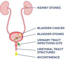
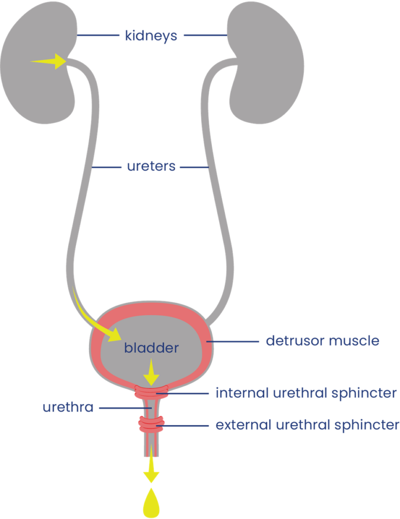
The urinary system helps the body filter and remove waste products and excess fluids. It consists of the kidneys, ureters, bladder, urethra, and bladder and sphincter muscles.

The kidneys filter the blood to produce urine, which is passed through narrow tubes called ureters to the bladder. The bladder is a sac that collects urine. Urine passes out of the body from the bladder through a tube called the urethra.

Filling and emptying of the bladder are partly controlled by the bladder muscles:

  • The bladder wall muscle (detrusor muscle) is smooth muscle that covers the outside of the bladder. When it contracts, it squeezes the bladder and pushes urine out through the urethra. When it is relaxed, the bladder is loose and can be filled with urine.
  • The bladder sphincter muscles (urethral sphincter or valve muscles) are two muscles which surround the exit of the bladder like a ring. When they tighten, they close off the urethra and hold urine in the bladder. When they relax, they allow urine to drain. The internal sphincter muscle is controlled unconsciously and the external sphincter muscle is controlled consciously.

Bladder function

When the bladder is not full, the bladder wall muscle is relaxed and urine produced by the kidneys passes through the ureters to fill the bladder. The bladder sphincter muscles are tightened so urine does not leak out.

When there is enough urine to stretch the bladder walls, a nerve signal is sent up the spinal cord to tell the brain that the bladder is full. Because the brain controls the external sphincter muscle, urine can be held until an appropriate time to empty.

When the bladder is to be emptied, signals are sent from the brain down the spinal cord to cause the coordinated squeezing of the bladder wall muscle and relaxation of the bladder sphincter muscles to allow urine to pass through the urethra and out of the body. Control of urination involves both bladder reflexes (in which emptying is triggered when the bladder is full) and voluntary control (in which urine can be held until a socially appropriate time to empty).

Neurogenic Bladder

Neurogenic bladder is bladder dysfunction caused by damage to the nerves, brain or spinal cord. After a spinal cord injury, nerve signals that normally allow the brain and bladder to communicate with one another cannot get through. This can affect bladder sensation and control.

Loss of bladder control

Signals from the brain are needed for the bladder muscles to contract and relax properly. If these signals cannot get through, the bladder muscles may contract too much, too little, or at the wrong times, depending on whether the person has spastic or flaccid bladder.

Reduced bladder sensation

Normally, signals that are sent up the spinal cord to the brain when the bladder is full. When the signals are interrupted, the ability to feel fullness and other sensations from the bladder may be reduced.

Bladder changes after SCI are different for everyone. Some people experience only mild changes to how the bladder works (such as greater sense of urgency when the bladder is full); while others experience complete loss of bladder sensation and control.

The symptoms of neurogenic bladder depend on the characteristics of the SCI, such as the level and completeness of the injury. There are two main types of neurogenic bladder after SCI, spastic bladder and flaccid bladder (see below).

Spastic Bladder

Spastic bladder (also called “reflex bladder” or “overactive bladder”) happens when the spinal cord is injured above T12. Spastic bladder happens because the brain can no longer control reflexes in the bladder muscles. This leads to tension in the bladder wall muscle when it is supposed to be relaxed and spasms of the bladder muscles which cause emptying.

Usually, the bladder sphincter muscles are also overactive and cannot coordinate very well with the bladder wall muscle. This is called detrusor dyssynergia or detrusor sphincter dyssynergia (DSD). When this happens, the bladder sphincter muscle tightens while the bladder wall muscle contracts, like squeezing a balloon that is tied off. This can cause high pressures within the bladder that can damage the bladder and kidneys.

Symptoms of spastic bladder:

  • Loss of control of bladder emptying (incontinence), leading to random emptying (accidents), inability to empty when you want to and leaking
  • Reflex emptying in response to things like touching the thigh or abdomen
  • People with some bladder sensation may experience sudden strong urges or a frequent need to urinate
  • Incomplete emptying of the bladder caused by poor coordination of the bladder wall muscle and bladder sphincter muscles (detrusor dyssynergia)
  • Reduced or complete loss of bladder sensation

Flaccid bladder

Flaccid bladder (also called “non-reflex bladder” or “underactive bladder”) happens when the spinal cord is injured below T12-L1 (i.e cauda equina injuries). Flaccid bladder happens because there is a loss of both input from the brain and reflexes from the spinal cord. This causes the bladder wall muscle to stay loose and floppy all the time. When this happens, the bladder wall muscle cannot squeeze the bladder to empty urine.

Usually, the external sphincter muscle is also overly relaxed, causing leaking during activities like transfers and coughing. However, the internal sphincter muscle is often in spasm and does not relax enough to allow urine to pass out of the body easily.

Symptoms of flaccid bladder:

  • Inability to empty the bladder, including loss of reflex emptying
  • Incomplete bladder emptying, leading to some urine remaining in the bladder after emptying (urinary retention)
  • Damage to the walls of the bladder when they are overstretched
  • Backflow of urine back to the kidneys (reflux), which can damage the kidneys
  • Reduced or complete loss of bladder sensation

Bladder examination

Bladder changes are diagnosed primarily through a bladder examination. A bladder examination typically involves several components:

  • Your health provider will ask you questions about your medical history, symptoms, bladder routine, and current treatments.
  • You may be asked to complete a “urinary diary” and/or detailed questionnaires about your bladder care. This often involves recording how often you empty your bladder, how much urine is produced each time, and details about your fluid intake (what you drink, when and how much).
  • A physical examination may involve an inspection of the abdominal, pelvic and genital areas, as well as neurological testing of your reflexes, muscle strength, and sensation.

Other testing

Other testing may also be done if your health providers need further information.

Picture of dipstick test strip beside black storage bottle with label for interpreting results

The dipstick or urine test strip is a basic diagnostic tool for identifying presence of substances or infection in urine.3

Urine culture

A urine culture and sensitivity test involves collecting urine in a sterile container to test for infection. Urine samples are usually collected mid-stream while emptying so the test is more accurate. If the sample is collected from an indwelling catheter, the catheter should be changed first. Samples are never taken from a urine drainage bag.

Blood tests

Blood tests may be done if there is concern about kidney function, kidney damage, or an infection. This usually involves testing for blood urea nitrogen (BUN) and creatinine.

Ultrasound

Ultrasound is an imaging technique that uses sound waves to visualize deep tissues. Ultrasound imaging may be done over the kidneys (known as renal ultrasound) to detect damage, kidney stones and infections.

Urodynamic testing

Urodynamic testing includes special tests that can be used to look at bladder pressures and urine flow. It can test how the bladder acts when it fills and empties, how well it coordinates, and the pressure within the bladder. This test may involve urinating into a special container that can measure the flow and volume of urine, insertion of a catheter to measure the leftover urine, and inserting water into the bladder to measure your ability to prevent emptying. It may also involve placing a small catheter into the rectum that measures the electrical activity of muscles.

Typical urodynamic measures

Bladder Capacity: The amount of urine the bladder can hold.

Voiding Efficiency: The amount of urine voided compared to the amount in the bladder before voiding. More efficiency means less urine is left in the bladder.

Bladder Compliance: The ability of the bladder to stretch in response to an increased amount of urine in the bladder. Without enough stretch there will be large increases in pressure, which is damaging to the urinary tract.

Imaging

Other imaging, such as x-ray, computed tomography (CT), and magnetic resonance imaging (MRI) are sometimes used for further investigation of bladder problems.

Cystoscopy

Cystoscopy (sometimes known as a “bladder scope”) is the use of a very small camera that can be inserted into the urethra to look at the urinary tract. Cystoscopy can be used to identify bladder stones, bladder health issues or damage including bladder cancer. It can also perform therapeutic procedures if needed such as removing tissue or stones.

Early bladder care

In the early hospital phase right after injury, the circulatory system is stabilizing, and the prevention of infections and other complications is the priority. During this phase, an indwelling catheter is placed in the bladder to constantly drain urine from the bladder. The catheter will be changed regularly and maintained in a sterile way by your nurse.

After the acute phase, bladder care will involve transitioning to more long-term bladder care techniques and developing a suitable bladder routine.

Bladder Routine

A bladder routine is a regular routine of bladder techniques and treatments that are done every day to maintain bladder function and health. This usually involves techniques to regularly empty the bladder, prevent leaks, and avoid serious complications long-term.

Every person’s routine is different and often involves trial and error to find the methods that best meet your unique symptoms, abilities, preferences, and lifestyle. There is a wide range of different techniques and treatments that may make up your routine, including catheters, medications, and methods of stimulation like electrical stimulation. Keep in mind that spastic bladder and flaccid bladder happen for different reasons and are managed differently.

Other things to consider when developing a bladder routine:

  • Timing and amount of fluids
  • Caffeine and alcohol consumption
  • Scheduling of bladder emptying (such as how long between catheterizations, before going to bed or certain activities, after drinking fluids)
  • What type of equipment to use, such as type of catheter and collection bag for different situations
  • What to do if you have a bladder infection or other new health problem
  • Regular assessment of bladder care with your health team

Spastic bladder management

The goals of spastic bladder management are to reduce overactivity in the bladder wall muscle which causes accidents, leaking, and wetness; as well as preventing high pressures within the bladder. This may include treatments such as:

Flaccid bladder management

The goals of flaccid bladder management are to regularly empty the bladder to prevent overfilling and increased pressure in the bladder; and to prevent leaking and wetness. This may include treatments such as:

Urinary catheters are pieces of equipment that are used to drain urine from the bladder. There are many different ways that catheters are used.

Intermittent catheterization

Intermittent catheterization is when a catheter is inserted and removed through the urethra to drain the bladder at regular intervals throughout the day. Bladder emptying with intermittent catheterization must be done hygienically and on a regular schedule.

Intermittent catheterization is usually used by people who have enough hand function to perform the procedure independently. It is the closest method to normal bladder function, where the bladder fills continuously for a period of time and then empties all at once.

A diagram of a yellow indwelling (Foley) catheter labelling the drainage port, size, balloon port, and bladder opening

A diagram of an indwelling (Foley) catheter.4

Indwelling catheters

Indwelling catheters (such as Foley catheters) are catheters that are inserted directly into the bladder and remain in place to continually drain the bladder. Indwelling catheters may be inserted through the urethra (urethral catheters) or through a surgically created hole through the abdomen (suprapubic catheters).

An upright condom catheter

An example of a condom catheter, for male use.5

Indwelling catheters are usually used if inserting your own catheter independently is difficult or there are concerns about leaking between sessions of emptying.

Condom catheters (only for males)

Condom catheters are catheters that resemble a condom and are placed over the penis and connected through tubes to a collection device. Condom catheters are usually used by people that leak in between emptying or for individuals who have the ability to trigger emptying by causing a spasm of their bladder (reflex voiding).

One of the main concerns of condom catheters is incomplete bladder drainage, which can cause kidney damage. A careful medical examination is needed to ensure that condom catheters are a safe option for use.

Refer to our article on Urinary Catheters for more information!

Catching leaks

Some people may use medical “penis pouches” (loosely fitted bags that can be placed around the penis), pads, or other devices to catch small leaks in between catheterizations. These will depend on the person and their risk of other problems like pressure injuries, and should be discussed in detail with your health providers before use.

Reflex voiding is a technique that can be used by some people with spastic bladder to stimulate urination. Reflex voiding is usually done by tapping over the bladder lightly and repeatedly with the fingertips or the side of the hand to stimulate reflexes in the bladder muscles. This technique can be used to help improve bladder emptying during intermittent catheterization and when using condom catheters. However, only a small number of people can use this technique safely without increasing the pressure too high in the bladder. Speak to your health team for more information about this technique.

Many reflex voiding techniques are not recommended

Older techniques for reflex voiding such as the Valsalva maneuver (increasing abdominal pressure by holding the breath and bracing) and the Credé technique (applying manual pressure onto the bladder through the abdomen) are no longer recommended because they can cause too much pressure in the bladder, which can damage the kidneys.

Several medications may be used to help manage bladder problems after SCI. These may help to relax overactive muscles or cause the bladder muscles to contract, depending on the type of bladder change experienced. A number of other medications may also be used for different aspects of bladder treatment after SCI.

Inserting liquid medications into the bladder

Some medications may be dissolved in a liquid solution and introduced into the bladder through a catheter after emptying. The solution is then left in the bladder until the next urination. This is called an intravesical instillation. Intravesical instillations may be used because their effects are more specific to the bladder, instead of throughout the whole body as with oral medications.

Anticholinergic medications

A pill box labelled with all the days of the week with pills in it.Anticholinergic medications (sometimes called antimuscarinic medications) are used to relax muscle spasms in the bladder wall muscle. This can help to reduce pressure within the bladder, increase the ability of the bladder to hold urine, and help reduce incontinence.

There are many different types of anticholinergic medications, with the most common being:

  • oxybutynin (Ditropan, Ditropal XL, Oxytrol, Uromax)
  • tolterodine (Detrol)
  • fesoterodine (Toviaz)
  • trospium chloride (TCL, Trosec)
  • propiverine hydrochloride (Mictonorm)
  • darifenacin (Enablex)
  • solifenacin (Vesicare)

These can be taken by mouth or administered directly into the bladder in a liquid form.

Alpha-adrenergic blockers

Alpha-adrenergic blockers are medications that are used to encourage the bladder sphincter muscles to relax to allow urine to flow out of the body. This can help with bladder emptying and help prevent urinary retention. Common alpha-adrenergic blockers that may be used include tamulosin, mosixylyte, terazosin, and phenoxybenzamine.

Botulinum toxin injections

A fluid filled syringe with pills surrounding it.Injecting small doses of some strains of botulinum toxin (Botox) into muscles can help to reduce muscle spasms. Injections into the bladder wall muscle or the external sphincter muscle can help to relax these muscles to help prevent leaking and incontinence or to improve bladder emptying. The effects of these injections can last for 6 to 12 months.

Read our article on Botulinum Toxin for more information!

Other medications

  • Capsaicin is commonly found in hot peppers.7

    Capsaicin, a chemical commonly found in hot peppers, and its derivative resiniferatoxin, may be administered as a liquid into the bladder to help reduce urinary frequency, leaking, and bladder pressures related to bladder wall muscle overactivity, and increase bladder capacity.

  • Nociceptin/orphanin phenylalanine glutamine is another medication with effects similar to capsaicin and resiniferatoxin. It may also be given into the bladder to reduce overactivity in the bladder wall muscle.
  • Medications that are normally used to treat spasticity may also help with bladder problems related to spastic bladder. For example, baclofen and clonidine may help with bladder function after SCI.
  • Phosphodiesterase-5 (PDE5) inhibitors such as tadalafil and vardenafil may help to reduce overactivity in the bladder wall muscle and increase bladder capacity.
  • 4-Aminopyridine (fampridine) improves the transfer of nerve signals, which may help individuals regain sensation and control of the bladder sphincter muscles to improve emptying.

Bladder surgery is usually only considered if other less-invasive treatments are not effective. Surgical procedures that may be used include the Mitrofanoff procedure, bladder augmentation, sphincterotomy (for males), and urethral stents.

Mitrofanoff procedure

The Mitrofanoff procedure involves the use of the appendix or part of the intestine to create a channel between the abdomen and bladder. The channel self-seals shut when the catheter is removed. This channel can be used for insertion of a catheter for intermittent catheterization. The urine can then be drained into a cup or toilet. This may be useful for people who have difficulty self-catheterizing directly into the urethra and is often used for women (who have greater difficulty inserting catheters).

A cartoon diagram of the surgical Mitrofanoff procedure, taking the appendix and connecting it to the bladder.

The Mitrofanoff procedure involves connecting parts of the intestine to the bladder.9

Bladder augmentation

Bladder augmentation (also called augmentation cystoplasty) is a procedure in which the bladder is made bigger to create more room to hold urine. This is done by removing a segment of the intestine and stitching this tissue to an incision into the bladder to make the bladder bigger. Bladder augmentation may help to reduce pressure in the bladder and help to prevent incontinence related to spastic bladder.

Bladder augmentation- A simplified step by step diagram of how parts of the intestine are taken to enlarge the bladder

Bladder augmentation is a surgical procedure done to enlarge the bladder by using parts of the intestine.10

Sphincterotomy (for males)

Sphincterotomy is a surgical procedure where the internal sphincter muscle (the circular muscle that surrounds the outlet of the bladder) is cut to weaken the muscle. This is done to improve bladder emptying if this muscle is causing difficulties emptying. After a sphincterotomy, bladder emptying will happen; therefore, you must wear a collection device.

Urethral stents

Urethral stents are prosthetic tubes (usually coils of metal) with openings on both sides that are be inserted into the opening of the bladder to hold it open. This is done to allow for improved bladder emptying for people with difficulty emptying due to overactivity in the bladder sphincter muscles.

Electrical stimulation

Electrical stimulation can be used to help normalize the activity of the bladder muscles and control of bladder emptying. This may involve the implantation of a stimulator and electrodes that stimulate the sacral nerves that send brain signals to the bladder. This is sometimes referred to as neuromodulation.

Commercially available electrical bladder stimulation systems may be used for this purpose. However, these systems may be expensive and is not available in all locations.

Refer to our articles on Neuromodulation for more information!

Acupuncture

Acupuncture and electroacupuncture have also been suggested as treatment options to help with bladder function by influencing nerve signals related to bladder function.

Refer to our article on Acupuncture for more information!

Watch SCIRE’s video on bladder care to understand more about managing your bladder as you age.11

Typically, people will experience changes in urination as they age. Some may need to go to the bathroom more often because the bladder can’t store as much urine, or find that the flow of urine is weaker. Others may experience more urine leaking because the muscles that keep the bladder closed are weaker or the bladder contracts when it is not supposed to. The kidneys that filter your blood and produce urine may not function as well, and the risk of developing kidney stones increases.

Women, both during and post-menopause may experience an increase in the frequency of UTIs.

The aging bladder in SCI

For people aging with SCI, the long-term use of indwelling catheters to manage the bladder increases risk for bladder stones, bladder cancer, UTIs, and urinary tract deterioration. Long-term use of intermittent catheterization can increase risk for urethral strictures. People with neurogenic bladder can experience high pressures in the urinary tract and urine back up to the kidneys. Over time, this damages the structures of the urinary system and increases the risk for stones.

Bladder changes that people aging with SCI may experience:

  • Increased UTIs
  • Increased urine leaking (incontinence)
  • Urethral strictures
  • Bladder/kidney stones
  • Increased risk of bladder cancer
  • Damage to urinary tracts and kidneys
  • Reduced kidney function

Bladder changes may also trigger spasticity and autonomic dysreflexia. Other aspects of aging like pain, osteoarthritis, decreased strength and mobility, and thinning of the skin, may also affect the ability to carry out bladder routines.

All of the changes from aging listed above could lead to a need to reassess bladder management strategies.

Refer to the “What other complications are related to bladder changes?” section for more information on specific bladder problems.

Managing bladder changes with age

If you experience changes to your bladder function as you age with SCI, consult regularly with a health care provider about your bladder management. Your family doctor, physiatrist, or urologist may be familiar with the check-ins necessary for bladder care as you age with a SCI.

Strategies to consider for the management of bladder changes may include:

  • Changing the catheterization method to reduce UTIs, avoid urinary tract damage, or accommodate reduced hand and wrist function
  • Screenings for bladder/kidney stones and urinary tract/kidney damage
  • Regular screening for bladder cancer if indwelling catheter has been used for 5-10 years
  • Consulting with professionals to determine the root cause of recurrent UTIs
  • Antibiotic medication to treat UTIs (only use if experiencing symptoms to avoid developing antibiotic resistance)
  • Supplements to prevent UTIs (i.e. D-mannose)
  • Botulinum toxin (Botox) injections to relax bladder muscles
  • Surgical procedures to make the bladder bigger, improve emptying, or make catheterization easier
  • Additional caregiver help
  • Quit or avoid smoking (smoking can up to quadruple risk for bladder cancer)

Many people with SCI change their bladder management as they age. In one study, half of participants changed their bladder management methods over 20 years. Over time, men who used condom drainage and women who used straining to manage their bladder were most likely to change their management methods. The use of indwelling catheters increased for men. A change to intermittent catheters or suprapubic indwelling catheters for bladder management increased for both men and women.

There is research looking at the potential for epidural and transcutaneous nerve stimulation to improve bladder function and management. This may be available depending on your location.

When do I need to review or change my bladder routine?

  1. Your current routine is not working anymore.
  2. The time and energy you spend on bladder management is limiting time spent with family/friends or doing things you love.

Questions to ask yourself to manage bladder changes

  • What has changed (issues, challenges, barriers)?
  • When did the change start?
  • What is the root cause of the change? (medical illness, injury, surgery, mobility, weakness, cognitive changes, social changes, financial changes)
  • What treatments or recommendations have been tried so far?
  • What has worked and what has not?
  • When was the most recent review of bladder with a family doctor or specialist such as a urologist or physiatrist?

Refer to the “Medications and injections”, “Bladder surgery and stents”, and “What other complications are related to bladder changes?” sections above for more information.) 

Bladder changes are common after SCI. Bladder care is an important part of self-management after SCI to prevent complications and maintain good health and quality of life.

Bladder care after SCI involves developing a regular bladder routine that meets your unique bladder needs. This may include a variety of techniques and treatments, such as catheters, medications, injections and other treatments. Speak to your health team about which bladder management options are best for you. Regular follow up with your doctor is recommended yearly.

For a review of how we assess evidence at SCIRE Community and advice on making decisions, please see SCIRE Community Evidence.

This page has been adapted from the SCIRE Professional “Bladder Management” Module:

Hsieh J, McIntyre A, Iruthayarajah J, Loh E, Ethans K, Mehta S, Wolfe D, Teasell R. (2014). Bladder Management Following Spinal Cord Injury. In Eng JJ, Teasell RW, Miller WC, Wolfe DL, Townson AF, Hsieh JTC, Connolly SJ, Noonan VK, Loh E, McIntyre A, editors. Spinal Cord Injury Rehabilitation Evidence. Version 5.0: p 1-196.
Available from: scireproject.com/evidence/bladder-management/

Biering-Sorensen F, Bagi P, Hoiby N. Urinary tract infections in patients with spinal cord lesions: Treatment and prevention. Drugs 2001;61(9):1275-1287.

Biering-Sorensen F. Urinary tract infection in individuals with spinal cord lesion. Curr Opin Urol 2002;12(1):45-49.

Chancellor MB, Bennett C, Simoneau AR, Finocchiaro MV, Kline C, Bennett JK et al. Sphincteric stent versus external sphincterotomy in spinal cord injured men: Prospective randomized multicenter trial. J Urol 1999;161(6):1893-1898.

Chancellor MB, Karasick S, Strup S, Abdill CK, Hirsch IH, Staas WE. Transurethral balloon dilation of the external urinary sphincter: Effectiveness in spinal cord-injured men with detrusor-external urethral sphincter dyssynergia. Radiology 1993b;187(2):557-560.

Chancellor MB, Karusick S, Erhard MJ, Abdill CK, Liu JB, Goldberg BB, Staas WE. Placement of a wire mesh prosthesis in the external urinary sphincter of men with spinal cord injuries. Radiology 1993c;187(2):551-555.

Chartier-Kastler E, Amarenco G, Lindbo L, Soljanik I, Andersen HL, Bagi P, Gjodsbol K, Domurath B. A prospective, randomized, crossover, multicenter study comparing quality of life using compact versus standard catheter for intermittent self-catheterization. J Urol 2013;190:942-947.

Cheng P-T, Wong M-K, Chang P-L. A therapeutic trial of acupuncture in neurogenic bladder of spinal cord injured patients-A preliminary report. Spinal Cord 1998;36(7):476-480.

Creasey GH, Grill JH, Korsten M, HS U, Betz R, Anderson R et al. An implantable neuroprosthesis for restoring bladder and bowel control to patients with spinal cord injuries: A multicenter trial. Arch Phys Med Rehabil 2001;82(11):1512-1519.

Das A, Chancellor MB, Watanabe T, Sedor J, Rivas DA. Intravesical capsaicin in neurologic impaired patients with detrusor hyperreflexia. J Spinal Cord Med 1996;19(3):190-193.

DeSeze M, Wiart L, de Seze MP, Soyeur L, Dosque JP, Blajezewski S et al. Intravesical capsaicin versus resiniferatoxin for the treatment of detrusor hyperreflexia in spinal cord injured patients: A double-blind, randomized, controlled study. J Urol 2004;171(1):251-255.

DeSeze M, Wiart L, Joseph PA, Dosque JP, Mazaux JM, Barat M. Capsaicin and neurogenic detrusor hyperreflexia: A double-blind placebo-controlled study in 20 patients with spinal cord lesions. Neurourol Urodyn 1998;17(5):513-523.

DeVivo, M.J. Sir Ludwig Guttman Lecture: Trends in SCI rehabilitation outcomes from model systems in the United States:1973-2006. Spinal Cord 2007;45(11):713-721.

Evans RJ. Intravesical therapy for overactive bladder. Current Urology Reports 2005;6:429-433.

Farag FF, Martens FM, Rijkhoff NJ, Heesakkers JP. Dorsal genital nerve stimulation in patients with detrusor overactivity: A systematic review. Curr Urol Rep 2012;12(5):385-388.

Foxman B. Epidemiology of urinary tract infections: Incidence, morbidity, and economic costs. Dis Mon;49(2):53-70.

Gobeaux N, Yates DR, Denys P, Even‐Schneider A, Richard F, Chartier‐Kastler E. Supratrigonal cystectomy with hautmann pouch as treatment for neurogenic bladder in spinal cord injury patients: Long‐term functional results. Neurourology and urodynamics 2012;31(5):672-676.

Goldman HB, Amundsen CL, Mangel J, Grill J, Bennet M, Gustafson KJ, Grill WM. Dorsal genital nerve stimulation for the treatment of overactive bladder symptoms. Neurourol Urodyn 2008;27(6):499-503.

Greenstein A, Rucker KS, Katz PG. Voiding by increased abdominal pressure in male spinal cord injury patients–long term follow up. Paraplegia 1992;30(4):253-255.

Grijalva I, Garcia-Perez A, Diaz J, Aguilar S, Mino D, Santiago-Rodriguez E, et al. High doses of 4-aminopyridine improve functionality in chronic complete spinal cord injury patients with MRI evidence of cord continuity. Arch Med Res 2010;41:567-575.

Groah SL, Weitzenkamp DA, Lammertse DP, Whiteneck GG, Lezotte DC, Hamman RF. Excess risk of bladder cancer in spinal cord injury: evidence for an association between indwelling catheter use and bladder cancer. Arch Phys Med Rehabil 2002;83(3):346-351.

Groah SL, Weitzenkamp DA, Lammertse DP, Whiteneck GG, Lezotte DC, Hamman RF. Excess risk of bladder cancer in spinal cord injury: evidence for an association between indwelling catheter use and bladder cancer. Arch Phys Med Rehabil 2002;83(3):346-351.

Gurung PM, Attar KH, Abdul-Rahman A, Morris T, Hamid R, Shah PJ. Long-term outcomes of augmentation ileocystoplasty inpatients with spinal cord injury: A minimum of 10 years of follow-up. BJU Int 2012;109(8):1236-1242.

Hackler RH. Long-term Suprapubic cystostomy drainage in spinal cord injury patients. Br J Urol 1982;54(2):120-121.

Hakenberg OW, Ebermayer J, Manseck A, Wirth MP. Application of the Mitrofanoff principle for intermittent self-catheterization in quadriplegic patients. Urology 2001;58(1):38-42.

Hansen J, Media S, Nohr M, Biering-Sorensen F, Sinkjaer T, Rijkhoff NJ. Treatment of neurogenic detrusor overactivity in spinal cord injured patients by conditional electrical stimulation. J Urol 2005;173(6):2035-2039.

Hassouna M, Elmayergi N, Abdelhady M. Update on sacral neuromodulation: Indications and outcomes. Curr Urol Rep 2003 Oct;4(5):391-398.

Hikita K, Honda M, Kawamoto B, Panagiota T, Inoue S, Hinata N. Botulinum toxin type A injection for neurogenic detrusor overactivity: Clinical outcome in Japanese patients. International J Urol 2013;20(1):94-99.

Hohenfellner M, Humke J, Hampel C, Dahms S, Matzel K, Roth S, Thuroff JW, Schultz-Lampel D. Chronic sacral neuromodulation for treatment of neurogenic bladder dysfunction: Long-term results with unilateral implants. Urology 2001;58(6):887-892.

Horvath EE, Yoo PB, Amundsen CL, Webster GD, Grill WM. Conditional and continuous electrical stiulation increase cystometric capacity in persons with spinal cord injury. Neurourol Urodyn 2010;29(3): 401-407.

Juma S, Mostafavi M, Joseph A. Sphincterotomy: Long-term complications and warning signs. Neurourol Urodyn 1995;14(1):33-41

Katsumi HK, Kalisvaart JF, Ronningen LD, Hovey Rm. Urethral versus suprapubic catheter: Choosing the best bladder management for male spinal cord injury patients with indwelling catheters. Spinal Cord 2010;48(4):325-329.

Katz PG, Greenstein A, Severs SL, Zampieri TA, Singh SK. Effect of implanted epidural stimulator on lower urinary tract function in spinal-cord-injured patients. Eur Urol 1991;20(2):103-106.

Kaufman JM, Fam B, Jacobs SC, Gabilondo F, Yalla S, Kane JP et al. Bladder cancer and squamous metaplasia in spinal cord injury patients. J Urol 1977;118(6):967-971.

Kim JH, Rivas DA, Shenot PJ, Green B, Kennelly M, Erickson JR et al. Intravesical resiniferatoxin for refractory detrusor hyperreflexia: A multicenter, blinded, randomized, placebo-controlled trial. J Spinal Cord Med 2003;26(4):358-363.

Kirkham AP, Knight SL, Craggs MD, Casey AT, Shah PJ. Neuromodulation through sacral nerve roots 2 to 4 with a Finetech-Brindley sacral posterior and anterior root stimulator. Spinal Cord 2002;40(6):272-281.

Kirkham APS, Shah NC, Knight SL, Shah PJR, Craggs MD. The acute effects of continuous and conditional neuromodulation on the bladder in spinal cord injury. Spinal Cord 2001;39(8):420-428.

Kutzenberger J, Domurath B, Sauerwein D. Spastic bladder and spinal cord injury: Seventeen years of experience with sacral deafferentation and implantation of an anterior root stimulator. Artif Organs 2005;29(3):239-241.

Lazzeri M, Calo G, Spinelli M, Guerrini R, Salvadori S, Beneforti P et al. Urodynamic effects of intravesical nociceptin/orphanin FQ in neurogenic detrusor overactivity: A randomized, placebo-controlled, double-blind study. Urology 2003;61(5):946-950.

Lazzeri M, Spinelli M, Beneforti, P, Zanollo A, Turini D. Intravesical resiniferatoxin for the treatment of detrusor hyperreflexia refractory to capsaicin in patients with chronic spinal cord diseases. Scandinavian J Urol and nephrology 1998;32(5):331-334.

Locke JR, Hill DE, Walzer Y. Incidence of squamous cell carcinoma in patients with long-term catheter drainage. J Urol 1985;133(6):1034-1035.

Lombardi G, Del PG. Clinical outcome of sacral neuromodulation in incomplete spinal cord injured patients suffering from neurogenic lower urinary tract symptoms. Spinal Cord 2009;47:486-491.

Nicolle LE, Bradley S, Colgan R, Rice JC, Schaeffer A, Hooton TM. Infectious Diseases Society of America guidelines for the diagnosis and treatment of asymptomatic bacteriuria in adults. Clin Infect Dis 2005;40(5):643-654.

Ord J, Lunn D, Reynard J. Bladder management and risk of bladder stone formation in spinal cord injured patients. Journal d’urologie 2003;170(5):1734-1737.

Pan D, Troy A, Rogerson J, Bolton D, Brown D, Lawrentschuk N. Long-term outcomes of external sphincterotomy in a spinal injured population. J Urol 2009;181:705-709.

Perkash I, Kabalin JN, Lennon S, Wolfe V. Use of penile prostheses to maintain external condom catheter drainage in spinal cord injury patients. Paraplegia 1992;30(5):327-332.

Perkash I. Efficacy and safety of terazosin to improve voiding in spinal cord injury patients. J Spinal Cord Med 1995;18(4):236-239.

Petersen T, Nielsen J, Schrøder H. Intravesical capsaicin in patients with detrusor hyper-reflexia: A placebo-controlled cross-over study. Scandinavian J Urol and nephrology 1999;33(2):104-110.

Popovic MR. Sacral root stimulation. Spinal Cord. 2002 Sep;40(9):431.

Sanford MT, Suskind AM. Neuromodulation in neurogenic bladder. Transl Androl Urol. 2016 Feb;5(1):117-26.

Sheriff MK, Foley S, McFarlane J, Nauth-Misir R, Craggs M, Shah PJ. Long-term suprapubic catheterisation: Clinical outcome and satisfaction survey. Spinal Cord 1998;36(3):171-176.

Sievert KD, Amend B, Gakis G, Toomey P, Badke A, Kaps HP, Stenzl A. Early sacral neuromodulation prevents urinary incontinence after complete spinal cord injury. Ann Neurol 2010;67:74-84.

Woodbury MG, Hayes KC, Askes HK. Intermittent catheterization practices following spinal cord injury: A national survey. Can J Urol 2008;15(3):4065-4071.

Wyndaele JJ, Madersbacher H, Kovindha A. Conservative treatment of the neuropathic bladder in spinal cord injured patients. Spinal Cord 2001;39(6):294-300.

Wyndaele JJ. Conservative treatment of patients with neurogenic bladder. European Urology Supplements 2008;7(8):557-565.

Evidence for “How does aging affect the bladder with SCI?” is based on:

Charlifue S, Jha A, Lammertse D. Aging with Spinal Cord Injury. Phys Med Rehabil Clin N Am. 2010;21(2):383-402. doi:10.1016/j.pmr.2009.12.002

Raz R. Hormone Replacement Therapy or Prophylaxis in Postmenopausal Women with Recurrent Urinary Tract Infection. J Infect Dis. 2001;183(s1):S74-S76. doi:10.1086/318842

West DA, Cummings JM, Longo WE, Virgo KS, Johnson FE, Parra RO. Role of chronic catheterization in the development of bladder cancer in patients with spinal cord injury. Urology. 1999;53(2):292-297. doi:10.1016/S0090-4295(98)00517-2

Zhang Z, Liao L. Risk factors predicting upper urinary tract deterioration in patients with spinal cord injury: a prospective study. Spinal Cord. 2014;52(6):468-471. doi:10.1038/sc.2014.63

Pavlicek D, Krebs J, Capossela S, et al. Immunosenescence in persons with spinal cord injury in relation to urinary tract infections -a cross-sectional study-. Immunity & Ageing. 2017;14(1):22. doi:10.1186/s12979-017-0103-6

Image Credits:

  1. The Urinary System ©SCIRE, CC BY-NC 4.0
  2. Modified from: Nephron Anatomy ©BruceBlaus, CC BY-SA 4.0
  3. Dipstick ©SCIRE, CC BY-NC 4.0
  4. Foley catheter EN ©Ikej Renesz, CC BY-SA 3.0
  5. Cewnik zewnetrzny 0211 ©Sobol2222 assumed (based on copyright claims), CC0 1.0
  6. Medications ©Steve Buissinne, CC0 1.0
  7. Syringe ©Arek Socha, CC0 1.0
  8. Chili ©PublicDomainPictures, CC0 1.0
  9. Mitrofanoff ©Aphelpsmd, CC BY-SA 4.0
  10. Ileocystoplasty JPEG ©Aphelpsmd, CC BY-SA 4.0
  11. Aging Bladder Thumbnail ©SCIRE, CC BY-NC 4.0
  12. Urinary System Aging Changes ©SCIRE, CC BY-NC 4.0


Disclaimer: This document does not provide medical advice. This information is provided for educational purposes only. Consult a qualified health professional for further information or specific medical advice. The SCIRE Project, its partners and collaborators disclaim any liability to any party for any loss or damage by errors or omissions in this publication.

Pain After Spinal Cord Injury

By | | No Comments

Author: SCIRE Community Team | Reviewer: Patricia Mills | Published: 12 April 2017 | Updated: 18 October 2017, 10 October 2024

This page provides information about pain and outlines common treatments for pain after spinal cord injury (SCI).

Key Points

  • Pain is a common health concern after spinal cord injury.
  • Pain can come from any part of the body, including the muscles, joints, organs, skin, and nerves.
  • Nerve pain from an SCI is called neuropathic pain, and is a common cause of chronic pain after SCI.
  • There are a wide range of treatments for pain, including mind-body treatments, physical treatments, medications, and surgeries.
  • Managing pain after SCI can be challenging. You may need to try several strategies before you find what works best for you.

Pain is very common after SCI. Everyone experiences some form of pain after SCI and many people experience pain that is long-lasting and severe.

Pain can be very distressing and can get in the way of work, staying healthy, mood, and sleep. Because of this, pain is often considered to be the most challenging health problem to manage after SCI.

Pain after SCI can arise from any part of the body, but it is often nerve pain from the injury to the spinal cord itself that causes the most severe and troubling pain after SCI.

Listen to Matt’s experience with his gradual pain reduction after suffering from constant pain.

Left hand holding right wrist. Right wrist is highlighted red to signify pain.

Wrist injuries are a common source of pain after SCI.1

Muscle, joint, and bone pain

Pain from the muscles, joints, and bones is called musculoskeletal pain. This type of pain is felt in areas where there is normal sensation, such as above the level of SCI in an individual who has a complete injury, and also below the level of SCI in an individual with an incomplete injury and preservation of sensation below the level of injury. Musculoskeletal pain may feel ‘dull’, ‘achy’, or ‘sharp’ and usually happens during certain movements or positions. After SCI, musculoskeletal pain often comes from shoulder and wrist injuries, neck and back strain, or muscle spasms.

Diagram showing areas of the body that correspond to pain associated with a certain organ.

Visceral pain may be felt in certain areas of the body based on the organ involved.2

Internal organ pain

Pain from the internal organs (like the stomach, bladder, or heart) is called visceral pain (pronounced ‘VISS-err-el’). This type of pain can also be felt after SCI from areas with normal sensation. Visceral pain is usually felt in the abdomen, pelvis, or back but it is often hard to pinpoint exactly where it is coming from. This type of pain often feels ‘dull’, ‘tender’, or like ‘cramping’. Visceral pain is often caused by problems like constipation, bladder overfilling, or bladder infections.

Nerve pain (Neuropathic pain)

Pain from the nerves is called neuropathic pain. Neuropathic pain can be felt anywhere in the body, including below the level of SCI, even when there is no other feeling in the area. Neuropathic pain often has unique and unusual qualities compared to other types of pain:

  • It may feel like it is ‘hot’, ‘burning’, ‘tingling’, ‘pricking’, ‘sharp’, ‘shooting’, ‘squeezing’, or like ‘painful cold’, ‘pins and needles’, or ‘an electric shock’
  • It may happen spontaneously (‘out of the blue’)
  • It may happen in response to things that do not normally cause pain (like the brush of clothing on the skin)
  • It may be felt in areas far away from where the damaged nerve is (such as pain in the hand from a nerve injury in the neck)
    Three silhouettes of a person's body with a brain and spinal cord. Left image shows the bottom half of the body colored purple. Centre image shows a band around the middle of the body in red. Right image shows all the body above the centre, including the arms in green.

    Areas for nerve pain after SCI: below the level of injury (left), at the level of injury (centre), and other neuropathic pain (above the level of injury) (right).3

There are three main types of nerve pain after spinal cord injury:

At-level SCI pain is nerve pain felt at or near the level of SCI, usually as a band of pain around the torso or neck, or along the arms or legs.

Below-level SCI pain is nerve pain felt in any area below the SCI (including areas without other sensation).

Other neuropathic pain is nerve pain that is unrelated to the SCI and is felt above the level of the SCI. For example, an injury to nerves outside of the spine like nerve compression at the level of the wrist (i.e., carpal tunnel syndrome).

What is chronic pain?

Chronic pain, or persistent pain, is pain that is present for a long time (usually 6 months or more). Chronic pain is very different from pain experienced right after an injury (called acute pain). Long term or unrelieved pain can change how pain is experienced in the nervous system. This can lead to pain that is very complex and often challenging to treat. Chronic pain requires a very different approach to how it is understood and managed.

Pain happens differently depending on where it comes from in the body.

Image of a person's body from the shoulder up showing a nerve from the arm to the spinal cord, the spinal cord in the centre of the body connecting to the brain. A blue arrow on the skin is captioned 'nociceptors activated'. Blue arrows move up the nerve towards the spinal cord and up the spinal cord to the brain, captioned 'signal travels through spinal cord'. The arrows reach the brain captioned 'brain interprets the signal as pain'.

Pathway of pain signals from the body tissues.4

Pain from the body tissues

Nociceptors are special sensors in the body tissues (like the skin and muscles) that detect possible damage to the body.

When nociceptors are activated, they send signals through the nerves and spinal cord to the brain.

In the brain, these signals are recognized and interpreted together with other nerve signals from the brain and body, resulting in the experience of pain.

Pain from the nerves

Image of a person's body from the shoulder up showing a nerve from the arm to the spinal cord, the spinal cord in the centre of the body connecting to the brain. A blue lightning bolt points at the spinal cord captioned 'damage to spinal cord causes danger signals to be sent'. Blue arrows move up the spinal cord to the brain, captioned 'signal travels through spinal cord'. The arrows reach the brain captioned 'brain interprets the signal as pain'.

Pathway of pain signals from the nerves.5

Pain from the nerves is different. When the nerves themselves are injured, there are no nociceptors involved. Instead, the signals about potential damage come from somewhere along the pathway of nerves from the body to brain.

Damage to the nerves (including the spinal cord) can cause signals related to pain to be sent inappropriately, resulting in many of the unique features of neuropathic pain.


Pain can be turned up or down

The pain pathway is complex. Pain signals are not static but can be turned up or down (or modulated) by other nerve signals from both the body and brain. In other words, the pain experience can change depending on other factors, such as worsening during a urinary tract infection, or improving with distraction during enjoyable activities.

Nerve signals from the body, such as those involved in touch, can alter pain signals. This is like how rubbing the skin over a sore area of the body makes it feel better. Nerve signals from the brain, like those involved in emotions and thoughts, can also affect feelings of pain. For example, fear can make pain worse but feeling calm or even distracted can reduce pain.

This happens because of the many different nerve connections involved in the experience of pain.

Since pain is a personal experience, the only way to measure pain is by asking you about your pain. One of the most common ways of measuring pain is using a simple scale from 0 to 10 (0 is “no pain” and 10 is “the worst pain”). There are also a number of questionnaires and other rating scales used to measure pain.

Other common questions about pain may include:

  • Where is the pain located?
  • What does the pain feel like? (Is it sharp, dull, or achy? or like tingling, pins and needles, or burning?)
  • What makes the pain worse or better?
  • How does the pain change throughout the day?
  • How easily is pain provoked and how long does it last once started?
  • How much does the pain interfere with your life?

These questions can help your healthcare team identify new pains, monitor changes over time, and determine if treatments are working.

Six cartoon faces showing a spectrum of expressions from happy at a rating of 0 to sad at a rating of 10.

The Wong-Baker FACES® Pain Rating Scale is a tool to measure pain intensity.6

There are many different treatment options for pain after SCI, ranging from conventional pain-relieving medications to a number of complementary and alternative medicine.

Treatments for pain after SCI may include:

  • Addressing the cause of the pain (such as emptying the bladder or relieving constipation)
  • Psychological and mind-body therapies
  • Personal pain management strategies (such as relaxation and distraction)
  • Physical treatments (such as physical therapy, massage, and heat)
  • Electrical and magnetic treatments (such as TENS)
  • Exercise
  • Medications
  • Surgery
  • Other treatments

Finding the right treatment often involves trial and error to find what works best. It is important to discuss your treatment options with your health providers, including possible side effects and risks, other options, and your personal preferences.

Medications are often the first treatments for managing pain after SCI. Speak with your health providers for detailed information about any medication you are considering taking.

A pile of different pills and capsules.

Medications are one of the many ways pain can be managed after SCI.7

Medications for muscle, joint, and bone pain

Except for spasticity (muscle spasms below the level of SCI), most musculoskeletal pain after SCI is treated with common medications such as over-the-counter pain relievers. Because of this, the research evidence supporting the use of these medications is often based on research done in people without SCI and on expert opinion.

Acetaminophen

Acetaminophen (Paracetamol) works to reduce pain and fever through mechanisms in the nervous system that are not well understood. Acetaminophen is usually taken by mouth and is a common first treatment for musculoskeletal pain after SCI.

Non-steroidal anti-inflammatory drugs

Non-steroidal anti-inflammatory drugs (NSAIDs) like aspirin, ibuprofen, naproxen, and diclofenac reduce pain and inflammation by affecting chemicals in the inflammatory response. NSAIDs may be taken by mouth, or in some cases, applied to the skin over small areas. NSAIDs can sometimes worsen stomach problems, so they are used as a second-line treatment after SCI.

Corticosteroid injectionsSilhouette of a syringe and injection bottle

Corticosteroids mimic the effects of the hormone cortisol to reduce inflammation. Corticosteroids are injected into painful joints to relieve pain caused by inflammation, on an as-needed basis.

Antispasticity medications

Antispasticity medications such as Baclofen and Botulinum toxin (Botox) may be used to help relax painful muscle spasms caused by spasticity. Medications like Baclofen are usually taken by mouth. Medications can also be injected into the affected muscles (in the case of Botox) or into the spinal canal (in the case of Baclofen, via a pump that is surgically implanted).

Opioids

Opioid medications are a type of narcotic pain medication that binds to opioid receptors in the body, reducing pain messages sent to the brain. Opioids may be used for muscle, joint, and bone pain and sometimes for neuropathic pain after SCI. However, opioids can worsen constipation, induce sleep disordered breathing, and may be linked to dependence when used long-term. Therefore, although they are effective for managing pain in the short term, the goal is usually to get off opioids once the acute pain is controlled and avoid their use for chronic pain management.

See what Matt has to say about his initial thoughts about medications following an SCI.


Matt describes his experience with the withdrawal effects of stopping medication.

Medications for neuropathic pain

Silhouette of a person's back with lightning bolts coming out of the spine to signify pain

Neuropathic pain is treated with different types of medications than musculoskeletal pain. The strongest evidence supports using the anticonvulsants Gabapentin and Pregabalin and the antidepressants Amitriptyline, Nortriptyline, and Desipramine (all the same class of drug) for treating neuropathic pain after SCI. There are also many other medications that need further study for pain after SCI.

Anticonvulsants

Anticonvulsants, originally used for epileptic seizures, are thought to reduce neuropathic pain by calming hyperactive nerve cells in the spinal cord.

Antidepressants

Normally used to treat depression, certain types of antidepressants, for example, a class of drugs called tricyclic antidepressants such as Amitriptyline, are also used for neuropathic pain. Antidepressants increase the availability of the chemicals norepinephrine and serotonin in the body that may help to control pain signals in the spinal cord.

Anesthetic medications

Anesthetic medications like Lidocaine and Ketamine provide short-term pain relief by blocking the transmission of nerve signals involved in sensation and pain. These may be applied directly to the skin or given by injection, catheter, or intravenous line.

Clonidine

Clonidine is a drug that is normally used for lowering blood pressure. Clonidine may also stimulate parts of the spinal cord that decrease pain signals.

Capsaicin

Capsaicin is a chemical compound found in hot peppers that may reduce pain. Capsaicin reduces the action of a molecule called substance P that transmits pain signals in the body. Capsaicin is applied to the skin to reduce pain in small areas.

Cannabinoid medications

Cannabinoid medications like Nabilone contain chemicals called cannabinoids that are present in cannabis (marijuana). Cannabinoids also occur naturally in the body and play a role in reducing pain signals in the nervous system. Cannabinoid medications may be taken by mouth or inhaled.

Physical treatments like exercise, massage, and electrotherapy may be used as part of physical or occupational therapy sessions or at home. Research evidence suggests that regular exercise, shoulder exercise, acupuncture, and TENS may help reduce some types of pain after SCI. However, many of the other physical treatments have not been studied extensively among people with SCI and we do not know for sure how effective they are.

Regular exercise

Regular exercise, such as aerobic exercise, strength training, and exercise programs, can help a person stay healthy, reduce stress, and improve mood, which can help to treat pain.

Read our content on Movement and Exercise for more information!

Exercise for shoulder pain

A man in a wheelchair swinging his racquet to hit a tennis ball.

Exercise provides various health benefits including pain reduction.10

Exercise is often used to treat pain from shoulder injuries. Shoulder exercise focuses on strengthening, stretching, and improving movement of the shoulder joint.

Read our article on Shoulder Injury and Pain for more information!

Massage

Massage is commonly used to help manage muscle pain.

Manual therapy

Hands-on techniques that involve mobilizing the soft tissues and joints to restore movement and reduce pain may be used for musculoskeletal pain. Manipulation techniques (‘thrust’ techniques) are not usually done after SCI because they can increase the risk of broken bones.

Heat

Heat is a common treatment for pain in the muscles and joints. Heat may reduce pain by stimulating sensory pathways that dampen pain signals. Heat should be used cautiously (or not used at all) in areas of reduced sensation or sensitive skin to avoid burns.

Person's lower leg with several acupuncture needles inserted into the skin

Thin needles are inserted in specific acupuncture points to treat pain.11

Acupuncture and dry needling

Acupuncture is an alternative practice derived from traditional Chinese medicine that involves the insertion of needles into specific points on the body. Acupuncture may help to stimulate the release of chemicals in the nervous system that reduce pain.

Dry needling (sometimes called intramuscular stimulation) is a technique for releasing muscle tension by stimulating sensitive points with an acupuncture needle.

Read our article on Acupuncture for more information!

Transcutaneous electrical nerve stimulation (TENS)

Transcutaneous electrical nerve stimulation (TENS) is the most common form of electrotherapy used in rehabilitation settings. TENS delivers electrical stimulation through electrodes placed on an area where pain is felt. The electrical stimulation may help to block pain signals in the spinal cord.

Read our article on TENS for more information!

Epidural stimulation 

Epidural stimulation or spinal cord stimulation involves the surgical placement of electrodes on the spinal cord. While the mechanism is unclear, it is thought that the electric currents produced by the electrodes stimulate areas of the spinal cord to interrupt the pain signals being sent to the brain. Weak evidence suggests that only some individuals receive pain reduction, with the greatest reduction seen in individuals with an incomplete SCI.

A study reported that satisfaction for epidural stimulation in pain reduction significantly drops off over time, with only 18% of participants being satisfied after 3 years. The research for epidural stimulation in pain reduction is still limited, with relatively few studies specifically focused on individuals with SCI.

Read our article on Epidural Stimulation for more information!

Future treatment options

Transcranial electrical stimulation and transcranial magnetic stimulation are treatment options that have been researched extensively but are not regularly available at this time. These treatments are both supported by strong evidence to be effective for treating neuropathic pain after SCI.

A woman with a magnetic coil placed above the head.

TMS is a non-invasive technique that may be used to treat neuropathic pain.12

Transcranial electrical stimulation

Transcranial electrical stimulation involves electrodes placed on the scalp to deliver electrical stimulation to areas of the brain that may help to reduce pain.

Transcranial magnetic stimulation

Transcranial magnetic stimulation (TMS) involves the use of an electromagnetic coil placed over the head to produce magnetic pulses that stimulate areas of the brain to reduce pain.

Psychological and mind-body therapies are used to address the many non-physical contributors to pain. These can range from treatment from a psychologist or physician to a number of complementary therapies. These treatments have an important and often underused role in pain management. Most of the psychological and mind-body therapies have not been studied extensively for pain after SCI and need further study before we know how effective they are.

Cognitive behavioural therapy (CBT)

Cognitive behavioural therapy (CBT) is a psychological therapy that is usually done with a therapist or other health provider. Cognitive behavioural therapy aims to change personal beliefs and coping skills through practices involving thoughts, emotions, and behaviours.

A man is connected to electrical sensors attached to the head. Another man beside him is pointing to a computer screen.

Biofeedback provides information about your body’s responses.13

Biofeedback

Biofeedback involves electrically monitoring bodily functions so the individual can learn to regain voluntary control of this function. Electroencephalography (EEG), a non-invasive technology measuring electrical brain activity, has been used to provide feedback on brain states related to chronic pain.

Visual imagery

Visual imagery techniques guide individuals through a series of images to change perceptions and behaviours related to pain.

Hypnosis

Hypnosis is an alternative treatment for chronic pain.

Other treatments

Other psychological and behavioural treatments for chronic pain after SCI, such as meditation, mindfulness, and relaxation techniques, have not yet been studied. Treatments for substance abuse, depression, anxiety, and post-traumatic stress disorder may also have an important role in pain management.

Surgery for pain is not common and is usually only considered when other treatments have not worked. The risks of surgery should be discussed carefully with your health team before going forward with any procedure. Research on surgery is challenging to conduct and each case is different so support is often based on weak evidence and expert opinion.

A silhouette of surgeon performing surgery on a person.Surgery for the cause of the pain

If the pain has a clear physical cause (such as a spinal instability or a torn muscle) surgery to correct the problem may help to reduce pain. This is done on a case-by-case basis depending on the problem.

Dorsal rhizotomy (DREZ procedure)

Dorsal rhizotomy (DREZ procedure) is a surgical procedure where parts of the nerves close to the spinal cord are cut to interrupt pain signals from being sent to the brain. This is a permanent procedure that can be used for the management of neuropathic pain after SCI.

Myelotomy

Dorsal longitudinal T-myelotomy is a surgical procedure where a small cut is made down the length of a thoracic spinal cord segment to disrupt nerve signals that cause spasticity and pain.

Watch SCIRE’s video about managing pain as you age.

Most people are familiar with the increase in aches and pains as they age. The gradual weakening and degeneration of the muscles, bones, ligaments, and tendons that comes with aging can eventually result in pain. However, the early stages of this degeneration do not usually have obvious symptoms. Pain can also happen because of other health conditions/disease (e.g. cancer, arthritis). Levels of pain can also be affected by mood, stress, and social support of family and friends.

Aging and pain in SCI

Nerve (or neuropathic) pain is the most common type of pain after an SCI. There is some evidence that in general, neuropathic pain is stable as people with SCI age. However, experiences of neuropathic pain for people with SCI are incredibly varied and individual. Over time, one may experience increases, decreases, or new neuropathic pain.

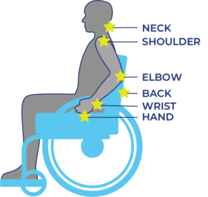
Musculoskeletal pain is caused by problems in the muscles, joints or bones. It is a common problem for all people as they get older, including those with SCI. Most people aging with SCI experience increased musculoskeletal pain in the upper extremities (shoulder and arm). Other common pain spots include the elbows, wrists, and hands. Issues with posture and seating can cause neck and back pain.

Overuse injuries develop with age from many years of transfers, pressure relief maneuvers, wheelchair use, and other movements that require weight-bearing and repetitive strain. Because the upper extremities are not designed for such a high physical load, people develop injuries (e.g., tendonitis, bursitis) and pain from overuse.

Research shows that on average, people with SCI experience more arthritis and joint breakdown in the shoulder than the general population.

Pain is also commonly caused by other aging-related health conditions like osteoarthritis of joints beyond that described of the shoulder, skin breakdown, and constipation, etc.

Refer to our article on Shoulder Injury and Pain for more information!

Managing changes in pain with age

To manage changes in musculoskeletal pain from aging with SCI, consult with your doctor, as well as physical and occupational therapists. They can conduct an injury assessment and help find ways to reduce and prevent pain.

Strategies may include:

  • Modifying and optimizing movements and wheelchair skills to prevent injury and reduce pain.
  • Strength exercises and stretching to stabilize the shoulder joint and improve muscle imbalances.
  • Changes in use of assistive devices and technology to prevent further injury/pain and rest the affected joints/muscles.
  • Changes in the work and home environment to reduce effort and pain in daily activities.
  • Changes in wheelchair setup for propulsion efficiency and ease.
  • Medication to relieve pain.
  • Cognitive behavioral therapy and mindfulness.

If experiencing a change in neuropathic pain, consult with a health care provider to determine together what may be causing the change and how to manage it.

Refer to the “What physical treatments are used for pain after SCI?”, and “What psychological and mind-body therapies are used for pain after SCI?” sections above for more information on managing pain.

Pain is a common health concern following spinal cord injury and can come from various parts of the body such as: muscle, joints, organs, skin, and nerves. Options such as physical treatment, psychological treatment, medication related treatment, or surgical treatments can be implemented for pain management.

While managing pain can be challenging, working with your health professionals to find a plan that works for you is an effective strategy for adjusting to life with an SCI.

For a review of how we assess evidence at SCIRE Community and advice on making decisions, please see SCIRE Community Evidence.

This page has been adapted from the SCIRE Professional “Pain Management” Module:

Mehta S, Teasell RW, Loh E, Short C, Wolfe DL, Hsieh JTC (2014). Pain Following Spinal Cord Injury. In Eng JJ, Teasell RW, Miller WC, Wolfe DL, Townson AF, Hsieh JTC, Connolly SJ, Noonan VK, Loh E, McIntyre A, editors. Spinal Cord Injury Rehabilitation Evidence. Version 5.0: p 1-79.

Available from: https://scireproject.com/evidence/pain-management/

Merskey H, Bogduk N, IASP Task Force on Taxonomy. Part III: Pain Terms, a Current List with Definitions and Notes on Usage. In: Merskey H, Bogduk N, (eds). Classification of chronic pain. Seattle: IASP Press;1994: 209–214.

Bryce TN, Biering-Sorensen F, Finnerup NB, Cardenas DD, Defrin R, Lundeberg T, Norrbrink C, Richards JS, Siddall P, Stripling T, Treede RD, Waxman SG, Widerström-Noga E, Yezierski RP, Dijkers M. International spinal cord injury pain classification: Part I. Background and description. March 6-7, 2009. Spinal Cord. 2012;50(6):413-7.

Loh E, Guy SD, Mehta S, Moulin DE, Bryce TN, Middleton JW, Siddall PJ, Hitzig SL, Widerström-Noga E, Finnerup NB, Kras-Dupuis A, Casalino A, Craven BC, Lau B, Côté I, Harvey D, O’Connell C, Orenczuk S, Parrent AG, Potter P, Short C, Teasell R, Townson A, Truchon C, Bradbury CL, Wolfe D. The CanPain SCI clinical practice guidelines for rehabilitation management of neuropathic pain after spinal cord: introduction, methodology and recommendation overview. Spinal Cord. 2016 Aug;54 Suppl 1:S1-6.

Ragnarsson KT. Management of pain in persons with spinal cord injury. Spinal Cord Med 1997;20:186-99.

Rose M, Robinson JE, Ells P, Cole JD. Letter to the editor. Pain following spinal cord injury: Results from a postal survey. Pain 1988;34:101-2.

Widerström-Noga EG, Turk DC. Types and effectiveness of treatments used by people with chronic pain associated with spinal cord injuries: Influence of pain and psychosocial characteristics. Spinal Cord 2003;41:600-9.

Norrbrink Budh C, Lundeberg T. Non-pharmacological pain-relieving therapies in individuals with spinal cord injury: A patient perspective. Complement Ther Med. 2004;12:189-97.

Mehta S, Guy S, Lam T, Teasell R, Loh E. Antidepressants Are Effective in Decreasing Neuropathic Pain After SCI: A Meta-Analysis. Topics in Spinal Cord Injury Rehabilitation. 2015;21(2):166-173. doi:10.1310/sci2102-166.

Wollaars MM, Post MW, van Asbeck FW, Brand N. Spinal cord injury pain: the influence of psychologic factors and impact on quality of life. Clin J Pain. 2007 Jun;23(5):383-91.

Craig A, Tran Y, Siddall P, Wijesuriya N, Lovas J, Bartrop R, Middleton J. Developing a model of associations between chronic pain, depressive mood, chronic fatigue, and self-efficacy in people with spinal cord injury. J Pain. 2013 Sep;14(9):911-20. doi: 10.1016/j.jpain.2013.03.002. Epub 2013 May 23.

Raichle KA, Hanley M, Jensen MP, Cardenas DD. Cognitions, coping, and social environment predict adjustment to pain in spinal cord injury. J Pain. 2007 Sep;8(9):718-29. Epub 2007 Jul 5.

Davidoff G, Roth E, Guarracini M, Silwa J, Yarkony G. Function-limiting dysesthetic pain syndrome among traumatic spinal cord injury patients: a cross-sectional study. Pain 1987; 29: 39–48.

Cohen M, McArthur D, Vulpe M, Schandler S, Gerber K. Comparing chronic pain from spinal cord injury to chronic pain of other origins. Pain 1988; 35: 57–63.

Rose M, Robinson J, Ells P, Cole J. Pain following spinal cord injury: results from a postal survey. Pain 1988; 34: 101–102.

Britell C, Mariano A. Chronic pain in spinal cord injury. Phys Med Rehabil 1991; 5: 71–82.

Mariano A. Chronic pain and spinal cord injury. Clin J Pain. 1992; 8: 87–92.

Cairns D, Adkins R, Scott M. Pain and depression in acute traumatic spinal cord injury: origins of chronic problematic pain? Arch Phys Med Rehabil 1996; 77: 329–335.

Modirian E, Pirouzi P, Soroush M, Karbalaie-Esmaeili S, Shojaei H, Zamani H. Chronic pain after spinal cord injury: results of a long-term study. Pain Med 2010; 11: 1037–1043

Burke DC, Woodward JM. Pain and phantom sensations in spinal paralysis. In Vinken PJ, Bruyn GW (Eds). Handbook of Clinical Neurology (pp. 489-499). Amsterdam, North Holland Publishing Co. 1976.

Rose M, Robinson JE, Ells P, Cole JD. Letter to the editor. Pain following spinal cord injury: Results from a postal survey. Pain 1988;34:101-2.

Turner JA, Cardenas DD, Warms CA, McClellan CB. Chronic pain associated with spinal cord injuries: A community survey. Arch Phys Med Rehab 2001;82:501-9.

Ahn SH, Park HW, Lee BS, Moon HW, Jang SH, Sakong J, et al. Gabapentin effect on neuropathic pain compared among patients with spinal cord injury and different durations of symptoms. Spine 2003;28:341-6.

Moulin DE, Clark AJ, Gilron I, Ware MA, Watson CPN, Sessle BJ, et al. Pharmacological management of chronic neuropathic pain. Consensus statement and guidelines from the Canadian pain society. Pain Res Manag 2007;12:13-21.

To TP, Lim TC, Hill ST, Frauman AG, Cooper N, Kirsa SW, et al. Gabapentin for neuropathic pain following spinal cord injury. Spinal Cord 2002;40:282-5.

Tai Q, Kirshblum S, Chen B, Millis S, Johnston M, DeLisa JA. Gabapentin in the treatment of neuropathic pain after spinal cord injury: A prospective, randomized, double-blind, crossover trial. J Spinal Cord Med 2002;25:100-5.

Levendoglu F, Ogun CO, Ozerbil O, Ogun TC, Ugurlu H. Gabapentin is a first line drug for the treatment of neuropathic pain in spinal cord injury. Spine 2004;29:743-51.

Siddall PJ, Cousins MJ, Otte A, Griesing T, Chambers R, Murphy TK. Pregabalin in central neuropathic pain associated with spinal cord injury: A placebo-controlled trial. Neurol 2006;67:1792-800.

Arienti C, Daccò S, Piccolo I, Redaelli T. Osteopathic manipulative treatment is effective on pain control associated to spinal cord injury. Spinal Cord 2011;49:515-9.

Cardenas DD, Nieshoff EC, Suda K, Goto S, Sanin L, Kaneko T, Spom J, Parsons B, Soulsby M, Yang R, Whalen E, Scavone J, Suzuki M, Knapp L. A randomized trial of pregabalin in patients with neuropathic pain due to spinal cord injury. Neurology. 2013;80:533-39.

Sandford PR, Lindblom LB, Haddox JD. Amitriptyline and carbamazepine in the treatment of dysesthetic pain in spinal cord injury. Arch Phys Med Rehab 1992;73:300-1.

Cardenas DD, Warms CA, Turner JA, Marshall H, Brooke MM, Loeser JD. Efficacy of amitriptyline for relief of pain in spinal cord injury: results of a randomized controlled trial. Pain 2002;96:365-73.

Vranken JH, Hollmann MW, Van der Vegt MH, Kruis MR, Heesen M, Vos K, et al. Duloxetine in patients with central neuropathic pain caused by spinal cord injury or stroke: A randomized, double-blind, placebo-controlled trial. Pain 2011;152:267-73.

Rintala DH, Holmes SA, Courtade D, Fiess RN, Tastard LV, Loubser PG. Comparison of the effectiveness of amitriptyline and gabapentin on chronic neuropathic pain in persons with spinal cord injury. Arch Phys Med Rehab 2007;88:1547-60.

Hocking G, Cousins MJ. Ketamine in chronic pain management: An evidence-based review. Anesth Analg 2003;97:1730-9.

Loubser PG, Donovan WH. Diagnostic spinal anaesthesia in chronic spinal cord injury pain. Paraplegia 1991;29:36.

Kvarnstrom A, Karlsten R, Quiding H, Gordh T. The analgesic effect of intravenous ketamine and lidocaine on pain after spinal cord injury. Acta Anaesthesiol Scand 2004;48:498-506.

Eide PK, Stubhaug A, Stenehjem AE. Central dysethesia pain after traumatic spinal cord injury dependent on N-Methyl-Diaspartate receptor activation. Neurosurgery 1995;37:1080-7.

Herman RM, D’Luzansky SC, Ippolito R. Intrathecal baclofen suppresses central pain in patients with spinal lesions: A pilot study. Clin J Pain 1992;8:338-45.

Boviatsis EJ, Kouyialis AT, Korfias S, Sakas DE. Functional outcome of intrathecal baclofen administration for severe spasticity. Clin Neurol Neurosurg 2005;107:289-95.

Plassat R, Perrouin Verbe B, Menei P, Menegalli D, Mathé JF, Richard I. Treatment of spasticity with intrathecal baclofen administration: Long-term follow-up, review of 40 patients. Spinal Cord 2004;42:686-93.

Loubser PG, Akman NM. Effects of intrathecal baclofen on chronic spinal cord injury pain. J Pain Symptom Manage 1996;12:241-7.

Uchikawa K, Toikawa H, Liu M. Subscapularis motor point block for spastic shoulders in patients with cervical cord injury. Spinal Cord 2009;47:249-51.

Marciniak C, Rader L, Gagnon C. The use of botulinum toxin for spasticity after spinal cord injury. Am J Phys Med Rehab 2008;87:312-7.

Attal N, Guirimand F, Brasseur L, Gaude V, Chauvin M, Bouhassira D. Effects of IV morphine in central pain: a randomized placebo-controlled study. Neurol 2002;58:554-63.

Norrbrink C, Lundeberg T. Tramadol in neuropathic pain after spinal cord injury: A randomized, double-blind, placebo-controlled trial. Clin J Pain 2009;25:177-84.

Eide PK, Stubhaug A, Stenehjem AE. Central dysethesia pain after traumatic spinal cord injury dependent on N-Methyl-Diaspartate receptor activation. Neurosurgery 1995;37:1080-7.

Hagenbach U, Luz S, Ghafoor N, Berger JM, Grotenhermen F, Brenneisen R et al. The treatment of spasticity with delta9-tetrahydrocannabinol in persons with spinal cord injury. Spinal Cord 2007;45:551-62.

Rintala DH, Fiess RN, Tan G, Holmes SA, and Bruel BM. Effect of dronabinol on central neuropathic pain after spinal cord injury: A pilot study. Am J Phys Med Rehab 2010;89:840-8.

Siddall PJ, Molloy AR, Walker S, Mather LE, Rutkowski SB, Cousins MJ. The efficacy of intrathecal morphine and clonidine in the treatment of pain after spinal cord injury. Anesth Analg 2000;91:1493-8.

Uhle EI, Becker R, Gatscher S, Bertalanffy H. Continuous intrathecal clonidine administration for the treatment of neuropathic pain. Stereotact Funct Neurosurg 2000;75:167-75.

Sandford PR, Benes PS. Use of capsaicin in the treatment of radicular pain in spinal cord injury. J Spinal Cord Med 2000;23:238-43.

Norrbrink Budh C, Lundeberg T. Non-pharmacological pain-relieving therapies in individuals with spinal cord injury: A patient perspective. Complement Ther Med. 2004;12:189-97.

Chase T, Jha A, Brooks CA, Allshouse A: A pilot feasibility study of massage to reduce pain in people with spinal cord injury during acute rehabilitation. Spinal Cord 2013;51:847-51.

Norrbrink C, Lundeberg T. Acupuncture and massage therapy for neuropathic pain following spinal cord injury: An exploratory study. Acupunc Med 2011;29:108-15.

Arienti C, Daccò S, Piccolo I, Redaelli T. Osteopathic manipulative treatment is effective on pain control associated to spinal cord injury. Spinal Cord 2011;49:515-9.

Dyson-Hudson TA, Shiflett SC, Kirshblum SC, Bowen JE, Druin EL. Acupuncture and trager psychophysical integration in the treatment of wheelchair user’s shoulder pain in individuals with spinal cord injury. Arch Phys Med Rehab 2001;82:1038-46.

Dyson-Hudson TA, Kadar P, LaFountaine M, Emmons R, Kirshblum SC, Tulsky D et al. Acupuncture for chronic shoulder pain in persons with spinal cord injury: a small-scale clinical trial. Arch Phys Med Rehab 2007;88:1276-83.

Yeh ML, Chung YC, Chen KM, Tsou MY, and Chen HH. Acupoint electrical stimulation reduces acute postoperative pain in surgical patients with patient-controlled analgesia: A randomized controlled study. Altern Ther Health Med 2010;16:10.

Ginis KAM, Latimer AE, McKechnie K, Ditor DS, Hicks AL, Bugaresti J. Using exercise to enhance subjective well-being among people with spinal cord injury: The mediating influences of stress and pain. Rehab Psychol 2003;48:157-64.

Nawoczenski DA, Ritter-Soronen JM, Wilson CM, Howe BA, Ludewig PM. Clinical trial of exercise for shoulder pain in chronic spinal injury. Phys Ther 2006;86:1604-18.

Serra-Ano P, Pellicer-Chenoll M, Garcia-Masso X, Morales J, Giner-Pascual M, Gonzalez LM: Effects of resistance training on strength, pain and shoulder functionality in paraplegics. Spinal Cord 2012;50:827-831.

Jensen MP, Barber J, Romano JM, Hanley MA, Raichle KA, Molton IR, et al. Effects of self-hypnosis training and EMG biofeedback relaxation training on chronic pain in persons with spinal-cord injury. Int J Clin Exp Hypn 2009;57,239-68.

Jensen MP, Barber J, Williams-Avery RM, Flores L, Brown MZ. The effect of hypnotic suggestion on spinal cord injury pain. J Back Musculoskeletal Rehab 2000;14:3-10.

Jensen MP, Gertz KJ, Kupper AE, Braden AL, Howe JD, Hakimian S, Sherlin LH: Steps toward developing an EEG biofeedback treatment for chronic pain. App Psychophysiol Biofeedback 2013;38:101-8.

Perry KN, Nicholas MK, Middleton JW. Comparison of a pain management program with usual care in a pain management center for people with spinal cord injury-related chronic pain. Clin J Pain 2010;26:206-16.

Heutink M, Post MWM, Bongers-Janssen HMH, Dijkstra CA, Snoek GJ, Spijkerman DCM, Lindeman E: The CONECSI trial: Results of a randomized controlled trial of a multidisciplinary cognitive behavioral program for coping with chronic neuropathic pain after spinal cord injury. Pain 2012;153:120-8.

Norrbrink Budh C, Kowalski J, Lundeberg T. A comprehensive pain management programme comprising educational, cognitive and behavioural interventions for neuropathic pain following spinal cord injury. J Rehab Med 2006;38:172-80.

Burns AS, Delparte JJ, Ballantyne EC, Boschen KA: Evaluation of an interdisciplinary program for chronic pain after spinal cord injury. Physical Medicine and Rehab 2013;5:832-8.

Soler MD, Kumru H, Pelayo R, Vidal J, Tormos JM, Fregni F, et al. Effectiveness of transcranial direct current stimulation and visual illusion on neuropathic pain in spinal cord injury. Brain 2010;133:2565-77.

Kumru H, Soler D, Vidal J, Tormos JM, Pascual-Leone A, Valls-Sole J. Evoked potentials and quantitative thermal testing in spinal cord injury patients with chronic neuropathic pain. Clinical Neurophysiology 2012;123:55-66.

Gustin SM, Wrigley PF, Gandevia SC, Middleton JW, Henderson LA, Siddall PJ. Movement imagery increases pain in people with neuropathic pain following complete thoracic spinal cord injury. Pain 2008;137:237-44.

Moseley GL. Using visual illusion to reduce at-level neuropathic pain in paraplegia. Pain 2007;130:294-8.

Capel ID, Dorrell HM, Spencer EP, Davis MW. The amelioration of the suffering associated with spinal cord injury with subperception transcranial electrical stimulation. Spinal Cord 2003;41:109-17.

Fregni F, Gimenes R, Valle AC, Ferreira MJ, Rocha RR, Natalle L et al. A randomized, sham-controlled, proof of principle study of transcranial direct current stimulation for the treatment of pain in fibromyalgia. Arthritis Rheum 2006;54:3988-98.

Soler MD, Kumru H, Pelayo R, Vidal J, Tormos JM, Fregni F, et al. Effectiveness of transcranial direct current stimulation and visual illusion on neuropathic pain in spinal cord injury. Brain 2010;133:2565-77.

Tan G, Rintala DH, Thornby JI, Yang J, Wade W, Vasilev C. Using cranial electrotherapy stimulation to treat pain associated with spinal cord injury. J Rehab Res Dev 2006;43:461-74.

Davis R, Lentini R. Transcutaneous nerve stimulation for treatment of pain in patients with spinal cord injury. Surg Neurol 1975;4:100-1.

Jette F, Cote I, Meziane HB, Mercier C: Effect of single-session repetitive transcranial magnetic stimulation applied over the hand versus leg motor area on pain after spinal cord injury. Neurorehabi Neural Repair 2013;27:636-643.

Defrin R, Grunhaus L, Zamir D, Zeilig G. The effect of a series of repetitive transcranial magnetic stimulations of the motor cortex on central pain after spinal cord injury. Arch Phys Med Rehab 2007;88:1574-80.

Cioni B, Meglio M, Pentimalli L, Visocchi M. Spinal cord stimulation in the treatment of paraplegic pain. J Neurosurg 1995;82:35-9.

Livshits A, Rappaport ZH, Livshits V, Gepstein R. Surgical treatment of painful spasticity after spinal cord injury. Spinal Cord 2002;40:161-6.

Falci S, Best L, Bayles R, Lammertse D, Starnes C. Dorsal root entry zone microcoagulation for spinal cord injury-related central pain: operative intramedullary electrophysiological guidance and clinical outcome. J Neurosurg Spine 2002;97:193-200.

Chun HJ, Kim YS, Yi HJ. A modified microsurgical DREZotomy procedure for refractory neuropathic pain. World Neurosurgery 2011;75:551-7.

Sindou M, Mertens P, Wael M. Microsurgical DREZotomy for pain due to spinal cord and/or cauda equina injuries: long-term results in a series of 44 patients. Pain 2001;92:159-71.

Spaic M, Petkovic S, Tadic R, Minic L. DREZ surgery on conus medullaris (after failed implantation of vascular omental graft) for treating chronic pain due to spine (gunshot) injuries. Acta Neurochirurgica 1999;141:1309-12.

Spaic M, Markovic N, Tadic R. Microsurgical DREZotomy for pain of spinal cord and cauda equina injury origin: Clinical characteristics of pain and implications for surgery in a series of 26 patients. Acta Neurochirurgica 2002;144:453-62.

Rath SA, Seitz K, Soliman N, Kahamba JF, Antoniadis G, Richter HP. DREZ coagulations for deafferentation pain related to spinal and peripheral nerve lesions: Indication and results of 79 consecutive procedures. Stereotact Funct Neurosurg 1997;68:161-7.

Sampson JH, Cashman RE, Nashold BS, Jr., Friedman AH. Dorsal root entry zone lesions for intractable pain after trauma to the conus medullaris and cauda equina. J Neursurg 1995;82:28-34.

Nashold BS, Jr., Vieira J, el-Naggar AO. Pain and spinal cysts in paraplegia: Treatment by drainage and DREZ operation. Br J Neurosurg 1990;4:327-35.

Friedman AH, Nashold BS. DREZ lesions for relief of pain related to spinal cord injury. J Neurosurgery 1986;65:465-9.

Image credits

  1. Wrist pain ©Injurymap, CC BY-SA 4.0
  2. ©OpenStax College, CC BY 3.0
  3. Image ©SCIRE, CC BY-NC 4.0
  4. Image ©SCIRE, CC BY-NC 4.0
  5. Image ©SCIRE, CC BY-NC 4.0
  6. ©Intermedichbo, CC BY-SA 4.0
  7. Assorted Medications ©NIAID, CC BY 2.0
  8. Treatment ©Royal@design, CC BY 3.0 US
  9. Back Pain ©Matt Wasser, CC BY 3.0 US
  10. _DSC0452_19632© ©Eric Neitzel, CC BY-NC-ND 2.0
  11. Her handiwork ©thepismire, CC BY-NC-ND 2.0
  12. Neuro-ms ©Baburov, CC BY-SA 4.0
  13. PhysiologyLab2009-07 ©Fredric Shaffer, CC0 1.0
  14. Surgery ©Healthcare Symbols, CC0 1.0
  15. Aging With Pain Thumbnail ©SCIRE, CC BY-NC 4.0
  16. Aging Pain Points ©SCIRE, CC BY-NC 4.0


Disclaimer: This document does not provide medical advice. This information is provided for educational purposes only. Consult a qualified health professional for further information or specific medical advice. The SCIRE Project, its partners and collaborators disclaim any liability to any party for any loss or damage by errors or omissions in this publication.