Adapted Driving

By | | No Comments

Author: Sharon Jang | Reviewer: Lisa Kristalovich | Published: 6 July 2022 | Updated: ~

Key Points

  • After spinal cord injury (SCI), many people are still able to drive.
  • In order to return to driving, an in-depth driving assessment needs to be conducted by a driving rehabilitation specialist or occupational therapist.
  • There are many different types of modifications that can be made to a vehicle based on your needs and limitations.

Wheelchair on beachBeing able to drive is an important skill that is helpful for day-to-day activities. Research has shown that being able to drive is related to many benefits, such as:

  • Improved happiness with life
  • Decreased depression
  • Increased access to health vehicle services in the community
  • Increased engagement in daily activities, such as running errands
  • A greater sense of independence

In addition, research has found that driving is associated with being able to work post-SCI. After SCI, one of the biggest barriers to working is a lack of transportation. Being able to drive on your own can address this issue, and promote working.

Many people can still drive after SCI. One study noted that many people with a C4 injury or below are able to independently drive. Although a formal driving assessment is often required before you are able to drive, some positive signs that you will be able to drive again include:

  • Stable SCI – there are no changes to your function
  • You don’t need narcotics to control your pain
  • Good vision/corrected vision
  • Controlled muscle spasms
  • Ability to transfer on and off a toilet

Research also shows that tetraplegics are able to drive as well as able-bodied individuals but have slower reaction times. Nonetheless, many people with SCI are able to drive.

Before getting on the road again, a formal driving assessment is often done by an occupational therapist or a driving rehabilitation specialist. During these assessments, the specialist will go over your medical history, driving history, and goals for driving. In addition, they will evaluate many aspects of your health and functioning, which include the following:

Vision

The specialist will assess if you are seeing things correctly with a vision test.

Physical abilities

Many aspects of your physical abilities will be assessed, including:

  • The strength and amount of movement in your limbs for controlling the vehicle
  • How much are you able to rotate your head and neck to check for vehicles
  • How quickly you are able to react to other vehicles, pedestrians, and other objects on the road (i.e., your reaction time)
  • Balance, which is used for getting in and out of the vehicle and being able to sit still while making turns
  • Hand-eye coordination
Cognition

Driving requires a lot of focus. Some tests will be done to evaluate how well and fast your brain is working. Some of these include:

  • Memory, which can influence remembering the rules of the road and navigating the road
  • Visual processing, or how fast you understand and interpret what you see happening on the road
  • Visual spatial abilities, or being able to identify where things are on the road and judging their distance
  • Visual perception, or your brain’s ability to make sense of what you see
  • Attention, which is required for paying attention to the road
  • Judgement and decision making, which are used in cases of knowing when to go/stop, when to switch lanes, etc.
Mood/behaviours

Mood and behaviours may also be evaluated during an assessment. Some traits may be red flags for driving, including being overly anxious on the road, being impulsive, and being highly irritable.

After you find out what kind of equipment you need to adapt your vehicle, you must learn to use it to drive in a safe manner. Driver rehab provides training and supervised practice using your newly modified vehicle. Some topics that may be covered in driver rehab include:

  • How to use your adaptive driving equipment or perform different driving techniques
  • Cognitive strategies to address issues with memory, attention, etc.
  • Visual strategies to address perception, sight, etc.
  • Anxiety management
  • A reintroduction to the driving environment

Often, you will need to participate in driver rehab sessions until you are able to demonstrate proficiency with using your vehicle modifications under typical driving conditions. In some areas of the world, a road test may be required to get your full license.

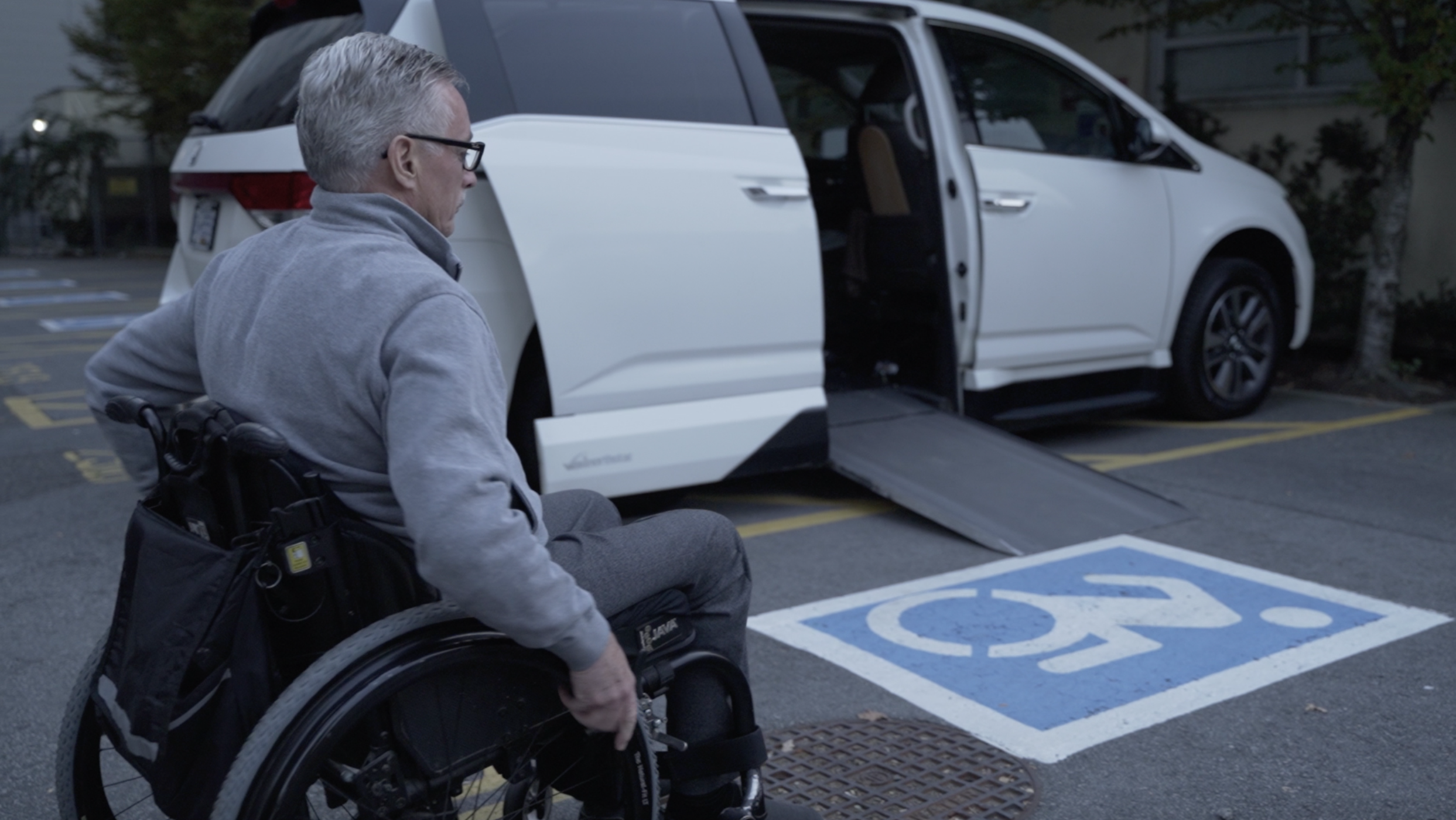
Many vehicles can be adapted for driving after SCI. However, the ideal vehicle for you is dependent on your wants and needs. For example, paraplegics tend to transfer into the driver seat of the vehicle, while among tetraplegics, half will transfer to the drivers seat and half will drive in their wheelchair. If you are driving in your wheelchair, you will need a larger vehicle to accommodate the wheelchair. However, if you are transferring into the vehicle seat, you might want a vehicle that is closer to the ground for an easier transfer and wheelchair loading. Larger vehicles like trucks and SUVs may require extra equipment to help with transfers and wheelchair loading.

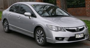
One study has looked at the measurements of various vehicles. In regards to the height between the ground and the driver seat, they found that the average height is:

  • 22 inches for a sedan
  • 28 inches for a mid-height vehicle (vans, small-medium SUVs)
  • 36 inches for a high-profile vehicle (large truck or SUV)

This study also found that the average difference in height between the driver’s seat and wheelchair seat is 3.7 inches, and ranged from -3.5 inches to 16 inches. This means that for some vehicles, the wheelchair seat may be above the vehicle seat, while in others, they can be up to 16 inches below the vehicle seat. Your ability to transfer is a consideration in what kind of vehicle to buy. Other considerations include how much space you want in your vehicle, where you will be driving your vehicle, and how/where you will be storing your wheelchair if you plan on transferring into the driver seat of the vehicle.

Collision warning braking support is available for some vehicles and can aid in collision prevention.

A vehicle can be adapted in many ways with the use of adaptive driving equipment, or technology used to make your vehicle more accessible. In general, driving is broken into 4 parts:

  • transferring in and out of the vehicle
  • loading your wheelchair
  • using primary controls (steering, accelerating, braking)
  • using secondary controls (e.g., controlling the windshield, signals, radio)

In addition, there are various safety features that can be added to the vehicle to help you drive if you have any limitations. Some driver rehabilitation centers will also complete a vehicle modification assessment. During this assessment, a driving specialist will help you select the equipment to get you and your wheelchair into the vehicle safely.

Transferring in and out of the vehicle

A ramp can be installed to allow for ease of vehicle entry/exit.

When getting in and out of your vehicle, the first consideration is whether you are able to transfer into the driver seat, or if you will stay in your wheelchair. Although it is possible to drive from your wheelchair, some additional considerations include:

  • the original driver seat in the vehicle has been designed to withstand a vehicle crash, and is in an optimal position to be used with the air bag and seatbelt
  • the seatbelt may not fit ideally when in your wheelchair due to the design of a wheelchair


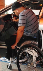
Transferring from a manual wheelchair into the driver seat and manually loading the wheelchair

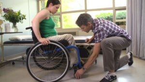
There are many ways to get into your vehicle from a wheelchair. The following is a general overview of the steps.

  1. Transfer into the seat. This can be done using a transfer board, hanging onto a grab bar/ handle, or placing a hand on the seat. Some people choose to transfer by placing their right leg into the vehicle before transferring, or they keep both their legs outside of the vehicle.
  2. Decide where you will place your wheelchair: in the front passenger seat, or the back seats. Those with weaker shoulder muscles should consider loading their wheelchair into the front seats.
  3. Remove the wheels from the wheelchair. This is commonly done by pressing the center button in the middle of the wheel. Place the tires in the vehicle.
  4. Some people remove the cushion and the side guard from the wheelchair. Place these in the vehicle.
  5. Load the wheelchair frame into the vehicle. Reclining the front seat can help you get the frame over your body and into the vehicle.

Driving from the driver seat

Swivel-style car seats can come out of the car or turn inside of the car.

If you have difficulties with transfering or loading your wheelchair there are many adaptations that can be used. Swivel seats are seats that turn and come out of the vehicle, giving you more space to transfer in. Alternatively, a transfer seat can be used. A transfer seat can move up or down in height, can turn, and can be moved in the vehicle for more space. This is done by placing the original driver seat on top of a motorized plate. However, it is important to note that swivel seats are only compatible with some SUV’s, trucks, and minivans, and transfer seats are only compatible with minivans or full sized vans. If you only need a bit of assistance getting in and out of a vehicle, additional grab bars can be installed into a vehicle.

Driving from your wheelchair

If it is decided that it is best for you to drive from your wheelchair, you will need a wheelchair accessible vehicle. To have enough height for a wheelchair to enter, the vehicle is raised up and the floor is lowered. A ramp is then installed. It may come out from the floor or fold out.  Once in your vehicle, it is important to make sure that your wheelchair is stiff enough to provide a stable driving platform, and will not move when you are driving.

Wheelchair tie downs should be used to secure the wheelchair when driving.

Your wheelchair will also need to be secured in place while driving. This can be done with a manual locking system and the help of another person. There are also automated docking systems which anchors your wheelchair without the help of another person. These systems have an additional piece that connects to your wheelchair. The part on your wheelchair clicks into the docking system on the floor of your vehicle. Automated docking systems are controlled electronically. A button installed in your vehicle releases the docking system lock. The part that attaches to your wheelchair weighs 10-19 lbs, and is permanently attached to your wheelchair. Many people using a manual wheelchair have a hard time managing the extra weight on the wheelchair, so this system is usually used with power wheelchairs.

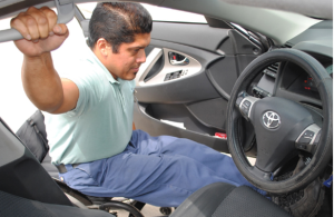
Primary Controls (steering, braking, acceleration)

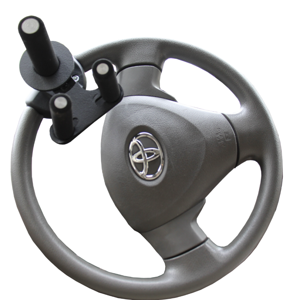
To help with steering and driving, different handles can be added onto the steering wheel. A spinner knob can be added to make it easier to control the steering wheel. For people with no hand function, a tri-pin add on may be helpful. A tri-pin handle consists of one larger straight prong, and two smaller straight prongs. The larger prong sits in your hand, and your wrist sits between the two smaller prongs. This allows you to use your shoulder and elbow muscles to steer.

Rods can be connected to the accelerator and brakes to allow for hand control driving.

To accelerate and brake, rods are connected to the pedals, and the rod is connected to a handle beside the steering wheel. The handle is pushed forward to brake. Different motions, including depressing, rocking, pulling, or twisting can be used to control the gas. These hand controls are not removable, but the pedals remain in place so an able-bodied person can drive. The vehicle can be shared!

With the advancement of technology, there electronic-based steering adaptations. Some of these technologies include:

  • Power-controlled levers and rods for accelerating/braking: similar to mechanical rods and levers, but with a motor built in to make the movement easier
  • Reduced effort steering: modifications made to the vehicle to reduce the strength required to turn the steering wheel
  • Using joysticks or other electronic wheels to drive the vehicle: a modification can be made to the vehicle so that it is controlled by a computer. The vehicle is then driven with a wheel or joystick that is connected to the computer.

Secondary Controls (windshield wipers, turn signals, etc)

Secondary controls on a button system.

Secondary controls are used to interact with other drivers on the road (such as signaling and using the horn), and to manage the vehicle (e.g., use the windshield wipers, changing the transmission gear, starting the vehicle, managing the heating/air conditioning etc). A lot of these functions can be adapted so that they are controlled with the push of a button. For example, buttons can be placed on the head rest so that they can be pressed with the head, or on the door so that it can be pressed with the elbow. Buttons can activate a single function, or can be used to trigger several functions. The multiple buttons can be programmed to the function of your desire, and can be connected to the steering wheel or other location that is convenient to you. These adaptations come in a variety of set-ups, and will require customization to your needs.

Funding considerations

There are often costs associated with the various parts of getting back on the road. In general, fees are required for the initial driving assessment, rehabilitation both in a clinic setting and on the road, and for adaptive equipment. In Canada, there is often no funding for these costs; this is often paid out of pocket unless you have an injury claim or other funding source. As a result, funding can be a big barrier to returning to driving.
For more information on the related fees, contact your local driving rehabilitation center for.

Considerations when looking to buy a vehicle to adapt

When looking to buy a vehicle to adapt after your injury, some things to consider include:

Transfer abilities

What are your transfer abilities? Will you be staying in your wheelchair to drive or will you transfer to the driver seat? If you are able to transfer, how easy is it for your to transfer to a higher surface? Do you need a ramp to get in and out of the vehicle?

Wheelchair storage

If you are planning on transferring out of your wheelchair, where will you store it? In the front seat or back?

Adaptive equipment required

Does the equipment you need only fit in a certain type of vehicle, such as a van? Can the vehicle accommodate the hand controls you need?

Passengers

If you plan on driving others, will there be enough space for passengers in the vehicle once it has been adapted?

Parking

Will the vehicle fit in the parking space you have?

Some driver rehabilitation centers will also complete a vehicle modification assessment. This assessment will help you select the equipment you need to get you and your wheelchair into the vehicle safely. There is usually a fee for a vehicle modification assessment.

Considerations when driving an adapted vehicle

Two studies interviewed people with disabilities who drove adapted vehicles. Some challenges that were identified by the drivers included:

Pain

Pain was experienced in the wrists when driving long distances, especially with a twist accelerator. Shoulder pain was also reported after driving for a long time. You may want to consider what position your arms are in, what movements are used, and if you can do this over a long period of time.

Trunk strength

Having a weak core resulted in some drivers needing to slow down or brace themselves when driving at high speeds or on winding roads. People with a higher spinal cord injury level often need extra trunk support, as they are unable to use their arms for support when hand controls are being used.

Fatigue

Driving can be tiring in comparison to driving able-bodied, as more focus is required for driving an adapted vehicle.

Accessibility of the environment

Some drivers found that the location they drove to was inaccessible, and they were unable to et out of their vehicle. For example, some garages had a step to get out of them, had a steep hill to the entrance, or if there is not enough space to open a ramp.

After an SCI, many people continue to drive with the use of adaptive driving equipment. There are many modifications that can be made to a vehicle to suit your needs and enable you to drive again. However, prior to hitting the road, you will need to be evaluated by a driving rehabilitation specialist or occupational therapist. This evaluation will help the clinician understand your needs and limitations, and help them determine the best adaptations for you. Although getting back to driving may be a lengthy process, it can be beneficial for your sense of independence, and partaking in activities that you want to do again.

For a review of how we assess evidence at SCIRE Community and advice on making decisions, please see SCIRE Community Evidence.

Evidence for “Why is driving after SCI important?” is based on:

Mtetwa, L., Classen, S., & van Niekerk, L. (2016). The lived experience of drivers with a spinal cord injury: A qualitative inquiry. South African Journal of Occupational Therapy, 46(3), 55–62.

Norweg, A., Jette, A. M., Houlihan, B., Ni, P., & Boninger, M. L. (2011). Patterns, predictors, and associated benefits of driving a modified vehicle after spinal cord injury: Findings from the national spinal cord injury model systems. Archives of Physical Medicine and Rehabilitation, 92(3), 477–483.

Evidence for “How do I know if I can drive?” is based on:

Anschutz, J. (2015). Driving After Spinal Cord Injury. Spinal Cord Injury Model System, (October). Retrieved from https://msktc.org/lib/docs/Factsheets/SCI_Driving.pdf

Kiyono, Y., Hashizume, C., Matsui, N., Ohtsuka, K., & Takaoka, K. (2001). Vehicle-driving abilities of people with tetraplegia. Archives of Physical Medicine and Rehabilitation, 82(10), 1389–1392.

Norweg, A., Jette, A. M., Houlihan, B., Ni, P., & Boninger, M. L. (2011). Patterns, predictors, and associated benefits of driving a modified vehicle after spinal cord injury: Findings from the national spinal cord injury model systems. Archives of Physical Medicine and Rehabilitation, 92(3), 477–483.

Peters, B. (2001). Driving performance and workload assessment of drivers with tetraplegia: An adaptation evaluation framework. Journal of Rehabilitation Research and Development, 38(2), 215–224.

Evidence for “What is a driving assessment based on?” is based on:

Anschutz, J. (2015). Driving After Spinal Cord Injury. Spinal Cord Injury Model System, (October). Retrieved from https://msktc.org/lib/docs/Factsheets/SCI_Driving.pdf

van Roosmalen, L., Paquin, G. J., & Steinfeld, A. M. (2010). Quality of Life Technology: The State of Personal Transportation. Physical Medicine and Rehabilitation Clinics of North America, 21(1), 111–125.

Evidence for “What kind of vehicle can I drive?” is based on:

Haubert, L. L., Mulroy, S. J., Hatchett, P. E., Eberly, V. J., Maneekobkunwong, S., Gronley, J. K., & Requejo, P. S. (2015). Vehicle transfer and wheelchair loading techniques in independent drivers with paraplegia. Frontiers in Bioengineering and Biotechnology, 3(139), 1-7.

van Roosmalen, L., Paquin, G. J., & Steinfeld, A. M. (2010). Quality of Life Technology: The State of Personal Transportation. Physical Medicine and Rehabilitation Clinics of North America, 21(1), 111–125.

Evidence for “What adaptations are available for my vehicle?” is based on:

Haubert, L. L., Mulroy, S. J., Hatchett, P. E., Eberly, V. J., Maneekobkunwong, S., Gronley, J. K., & Requejo, P. S. (2015). Vehicle transfer and wheelchair loading techniques in independent drivers with paraplegia. Frontiers in Bioengineering and Biotechnology, 3(139), 1-7.

van Roosmalen, L., Paquin, G. J., & Steinfeld, A. M. (2010). Quality of Life Technology: The State of Personal Transportation. Physical Medicine and Rehabilitation Clinics of North America, 21(1), 111–125.

Evidence for ” What are some considerations when using and buying an adapted vehicle?” is based on:

Christopher and Dana Reeve Foundation (2021). Vehicles and Driving. https://www.christopherreeve.org/living-with-paralysis/home-travel/driving

Hutchinson, C., Berndt, A., Gilbert-Hunt, S., George, S., & Ratcliffe, J. (2020). Modified motor vehicles: the experiences of drivers with disabilities. Disability and Rehabilitation, 42(21), 3043–3051. Retrieved from https://doi.org/10.1080/09638288.2019.1583778

Mtetwa, L., Classen, S., & van Niekerk, L. (2016). The lived experience of drivers with a spinal cord injury: A qualitative inquiry. South African Journal of Occupational Therapy, 46(3), 55–62.

Image credits
  1. Wheelchair holiday bea disabled summer ©LonelyTaws, Pixabay License
  2. Eye ©Veronika Krpciarova, CC BY 3.0 
  3. Stretch ©Andrejs Kirma, CC BY 3.0 
  4. Brain ©Amethyst Studio, CC BY 3.0 
  5. Mood ©shuai tawf, CC BY 3.0 
  6. Adapted wheel with spinner, ©SCIRE Community Team
  7. Honda Odyssey (2018-present) ©Kevauto, CC BY-SA 4.0
  8. Eighth-generation Civic sedan ©OSX, CC 0
  9. Ford F-150 crew cab – 05-28-2011 ©IFVEHICLE, CC 0
  10. Collision warning brake support ©Ford Motor Company, CC BY 2.0
  11. Adapted Van ©SCIRE Community Team
  12. Haubert, L. L., Mulroy, S. J., Hatchett, P. E., Eberly, V. J., Maneekobkunwong, S., Gronley, J. K., & Requejo, P. S. (2015). Vehicle transfer and wheelchair loading techniques in independent drivers with paraplegia. Frontiers in Bioengineering and Biotechnology, 3(139), 1-7.
  13. A disabled man in a wheelchair getting out of a vehicle ©CDC/Amanda Mills, CC 0
  14. BraunAbility Turny Evo Handicap Swivel Vehicle Seat Transfer Seat ©BraunAbility, 2020
  15. BraunAbility B&D Transfer Seat ©BraunAbility, 2020
  16. Special, vehicle, wheelchair ©CDC/Amanda Mills, CC 0
  17. QRT-360 ©Q’Straint, 2021
  18. Sure-Grip Tri-pin Spinner Knob ©Indemedical, 2021
  19. Adapted driving levers and rods. ©SCIRE Community Team
  20. Bever 8-touch Keypad ©Bever Mobility Products Inc
  21. Money ©Mahabbah, CC BY 3.0 


Disclaimer: This document does not provide medical advice. This information is provided for educational purposes only. Consult a qualified health professional for further information or specific medical advice. The SCIRE Project, its partners and collaborators disclaim any liability to any party for any loss or damage by errors or omissions in this publication.

Epidural Stimulation

By | | No Comments

Authors: Dominik Zbogar and Sharon Jang | Reviewer: Susan Harkema | Published: 14 February 2022 | Updated: ~

Key Points

  • Epidural stimulation is a treatment that sends electrical signals to the spinal cord.
  • Epidural stimulation requires a surgical procedure to implant electrodes close to the spinal cord.
  • One of the ways epidural stimulation works is by replacing the signals that would normally be sent from the brain to the spinal cord before spinal cord injury (SCI).
  • Epidural stimulation affects numerous systems. Stimulation aimed at activating leg muscles may potentially also affect bowel, bladder, sexual, and cardiovascular function.
  • Studies of epidural stimulation in spinal cord injury (SCI) generally do not include a comparison group without stimulation. The benefits of epidural stimulation that have been reported have been in small numbers of participants. So, while reports thus far are encouraging, more research is necessary.
  • Because it is in the research and development phase, epidural stimulation for spinal cord injury is not part of standard care nor is it a readily available treatment.

Neuromodulation is a general term for any treatment that changes or improves nerve pathways. Different types of neuromodulation can work at different sites along the nervous system (e.g., brain, nerves, spinal cord) and may or may not be invasive (i.e., involve surgery). Epidural stimulation (also known as epidural spinal cord stimulation or direct spinal cord stimulation) is a type of invasive neuromodulation that stimulates the spinal cord using electrical currents. This is done by placing an electrode on the dura (the protective covering around the spinal cord).

To read more about other types of neuromodulation used in SCI, access these SCIRE Community articles: Functional Electrical Stimulation (FES), Transcutaneous Electrical Nerve Stimulation (TENS), sacral nerve stimulation, and intrathecal Baclofen.

Watch our neuromodulation series videos! Our experts explain experimental to more commonplace applications, and individuals with SCI describe how neuromodulation has affected their lives.


What is “an Epidural”?

Epi- is a prefix and means “upon”, and the dura (full name: dura mater) is a protective covering of the spinal cord. So epidural means “upon the dura”, and in the context of epidural stimulation, this is where the electrodes that stimulate the spinal cord are placed. Yes, it is also possible to have sub-dural (under the dura) or endo-dural (within the dura) electrode placement. And, there are more layers between the dura and the spinal cord, not to mention the spinal cord itself where electrodes could be placed in what is called intraspinal microstimulation. The benefit of being beneath the dura and closer to the spinal cord is that there is a more direct stimulation. Having the electrode closer to the spinal cord allows more precision with the signal going more directly to the neurons.

The drawback is that more complications can arise with closer placement because the electrodes are in the spinal cord tissue. Such placement is currently rare, experimental, or non-existent but that will change as the technology advances. Intraspinal microstimulation has been tested in animal models and is in the process of being translated to humans.

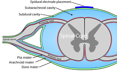
You are probably familiar with the term “epidural” already, as it is often mentioned in relation to childbirth. If a new mother says she had an epidural, what she usually means is that she had pain medication injected into the epidural space for the purpose of managing pain during birth.

We specifically discuss epidural spinal cord stimulation in this article. Spinal cord stimulation can also be applied transcutaneously. This type of spinal cord stimulation is non-invasive as the stimulating electrodes are placed on the skin. With transcutaneous stimulation, the signal has to travel a greater distance through muscle, fat, and other tissues, which means the ability to be precise with stimulation is hampered. However, it does allow for more flexibility in electrode placement and does not require surgery. There is research published or underway investigating the impact of transcutaneous stimulation in some of the areas discussed above, including hand, leg, and cardiovascular function.

Normally, input from your senses travels in the form of electrical signals through the nerves, up the spinal cord, and reaches the brain. The brain then tells the muscles or organs what to do by sending electrical signals back down the spinal cord. After a spinal cord injury, this pathway is disrupted, preventing electrical signals from traveling below the level of injury to reach where they need to go. However, the nerves, muscles, and organs can still respond below the injury to electrical signals.

Epidural stimulation works by helping the network of nerves in the spinal cord below the injury function better and take advantage of any leftover signals from the spinal cord. To do so, the stimulation must be fine-tuned to make sure the amount of stimulation is optimal for each person and a specific function, such as moving the legs.

Recent studies of the role of epidural stimulation on standing and walking have noted unexpected beneficial changes in some participants’ bowel, bladder, sexual, and temperature regulation function. This highlights both the potential for epidural stimulation to improve quality of life in multiple ways and that much research remains to be done to understand how epidural stimulation affects the body.


There may still be spared connections in the spinal cord with a complete injury.

How can someone with a complete injury regain movement control with epidural stimulation?

Being assessed with a complete injury implies that there is no spared function below the injury. However, scientists are finding that this may not be the case. Studies have found that even with a complete loss of sensory and motor function, there may be some inactive connections that are still intact across the injury site. These remaining pathways may be important for regaining movement or other functions. Another hypothesis is that epidural stimulation in combination with training may encourage stronger connections across the level of injury. Although these pathways may provide some substitution for the injured ones, they are not as effective as non-injured pathways across the injury level.

When it is decided that an individual will receive epidural stimulation, a health professional, such as a neurosurgeon, will perform an assessment of the spinal cord using magnetic resonance imaging (MRI) to determine the best place to implant the electrodes.

In most of the studies mentioned in this article, the electrodes were placed between the T9-L1 levels, though researchers are investigating the impact of epidural stimulation on hand function.


Xray image of wires connecting power and signal to electrodes (red circle) placed on a spinal cord.

There are two possible procedures. One approach is to have two surgeries. During the initial surgery, a hollow needle is inserted through the skin into the epidural space, guided using fluoroscopy, a type of X-ray that allows the surgeon to see where the needle is in real time. Potential spots on the spinal cord are tested using a stimulator. A clinician will look to see if stimulation over those areas of the spinal cord leads to a desired response. Once found, the electrode array is properly positioned over the dura and the surgery is completed. This begins a trial period where the response to epidural stimulation is monitored. During this time the electrode array is attached to an electrical generator and power supply, which is worn on a belt outside of the body. When it is shown that things are working as desired, the generator is implanted underneath the skin in the abdomen or buttocks. The generator can be rechargeable or non-rechargeable. A remote control allows one to turn the generator on or off and control the frequency and intensity of stimulation.

The second method is to only have one surgery and no trial period. This is possible due to increased knowledge in how to stimulate the spinal cord. Soon after surgery, the individual will be taught how and when to use the epidural stimulation system at home. If needed, the frequency (how often) and intensity (how strong) of the stimulation will be adjusted at follow-up appointments with the physician. In other cases, many practice sessions of learning the right way to stimulate may be needed before a person can stimulate at home.

If the epidural stimulation is used for leg control, movement training, standing, and stepping will be required to learn how to coordinate and control movement during stimulation. This is required for the recovery of voluntary movements, standing and/or walking.

Epidural stimulation can be used in all people with SCI, regardless of the level or completeness of injury. However, certain situations can make it an unsafe treatment in some. It is important to speak to a health professional about your health history before beginning any new treatment.

Epidural stimulation should not be used in the following situations:

  • By people with implanted medical devices like cardiac pacemakers
  • By people who are unable to follow instructions or provide accurate feedback
  • By people with an active infection
  • By people with psychological or psychiatric conditions (e.g., depression, schizophrenia, substance abuse)
  • By people who are unable to form clots (anticoagulopathy)
  • Near areas of spinal stenosis (narrowing of the spinal canal)

Epidural stimulation should be used with caution in the following situations:

  • By children or pregnant women
  • By people who require frequent imaging tests like ultrasound or MRI (some epidural stimulation systems are compatible)
  • By people using anticoagulant medications (blood thinners)

Epidural stimulation is generally well-tolerated, but there is a risk of experiencing negative effects.

The most common risks and side effects of epidural stimulation include:

  • Technical difficulties with equipment, such as malfunction or shifting of the electrodes that may require surgery to fix
  • Unpleasant sensations of jolting, tingling, burning, stinging, etc. (from improper remote settings)

Other less common risks and side effects of epidural stimulation include:

  • Damage to the nervous system
  • Leakage of cerebrospinal fluid
  • Increased pain or discomfort
  • Broken bones
  • Masses/lumps growing around the site of the implanted electrode

Risks specific to the surgery which involves the removal of part of the vertebral bone (laminectomy) include:

  • bleeding and/or infection at the surgical site
  • spinal deformity and instability

Proper training on how to use the equipment and using the stimulation according to the directions of your health provider can help decrease the risks of experiencing these side effects.

Neuromodulation methods to manage bladder function have usually involved stimulation of the sacral nerves (which are outside of the spinal cord), not with epidural spinal cord stimulation. This is reflected in the fact that almost no research exists regarding the effects of epidural stimulation on bowel and bladder function in the previous century.

New information on epidural stimulation relating to bladder function is coming. In the last several years, several studies (weak evidence) from a very small group of participants of participants (who were AIS A or B) have found consistent improvements in bladder function. Participants in these reports were fitted with epidural stimulators for reactivation of paralyzed leg muscles for walking and reported additional benefits of improvements in bladder and/or bowel function. However, other studies have shown small changes to bladder function and no changes to bowel function. Negative changes, such as decreased control over the bladder, have even been noticed by some participants in another study. These findings suggest that epidural stimulation may improve quality of life by safely increasing the required time between catheterizations. Fewer catheterizations and reduced pressure in the bladder would preserve lower and upper urinary tract health. More research is required, especially with respect to bowel function. It must be noted that walk training alone has been shown to improve bladder and bowel function. Epidural stimulation may provide additional improvement to bladder function in comparison to walk training alone. Neuromodulation methods to manage bladder function have usually involved stimulation of the sacral nerves (which are outside of the spinal cord), not with epidural spinal cord stimulation. This is reflected in the fact that almost no research exists regarding the effects of epidural stimulation on bowel and bladder function in the previous century.

For more information, visit our pages on Bowel and Bladder Changes After SCI!

Why does walk/stand training alone have a beneficial effect on bladder, bowel, and sexual function?

Relationships between the leg movement and nerves in the low back regions have been identified.

Some evidence suggests that walk/step training alone can create improvements in bladder/bowel function. Researchers hypothesize that the sensory information created through walking or standing provides stimulation to the nerves in the low back region, which contains the nerves to stimulate bowel, bladder, and sexual function. Research has shown that bending and straightening the legs can be enhanced by how full the bladder is and the voiding of urine.

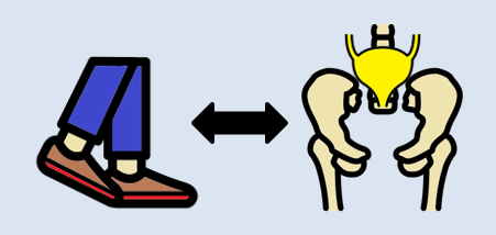
One of the consequences of SCI is the loss of muscle mass below the injury and a tendency to accumulate fat inside the abdomen (abdominal fat or visceral fat) and under the skin (subcutaneous fat). These changes and lower physical activity after SCI increase the risk for several diseases.

A single (weak-evidence) study measured body composition in four young males with complete injuries. Participants underwent 80 sessions of stand and step training without epidural stimulation, followed by another 160 sessions of stand/step training with epidural stimulation. This involved one hour of standing and one hour of stepping five days a week. After all, training was complete, all four participants had a small reduction in their body fat, and all participants but one experienced an increase in their fat free body mass (i.e., the weight of their bones, muscles, organs, and water in the body) in comparison to their initial values prior to stimulation. While all participants experienced a reduction of fat, the amount of fat loss was minimal, ranging from 0.8 to 2.4 kg over a period of a year.

The first use of epidural stimulation was as a treatment for chronic pain in the 1960s. Since then, it has been widely used for chronic pain management in persons without SCI. However, it is important to recognize that the chronic pain experienced by those without SCI is different from the chronic neuropathic pain experienced after SCI. This may explain, to some extent, why epidural stimulation has not been as successful in pain treatment for SCI. The mechanism by which electrical stimulation of the spinal cord can help with pain relief is unclear. Some research suggests that special nerve cells that block pain signals to the brain may be activated by epidural stimulation.

There are a few studies focused on the role of epidural stimulation in managing pain after SCI. A number of other studies included a mix of different people with and without SCI. Because chronic neuropathic pain after SCI may not be the same as the chronic pain others experience, studies that do not separate mixed groups raise questions about the validity of findings. The number of individuals with SCI in these studies is often small, most were published in the 1980s and 1990s and so are quite dated, and the research is classified as weak evidence.

The results of this body of research show that some people may receive some pain reduction. Those who saw the most reduction in pain were individuals with an incomplete SCI. Also, satisfaction with pain reduction drops off over time. One study showed only 18% were satisfied 3 years after implantation. A different study looking at the long-term use of epidural stimulation for pain reduction found seven of nine individuals stopped using this method.

In the only recent study in this area, one woman with complete paraplegia (weak evidence) experienced a reduction in neuropathic pain frequency and intensity, and a reduction in average pain from 7 to 4 out of 10, with 0 being no pain and 10 being the worst imaginable pain. This improvement remained up to three months later after implantation of the epidural stimulation device.

It should be noted that the studies for pain place electrodes in different parts of the spinal cord compared to the more recent studies for voluntary movement, standing and stepping.

Refer to our article on Pain After SCI for more information!

Using epidural stimulation to improve respiratory function is useful because it contracts the diaphragm and other muscles that help with breathing. Also, these muscles are stimulated in a way that imitates a natural pattern of breathing, reducing muscle fatigue. More common methods of improving respiratory function do not use epidural stimulation, but rather, directly stimulate the nerves that innervate the respiratory muscles. While such methods significantly improve quality of life and function in numerous ways, they are not without issues, including muscle fatigue from directly stimulating the nerves.

To date, most research into using epidural stimulation to improve respiratory function has been done in animals. Recently, research has been done in humans and weak evidence suggests that epidural stimulation may:

  • help produce a cough strong enough to clear secretions independently.
  • reduce frequency of respiratory tract infections.
  • reduce the time required caregiver support.
  • help individuals project their voice better and communicate more effectively.

Long term use of epidural stimulation shows that improvements remain over years and that minimal supervision is needed, making it suitable for use in the community.

Refer to our article on Respiratory Changes After SCI for more information!

The impact of epidural stimulation on sexual function has been a secondary focus in research studies looking at standing and walking. Currently, there are reports from one male and two females.

After a training program of walk training with epidural stimulation, one young adult male reported stronger, more frequent erections and the ability to reach full orgasm occasionally, which was not possible before epidural stimulation. However, this study looked at the effects of walk training and epidural stimulation together, which took place after several months of walk training without stimulation. Because the researchers did not describe what the individual’s sexual function was like after walk training, it is difficult to say how much benefit is attributed to epidural stimulation versus walk training.

In another study with two middle-aged females 5-10 years post-injury, one reported no change in sexual function and the other reported the ability to experience orgasms with epidural stimulation, which was not possible since her injury.

Refer to our article on Sexual Health After SCI for more information!

Botulinum toxin (Botox) injections and surgically implanted intrathecal Baclofen pumps are the most common ways to manage spasticity. Baclofen pumps are not without issues, however. Many individuals do not qualify for this treatment if they have seizures or blood pressure instability, and pumps require regular refilling.

Research in the 80s and 90s on the use of epidural stimulation for spasticity did not report very positive findings. It was noted that greater benefits were found in those with incomplete injury compared to those who were complete. Another paper concluded that (weak evidence) the beneficial effects of epidural stimulation on spasticity may subside for most users over a short period of time. This, combined with the potential for equipment failure and adverse events, suggested that epidural stimulation was not a feasible approach for ongoing management of spasticity.

More recently, positive results with epidural stimulation have been observed (weak evidence). This is likely due to improvements in technology, electrode placement, and stimulation parameters. Positive findings show that participants:

  • reported fewer spasms over 2 years
  • reported a reduction in severe spasms over 2 years
  • reported a reduction in spasticity
  • reported an improvement in spasticity over 1 year
  • were able to stop or reduce the dose of antispastic medication

For more information, visit our page on Botulinum Toxin and Spasticity!

In a study with a single participant (weak evidence) investigating walking, an individual implanted with an epidural stimulator also reported improvement in body temperature control, however details were not provided. More research is required to understand the role of epidural stimulation for temperature regulation.

In severe SCI, individuals may suffer from chronic low blood pressure and orthostatic hypotension (fall in blood pressure when moving to more upright postures). These conditions can have significant effects on health and quality of life. Some recent studies have looked at how epidural stimulation affects cardiovascular function to improve orthostatic hypotension. Overall, they show (weak evidence) that epidural stimulation immediately increases blood pressure in individuals with low blood pressure while not affecting those who have normal blood pressure. They also showed that there is a training effect with repeated stimulation. This means that after consistently using stimulation for a while, normal blood pressure can occur even without stimulation when moving from lying to sitting.

Moreover, researchers are starting to believe that changes in orthostatic hypotension and blood pressure can promote changes in the immune system (Bloom et al., 2020). In the body, the blood helps to circulate immune cells so they are able to fight infections in various areas. One case study found that after 97 sessions of epidural stimulation, the participant had fewer precursors for inflammation and more precursors for immune responses. Although these changes are exciting, researchers are still unsure why this happens, and whether these effects occur with all people who are implanted with an epidural stimulator.

Refer to our article on Orthostatic Hypotension for more information!

For individuals with tetraplegia, even some recovery of hand function can mean a big improvement in quality of life. Research into using epidural stimulation to improve hand function consists of one case study (weak evidence) involving two young adult males who sustained motor complete cervical spinal cord injury over 18 months prior.

The researchers reported improvements in voluntary movement and hand function with training while using epidural stimulation implanted in the neck. Training involved grasping and moving a handgrip while receiving stimulation. For 2 months, one man engaged in weekly sessions while the other trained daily for seven days. One participant was tested for a longer time as a permanent electrode was implanted, while the other participant only received a temporary implant. Both participants increased hand strength over the course of one session. Additional sessions brought additional gradual improvements in hand strength as well as hand control (i.e., the ability to move the hand precisely). These improvements carried over to everyday activities, such as feeding, bathing, dressing, grooming, transferring in and out of bed and moving in bed. Notably, these improvements were maintained when participants were not using epidural stimulation.

Being able to control your trunk (or torso) is important for performing everyday activities such as picking things up or reaching for items. One study found that using epidural stimulation can increase the amount of distance you are able to lean forward. The improvement in forward reach occurred immediately when the stimulation was turned on. The two participants in this study were also able to reach more side to side as well, but the improvement was minor.

Learning to make voluntary movements

Voluntary movements (i.e., being able to move your body when you want to) of affected limbs can occur with the use of epidural stimulation. Researchers are still unsure of the right training regimen to optimize results. For example, one study found that many sessions of step training with epidural stimulation are required for participants to slowly regain voluntary movement of the leg and foot with epidural stimulation when lying down. However, another study found that participants were able to voluntarily move their legs with stimulation and no stand training.

Voluntary movements (i.e., being able to move your body when you want to) of affected limbs can occur with the use of epidural stimulation. Researchers are still unsure of the right training regimen to optimize results. For example, one study found that many sessions of step and stand training with epidural stimulation are required for participants to slowly regain voluntary movement of the leg and foot with epidural stimulation when lying down. However, another study found that participants were able to voluntarily move their legs with stimulation and no stand training though the amount each participant was able to move their legs with epidural stimulation varied greatly. For example, one participant was able to voluntarily move their leg without any stimulation after over 500 hours of stand training with epidural stimulation while another participant from the same study was not able to voluntarily move their leg without stimulation after training. Overall, more than 25 people can move some or all of their leg joints voluntarily from the first time they receive epidural stimulation.

More recently, research shows that some with epidural stimulators can produce voluntary movements without stimulation on and without any intensive training program. In one study, participants did not do a consistent intensive training program, although many of them attended out-patient therapy or did therapy at home. Over the period of a year, 3 of 7 participants were able to voluntarily bend their knee, and bend and straighten their hips. Additionally, of those 3 participants, 2 were able to point their toes up and down. While the number of people able to make voluntary movements without stimulation is small, many more studies are underway.

Recent research indicates that epidural stimulation can influence walking function in individuals with limited or no motor function. While these findings are exciting, researchers are still learning how to use stimulation effectively to produce walking motions. Before being able to walk again, people must be able to make voluntary movements and be able to stand.

Learning to stand

Some studies have also found that with extensive practice (e.g., 80 sessions), independent standing (i.e., standing without the help of another person, but holding onto a bar) may be achieved without epidural stimulation. Gaining the ability to stand may also occur with stand training combined with epidural stimulation. However, the findings with regard to the effect of stand training with epidural stimulation have been mixed. For example, one study showed that stand training for 5 days a week over a 4 month period with epidural stimulation resulted in independent standing for up to 10 minutes in an individual with a complete C7 injury, while another study has suggested that independent standing for 1.5 minutes can be achieved with epidural stimulation and 2 weeks of non-step specific training in an individual with complete T6 injury.

Learning to walk

Earlier research has found that epidural stimulation can help with the development of walking-like movements, but these movements do not resemble “normal” walking. Instead, they resemble slight up and down movements of the leg. Recent studies have shown that with 10 months of practicing activities while lying down on the back and on the side, in addition to standing and stepping training, people are able to take a step without assistance from another person or body weight support. While some individuals in these studies have been able to regain some walking function, they are walking at a very slow pace, ranging from 0.19 meters per second to 0.22 meters per second. This is much slower than the 0.66 meters per second required for community walking. For example, of the 4 participants in one study, two were able to walk on the ground with a walker, one was only able to walk on a treadmill, and one was able to walk on the ground while holding the hands of another person. These differences in walking abilities gained by participants were not expected.

In late 2018, one researcher demonstrated that constant epidural stimulation was interfering with proprioception, or the body’s ability to know where your limbs are in space, which ultimately hinders the walking relearning process. The solution to this problem involves activating the stimulation in a specific sequence, rather than having it continuously on. With this method and a year’s worth of training, participants were able to begin walking with an assistive device (such as a walker or poles) without stimulation. However, these individuals had to intensively practice standing and walking with stimulation for many months to produce these results. In these studies, one case of injury was reported where a participant sustained a hip fracture during walking with a body weight support. Further studies on how to individualize therapy will be necessary as the response to treatment in these studies varied greatly from person to person depending on the frequency and intensity of the stimulation.

Is it the training or the epidural stimulation?

Most of the stand/walk training conducted in the studies is with the use of a body weight support treadmill.


Arm and leg movement and blood pressure have been seen to improve with epidural stimulation, but the role of rehabilitation in these recoveries is unclear. Rehabilitation techniques can have an effect on regaining motor function. For example, step/walk training alone can help improve the ability to make voluntary movements, walking and blood pressure among individuals with incomplete injuries.  In much of the current research, epidural stimulation is paired with extensive training (typically around 80 sessions) before and after the epidural stimulator is implanted. Furthermore, these studies do not compare the effects of epidural stimulation to a control group who receives a fake stimulation (a placebo) which would help to see if stimulation truly has an effect. Without this comparison, we are unable to clearly understand the extent of recovery that is attributable to epidural stimulation versus the effects of training. However, evidence now shows that voluntary movement and cardiovascular function can be improved from the first time epidural stimulation is used, if the stimulation parameters are specific for the function and person, which supports the role of epidural stimulation in improving function.

Access to new medical treatment for those requiring it cannot come soon enough. Experimental therapies are typically expensive and not covered by health care. Rigorous and sufficient testing is required before treatments become standard practice and receive health care coverage. Epidural stimulation for improving function in SCI is a unique example because epidural stimulation technology has been used widely to treat intractable back pain in individuals without SCI. The benefit of this is that, if/when epidural stimulation for individuals with SCI is shown to be safe and effective, the move from experimental clinical practice could happen relatively quickly as a number of hurdles from regulatory bodies have already been overcome. That said, current barriers to accessing epidural stimulation noted in a survey study of doctors include a lack of strong evidence research showing benefits, a lack of guidelines for the right stimulation settings, and an inability to determine who will benefit from it.

In Canada, the cost for an institution to install an epidural stimulation system for back pain in those without spinal cord injury, which is a common procedure, was $21,595 CAD. The cost incurred by a Canadian citizen undergoing implantation in Canada is $0 as it is covered by publicly funded health care.


In the United States, the cost for an institution to install an epidural stimulation system for back pain in those without spinal cord injury ranged between $32,882 USD (Medicare) and $57,896 USD (Blue Cross Blue Shield). The cost incurred for American citizens in the US will vary widely depending on their insurance coverage.

In contrast, for individuals with SCI, an epidural stimulation system is reported to cost over $100,000 USD in Thailand, and higher in other countries. Prospective clients should be aware that the epidural stimulation offered by these clinics may not be the same as that in the research reported in this article.

The recommended course for those wishing to try epidural stimulation is to register in a clinical trial. Regardless, persons interested in pursuing surgery at a private clinic or registering for clinical trials will find it useful to refer to the clinical trial guidelines published by ICORD (https://icord.org/research/iccp-clinical-trials-information/) for information on what they should be aware of when considering having an epidural stimulator implanted. Research studies that involve epidural stimulation can be found by searching the clinicaltrials.gov database.

Overall, there is evidence that epidural stimulation can improve function and health after SCI in numerous ways. However, because of the invasive nature of epidural stimulator implantation, research in this area involves few participants, no control groups, and no randomization, so it is classified as weak evidence. It is therefore important to keep in mind that while these recent reports are encouraging, more rigorous studies with more participants are needed to confirm the benefits and risks of this treatment to determine its place in SCI symptom management.

Epidural stimulation is not “plug and play” technology. Each implanted device needs to be tailored to the spine of the recipient. Some individuals respond to certain stimulation settings while others may respond better to other settings. Furthermore, over time, the need to change stimulation settings or even reposition the implant to maintain effectiveness may be required. Extensive physical training appears to be required for epidural stimulation to be most effective in improving standing or walking. The additional benefit of epidural stimulation to walk training is not always clear from the literature.

For a review of how we assess evidence at SCIRE Community and advice on making decisions, please see SCIRE Community Evidence.


Parts of this page have been adapted from the SCIRE Project (Professional) “Spasticity”, “Bladder Management”, and “Pain Management” chapters:

Hsieh JTC, Connolly SJ, McIntyre A, Townson AF, Short C, Mills P, Vu V, Benton B, Wolfe DL (2016). Spasticity Following Spinal Cord Injury. In Eng JJ, Teasell RW, Miller WC, Wolfe DL, Townson AF, Hsieh JTC, Connolly SJ, Curt A, Mehta S, Sakakibara BM, editors. Spinal Cord Injury Rehabilitation Evidence. Version 6.0.

Available from: scireproject.com/evidence/rehabilitation-evidence/spasticity/

Hsieh J, McIntyre A, Iruthayarajah J, Loh E, Ethans K, Mehta S, Wolfe D, Teasell R. (2014). Bladder Management Following Spinal Cord Injury. In Eng JJ, Teasell RW, Miller WC, Wolfe DL, Townson AF, Hsieh JTC, Connolly SJ, Noonan VK, Loh E, McIntyre A, editors. Spinal Cord Injury Rehabilitation Evidence. Version 5.0: p 1-196.

Available from: scireproject.com/evidence/rehabilitation-evidence/bladder-management/

Mehta S, Teasell RW, Loh E, Short C, Wolfe DL, Benton B, Hsieh JTC (2016). Pain Following Spinal Cord Injury. In Eng JJ, Teasell RW, Miller WC, Wolfe DL, Townson AF, Hsieh JTC, Connolly SJ, Loh E, McIntyre A, Querée M, editors. Spinal Cord Injury Rehabilitation Evidence. Version 6.0: p 1-92.

Available from: scireproject.com/evidence/rehabilitation-evidence/pain-management/


Evidence for “What is epidural stimulation” is based on the following studies:

International Neuromodulation Society. (2010). Neuromodulation: An Emerging Field.

Toossi, A., Everaert, D. G., Azar, A., Dennison, C. R., & Mushahwar, V. K. (2017). Mechanically Stable Intraspinal Microstimulation Implants for Human Translation. Annals of Biomedical Engineering, 45(3), 681–694. Retrieved from http://link.springer.com/10.1007/s10439-016-1709-0

Evidence for “How does epidural stimulation work?” is based on the following studies:

Evidence for “How are epidural stimulation electrodes implanted?” is based on the following studies:

Lu, D. C., Edgerton, V. R., Modaber, M., AuYong, N., Morikawa, E., Zdunowski, S., … Gerasimenko, Y. (2016a). Engaging Cervical Spinal Cord Networks to Reenable Volitional Control of Hand Function in Tetraplegic Patients. Neurorehabilitation & Neural Repair, 30(10), 951–962. Retrieved from http://www.ncbi.nlm.nih.gov/pubmed/27198185

Lu, D. C., Edgerton, V. R., Modaber, M., AuYong, N., Morikawa, E., Zdunowski, S., … Gerasimenko, Y. (2016b). Engaging Cervical Spinal Cord Networks to Reenable Volitional Control of Hand Function in Tetraplegic Patients. Neurorehabilitation & Neural Repair, 30(10), 951–962.

Evidence for “Are there restrictions or precautions for using epidural stimulation?” is based on the following studies:

Moore, D. M., & McCrory, C. (2016). Spinal cord stimulation. BJA Education, 16(8), 258–263. Retrieved from https://linkinghub.elsevier.com/retrieve/pii/S2058534917300975

Wolter, T. (2014). Spinal cord stimulation for neuropathic pain: current perspectives. Journal of Pain Research, 7, 651–663.

Evidence for “Are there risks and side effects of epidural stimulation?” is based on the following studies:

Eldabe, S., Buchser, E., & Duarte, R. V. (2015). Complications of Spinal Cord Stimulation and Peripheral Nerve Stimulation Techniques: A Review of the Literature. Pain Medicine, 17(2), pnv025. Retrieved from https://academic.oup.com/painmedicine/article-lookup/doi/10.1093/pm/pnv025

Taccola, G., Barber, S., Horner, P. J., Bazo, H. A. C., & Sayenko, D. (2020). Complications of epidural spinal stimulation: lessons from the past and alternatives for the future. Spinal Cord, 58(10), 1049–1059. Retrieved from http://dx.doi.org/10.1038/s41393-020-0505-8

Evidence for “Epidural stimulation and bladder and bowel function” is based on the following studies:

Herrity, A. N., Williams, C. S., Angeli, C. A., Harkema, S. J., & Hubscher, C. H. (2018). Lumbosacral spinal cord epidural stimulation improves voiding function after human spinal cord injury. Scientific Reports, 8(1), 1–11. Retrieved from http://dx.doi.org/10.1038/s41598-018-26602-2

Herrity, April N., Aslan, S. C., Ugiliweneza, B., Mohamed, A. Z., Hubscher, C. H., & Harkema, S. J. (2021). Improvements in Bladder Function Following Activity-Based Recovery Training With Epidural Stimulation After Chronic Spinal Cord Injury. Frontiers in Systems Neuroscience, 14(January), 1–14.

Hubscher, C. H., Herrity, A. N., Williams, C. S., Montgomery, L. R., Willhite, A. M., Angeli, C. A., & Harkema, S. J. (2018). Improvements in bladder, bowel and sexual outcomes following task-specific locomotor training in human spinal cord injury. Plos One, 1–26.

Darrow, D., Balser, D., Netoff, T. I., Krassioukov, A., Phillips, A., Parr, A., & Samadani, U. (2019). Epidural Spinal Cord Stimulation Facilitates Immediate Restoration of Dormant Motor and Autonomic Supraspinal Pathways after Chronic Neurologically Complete Spinal Cord Injury. Journal of Neurotrauma, 2336, neu.2018.6006. Retrieved from https://www.liebertpub.com/doi/10.1089/neu.2018.6006

Beck, L., Veith, D., Linde, M., Gill, M., Calvert, J., Grahn, P., … Zhao, K. (2020). Impact of long-term epidural electrical stimulation enabled task-specific training on secondary conditions of chronic paraplegia in two humans. Journal of Spinal Cord Medicine, 0(0), 1–6. Retrieved from https://doi.org/10.1080/10790268.2020.1739894

Evidence for “Epidural stimulation and body composition” is based on the following studies:

Terson de Paleville, D. G. L., Harkema, S. J., & Angeli, C. A. (2019). Epidural stimulation with locomotor training improves body composition in individuals with cervical or upper thoracic motor complete spinal cord injury: A series of case studies. The Journal of Spinal Cord Medicine, 42(1), 32–38.

Evidence for “Epidural stimulation and pain” is based on the following studies:

Guan, Y. (2012). Spinal cord stimulation: neurophysiological and neurochemical mechanisms of action. Current Pain and Headache Reports, 16(3), 217–225.

Marchand, S. (2015). Spinal cord stimulation analgesia. PAIN, 156(3), 364–365.

Tasker, R. R., DeCarvalho, G. T., & Dolan, E. J. (1992). Intractable pain of spinal cord origin: clinical features and implications for surgery. Journal of Neurosurgery.

Cioni, B., Meglio, M., Pentimalli, L., & Visocchi, M. (1995). Spinal cord stimulation in the treatment of paraplegic pain. Journal of Neurosurgery, 82(1), 35–39.

Warms, C. A., Turner, J. A., Marshall, H. M., & Cardenas, D. D. (2002). Treatments for chronic pain associated with spinal cord injuries: many are tried, few are helpful. Clinical Journal of Pain, 18(3), 154–163.

Reck, T. A., & Landmann, G. (2017). Successful spinal cord stimulation for neuropathic below-level spinal cord injury pain following complete paraplegia: a case report. Spinal Cord Series and Cases, 3, 17049.

Evidence for “Epidural stimulation and respiratory function” is based on the following studies:

Hachmann, J. T., Grahn, P. J., Calvert, J. S., Drubach, D. I., Lee, K. H., & Lavrov, I. A. (2017). Electrical Neuromodulation of the Respiratory System After Spinal Cord Injury. Mayo Clinic Proceedings, 92(9), 1401–1414. Retrieved from http://www.ncbi.nlm.nih.gov/pubmed/28781176

DiMarco, A. F., Kowalski, K. E., Geertman, R. T., & Hromyak, D. R. (2006). Spinal cord stimulation: a new method to produce an effective cough in patients with spinal cord injury. American Journal of Respiratory and Critical Care Medicine, 173(12), 1386–1389.

DiMarco, A. F., Kowalski, K. E., Geertman, R. T., & Hromyak, D. R. (2009). Lower thoracic spinal cord stimulation to restore cough in patients with spinal cord injury: results of a National Institutes of Health-sponsored clinical trial. Part I: methodology and effectiveness of expiratory muscle activation. Archives of Physical Medicine & Rehabilitation, 90(5), 717–725.

Harkema, S. J., Wang, S., Angeli, C. A., Chen, Y., Boakye, M., Ugiliweneza, B., & Hirsch, G. A. (2018). Normalization of Blood Pressure With Spinal Cord Epidural Stimulation After Severe Spinal Cord Injury. Frontiers in Human Neuroscience, 12, 83.

DiMarco, A. F., Kowalski, K. E., Hromyak, D. R., & Geertman, R. T. (2014). Long-term follow-up of spinal cord stimulation to restore cough in subjects with spinal cord injury. The Journal of Spinal Cord Medicine, 37(4), 380–388.

Evidence for “Epidural stimulation and sexual function” is based on the following studies:

Harkema, S., Gerasimenko, Y., Hodes, J., Burdick, J., Angeli, C., Chen, Y., … Edgerton, V. R. (2011). Effect of epidural stimulation of the lumbosacral spinal cord on voluntary movement, standing, and assisted stepping after motor complete paraplegia: A case study. The Lancet, 377(9781), 1938–1947.

Darrow, D., Balser, D., Netoff, T. I., Krassioukov, A., Phillips, A., Parr, A., & Samadani, U. (2019). Epidural Spinal Cord Stimulation Facilitates Immediate Restoration of Dormant Motor and Autonomic Supraspinal Pathways after Chronic Neurologically Complete Spinal Cord Injury. Journal of Neurotrauma, 2336, neu.2018.6006. Retrieved from https://www.liebertpub.com/doi/10.1089/neu.2018.600

Evidence for “Epidural stimulation and spasticity” is based on the following studies:

Nagel, S. J., Wilson, S., Johnson, M. D., Machado, A., Frizon, L., Chardon, M. K., … Howard, M. A. 3rd. (2017). Spinal Cord Stimulation for Spasticity: Historical Approaches, Current Status, and Future Directions. Neuromodulation: Journal of the International Neuromodulation Society, 20(4), 307–321.

Dekopov, A. V., Shabalov, V. A., Tomsky, A. A., Hit, M. V., & Salova, E. M. (2015). Chronic spinal cord stimulation in the treatment of cerebral and spinal spasticity. Stereotactic and Functional Neurosurgery.

Dimitrijevic, M. R., Illis, L. S., Nakajima, K., Sharkey, P. C., & Sherwood, A. M. (1986). Spinal cord stimulation for the control of spasticity in patients with chronic spinal cord injury: II. Neurophysiologic observations. Central Nervous System Trauma, 3(2), 145–152. Retrieved from http://ovidsp.ovid.com/ovidweb.cgi?T=JS&CSC=Y&NEWS=N&PAGE=fulltext&D=med2&AN=3490313

Midha, M., & Schmitt, J. K. (1998). Epidural spinal cord stimulation for the control of spasticity in spinal cord injury patients lacks long-term efficacy and is not cost-effective. Spinal Cord, 36(3), 190–192. Retrieved from https://www.nature.com/articles/3100532

Barolat, G., Singh-Sahni, K., Staas, W. E. J., Shatin, D., Ketcik, B., & Allen, K. (1995). Epidural spinal cord stimulation in the management of spasms in spinal cord injury: a prospective study. Stereotactic & Functional Neurosurgery, 64(3), 153–164.

Dekopov, A. V., Shabalov, V. A., Tomsky, A. A., Hit, M. V., & Salova, E. M. (2015). Chronic spinal cord stimulation in the treatment of cerebral and spinal spasticity. Stereotactic and Functional Neurosurgery.

Pinter, M. M., Gerstenbrand, F., & Dimitrijevic, M. R. (2000). Epidural electrical stimulation of posterior structures of the human lumbosacral cord: 3. Control Of spasticity. Spinal Cord, 38(9), 524–531. Retrieved from https://www.nature.com/articles/3101040

Evidence for “Epidural stimulation and temperature regulation” is based on the following studies:

Edgerton, V. R., & Harkema, S. (2011). Epidural stimulation of the spinal cord in spinal cord injury: current status and future challenges. Expert Review of Neurotherapeutics, 11(10), 1351–1353. Retrieved from http://ovidsp.ovid.com/ovidweb.cgi?T=JS&CSC=Y&NEWS=N&PAGE=fulltext&D=med7&AN=21955190

Harkema, S. J., Gerasimenko, Y., Hodes, J., Burdick, J., Angeli, C., Chen, Y., … Edgerton, V. R. (2011). Supplementary index: Effect of epidural stimulation of the lumbosacral spinal cord on voluntary movement, standing, and assisted stepping after motor complete paraplegia: A case study. The Lancet, 377(9781), 1938–1947. Retrieved from http://www.ncbi.nlm.nih.gov/pubmed/21601270

Evidence for “Epidural stimulation and cardiovascular function” is based on the following studies:

Bloom, O., Wecht, J. M., Legg Ditterline, B. E., Wang, S., Ovechkin, A. V., Angeli, C. A., … Harkema, S. J. (2020). Prolonged Targeted Cardiovascular Epidural Stimulation Improves Immunological Molecular Profile: A Case Report in Chronic Severe Spinal Cord Injury. Frontiers in Systems Neuroscience, 14(October), 1–11.

Evidence for “Epidural stimulation and hand function” is based on the following study:

Lu, D. C., Edgerton, V. R., Modaber, M., AuYong, N., Morikawa, E., Zdunowski, S., … Gerasimenko, Y. (2016a). Engaging Cervical Spinal Cord Networks to Reenable Volitional Control of Hand Function in Tetraplegic Patients. Neurorehabilitation & Neural Repair, 30(10), 951–962. Retrieved from http://www.ncbi.nlm.nih.gov/pubmed/27198185

Evidence for “Epidural stimulation and movement: trunk control” is based on the following studies:

Evidence for “Epidural stimulation and movement: voluntary movements” is based on the following studies:

Rejc, E., Angeli, C. A., Bryant, N., & Harkema, S. J. (2017). Effects of Stand and Step Training with Epidural Stimulation on Motor Function for Standing in Chronic Complete Paraplegics. Journal of Neurotrauma, 34, 1787–18023. Retrieved from www.liebertpub.com

Angeli, C. A., Edgerton, V. R., Gerasimenko, Y. P., & Harkema, S. J. (2014). Altering spinal cord excitability enables voluntary movements after chronic complete paralysis in humans. Brain, 137(Pt 5), 1394–1409. Retrieved from https://www.ncbi.nlm.nih.gov/pmc/articles/PMC3999714/

Peña Pino, I., Hoover, C., Venkatesh, S., Ahmadi, A., Sturtevant, D., Patrick, N., Freeman, D., Parr, A., Samadani, U., Balser, D., Krassioukov, A., Phillips, A., Netoff, T. I., & Darrow, D. (2020). Long-Term Spinal Cord Stimulation After Chronic Complete Spinal Cord Injury Enables Volitional Movement in the Absence of Stimulation. Frontiers in systems neuroscience14, 35. https://doi.org/10.3389/fnsys.2020.00035

Evidence for “Epidural stimulation and movement: walking and standing” is based on the following studies:

Grahn, P. J., Lavrov, I. A., Sayenko, D. G., Straaten, M. G. Van, Gill, M. L., Strommen, J. A., … Lee, K. H. (2017). Enabling Task-Specific Volitional Motor Functions via Spinal Cord Neuromodulation in a Human with Paraplegia. Mayo Clinic Proceedings, 92(4), 544–554. Retrieved from http://dx.doi.org/10.1016/j.mayocp.2017.02.014

Harkema, S. J., Gerasimenko, Y., Hodes, J., Burdick, J., Angeli, C., Chen, Y., … Edgerton, V. R. (2011). Supplementary index: Effect of epidural stimulation of the lumbosacral spinal cord on voluntary movement, standing, and assisted stepping after motor complete paraplegia: A case study. The Lancet, 377(9781), 1938–1947. Retrieved from http://www.ncbi.nlm.nih.gov/pubmed/21601270

Rejc, E., Angeli, C. A., Atkinson, D., & Harkema, S. J. (2017). Motor recovery after activity-based training with spinal cord epidural stimulation in a chronic motor complete paraplegic. Scientific Reports, 7(1), 13476. Retrieved from www.nature.com/scientificreports

Rejc, E., Angeli, C., & Harkema, S. (2015). Effects of Lumbosacral Spinal Cord Epidural Stimulation for Standing after Chronic Complete Paralysis in Humans. PLoS ONE [Electronic Resource], 10(7), e0133998. Retrieved from http://ovidsp.ovid.com/ovidweb.cgi?T=JS&CSC=Y&NEWS=N&PAGE=fulltext&D=med8&AN=26207623

Grahn, P. J., Lavrov, I. A., Sayenko, D. G., Straaten, M. G. Van, Gill, M. L., Strommen, J. A., … Lee, K. H. (2017). Enabling Task-Specific Volitional Motor Functions via Spinal Cord Neuromodulation in a Human with Paraplegia. Mayo Clinic Proceedings, 92(4), 544–554. Retrieved from http://dx.doi.org/10.1016/j.mayocp.2017.02.014

Gill, M. L., Grahn, P. J., Calvert, J. S., Linde, M. B., Lavrov, I. A., Strommen, J. A., … Zhao, K. D. (2018). Neuromodulation of lumbosacral spinal networks enables independent stepping after complete paraplegia. Nature Medicine, 24(11), 1677–1682. Retrieved from https://doi.org/10.1038/s41591-018-0175-7

Angeli, C. A., Boakye, M., Morton, R. A., Vogt, J., Benton, K., Chen, Y., … Harkema, S. J. (2018). Recovery of Over-Ground Walking after Chronic Motor Complete Spinal Cord Injury. New England Journal of Medicine, 379(13), 1244–1250. Retrieved from http://ovidsp.ovid.com/ovidweb.cgi?T=JS&CSC=Y&NEWS=N&PAGE=fulltext&D=medl&AN=30247091

van de Port, I. G., Kwakkel, G., & Lindeman, E. (2008). Community ambulation in patients with chronic stroke: How is it related to gait speed? Journal of Rehabilitation Medicine, 40(1), 23–27.

Wagner, F. B., Mignardot, J.-B., Le Goff-Mignardot, C. G., Demesmaeker, R., Komi, S., Capogrosso, M., … Courtine, G. (2018). Targeted neurotechnology restores walking in humans with spinal cord injury. Nature, 563(7729), 65–71. Retrieved from http://www.nature.com/articles/s41586-018-0649-2

Angeli, C. A., Edgerton, V. R., Gerasimenko, Y. P., & Harkema, S. J. (2014). Altering spinal cord excitability enables voluntary movements after chronic complete paralysis in humans. Brain, 137(Pt 5), 1394–1409. Retrieved from https://www.ncbi.nlm.nih.gov/pmc/articles/PMC3999714/

Carhart, M. R., He, J., Herman, R., D’Luzansky, S., & Willis, W. T. (2004). Epidural spinal-cord stimulation facilitates recovery of functional walking following incomplete spinal-cord injury. IEEE Transactions on Neural Systems & Rehabilitation Engineering, 12(1), 32–42. Retrieved from http://ovidsp.ovid.com/ovidweb.cgi?T=JS&CSC=Y&NEWS=N&PAGE=fulltext&D=med5&AN=15068185

Harkema, S. J., Wang, S., Angeli, C. A., Chen, Y., Boakye, M., Ugiliweneza, B., & Hirsch, G. A. (2018). Normalization of Blood Pressure With Spinal Cord Epidural Stimulation After Severe Spinal Cord Injury. Frontiers in Human Neuroscience, 12, 83.

Legg Ditterline, B. E., Aslan, S. C., Wang, S., Ugiliweneza, B., Hirsch, G. A., Wecht, J. M., & Harkema, S. (2020). Restoration of autonomic cardiovascular regulation in spinal cord injury with epidural stimulation: a case series. Clinical Autonomic Research, (0123456789), 2–5. Retrieved from https://doi.org/10.1007/s10286-020-00693-2

Evidence for “Costs and availability of epidural stimulation” is based on the following studies:

Solinsky, R., Specker-Sullivan, L., & Wexler, A. (2020). Current barriers and ethical considerations for clinical implementation of epidural stimulation for functional improvement after spinal cord injury. Journal of Spinal Cord Medicine, 43(5), 653–656.

Kumar, K., & Bishop, S. (2009). Financial impact of spinal cord stimulation on the healthcare budget: a comparative analysis of costs in Canada and the United States. Journal of Neurosurgery: Spine.

Image credits
  1. Image by SCIRE Community Team
  2. Image by SCIRE Community Team
  3. Image by SCIRE Community Team
  4. Image by SCIRE Community Team
  5. Adapted from image made by Mysid Inkscape, based on plate 770 from Gray’s Anatomy (1918, public domain).
  6. Pregnant woman holding tummy. [CC BY-SA 4.0 (https://creativecommons.org/licenses/by-sa/4.0)] via Google Images.
  7. Edited from Nervous system, Musculature. ©Servier Medical Art. CC BY 3.0.
  8. Neurons ©NIH Image Gallery. CC BY-NC 2.0.
  9. Image by SCIRE Community
  10. bladder by fauzan akbar from the Noun Project
  11. Large Intestine by BomSymbols from the Noun Project
  12. Feet by Matt Brooks from the Noun Project
  13. hip by priyanka from the Noun Project
  14. visceral fat by Olena Panasovska from the Noun Project
  15. Lightning by FLPLF from the Noun Project
  16. Lungs by dDara from the Noun Project
  17. Love by Jake Dunham from the Noun Project
  18. Male by Centis MENANT from the Noun Project
  19. Female by Centis MENANT from the Noun Project
  20. Image by SCIRE Community
  21. Temperature by Adrien Coquet from the Noun Project
  22. Heart by Nick Bluth from the Noun Project
  23. Image by SCIRE Community
  24. Hand by Sergey Demushkin from the Noun Project
  25. Torso by Ronald Vermeijs from the Noun Project
  26. Yoga posture by Gan Khoon Lay from the Noun Project
  27. Standing by Rafo Barbosa from the Noun Project
  28. Walking by Samy Menai from the Noun Project
  29. Image by SCIRE Community
  30. Canada by Yohann Berger from the Noun Project
  31. United States of America by Yohann Berger from the Noun Project


Disclaimer: This document does not provide medical advice. This information is provided for educational purposes only. Consult a qualified health professional for further information or specific medical advice. The SCIRE Project, its partners and collaborators disclaim any liability to any party for any loss or damage by errors or omissions in this publication.

Robotic Exoskeletons

By | | No Comments

Author: Sharon Jang | Reviewer: Riley Louie | Published: 7 April 2021 | Updated: ~

New technology has led to the creation of wearable robotic devices to improve leg movement for activities such as standing and walking. This page discusses the use of robotic exoskeletons in people after spinal cord injury (SCI).

Key Points

  • Robotic exoskeletons are electromechanical devices that are worn around the limbs to support activities such as standing and walking in SCI people.
  • In addition to improved mobility, robotic exoskeletons allow increased levels of activity for general health benefits related to regular exercise.
  • Limitations of using exoskeletons include their high costs, limited availability, and restricted use in real-world settings.
  • Moderate evidence shows that exoskeletons increase safety and decrease energy requirements during walking for people with thoracic SCI.

Robotic exoskeletons (also known as powered gait orthoses) are wearable electromechanical devices that enhance movement of weak or paralyzed legs. They are electrically powered at the joints, allowing the hips, knees, and ankles to move. The greatest advantage exoskeletons have over passive orthotics or braces is that they are programmed to enable coordinated movement without much effort from the user. This is especially so since exoskeletons carry their own weight as well as that of the user. Movements that most exoskeletons assist include sit-to-stand and walking.

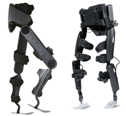
As of 2019, many different models of exoskeletons exist, and this number continues to grow as research and technology progresses. However, it is important to note that only three have been approved by the FDA for sale in North America and only two have been approved for home use, while the other is only approved for research and rehabilitation purposes. The two that are approved for home use are the ReWalk and the Indego, while the Ekso has been approved for research and rehabilitation purposes. The current models of exoskeletons have the ability to move from 0.2-2.6 km/hr, and weigh between 12-38 kg.

Exoskeletons are slowly moving into the community. Currently, most models can only move over flat, smooth surfaces. However, some can go up inclines. Moreover, newer models have the added function of enabling the user to sit in or wheel a wheelchair without having to take off the device. These features can increase the independence of people with SCI and enable/enhance the performance of activities such as standing, walking, and climbing stairs. However, these devices are still expensive, ranging from $70,000-120,000 USD.

In deciding if robotic exoskeletons are an appropriate option for you, a health care provider will perform an assessment where they account for factors such as your level of injury, risk for falls and fractures, and range of movement. Once exoskeletons are deemed appropriate, training will be required to learn how to properly use the device. A physiotherapist or caregiver may help you put on and take off the device. Generally, the feet are placed on footplates and the torso, hips, and legs are strapped into the exoskeleton. Some models extend upwards to include a backpack-like structure, which offers more trunk support and contains the computer and battery. Other models are secured only at the waist and below. In addition, some models can be programmed externally using a tablet. A pair of forearm crutches or a walker is often used to maintain balance. As someone with SCI walks, the built-in sensors and motorized joints (hip, knee, and/or ankle) constantly accommodate to encourage a rhythmic walking pattern. When the individual improves their own walking function and requires less assistance, the exoskeleton can be adjusted to provide less support.


A conceptual diagram of the Hardiman.

The term “exoskeleton” was originally borrowed from animal biology. An exoskeleton (such as a shell) is an outer cover that protects and supports the animal. In a similar way, robotic exoskeletons have been designed to help externally support and enhance human movement.

The earliest exoskeletons were developed with a military focus, aimed to help soldiers carry heavier loads, run faster, and jump higher. In 1968, the first exoskeleton, dubbed “the Hardiman” was developed in partnership with the US military. This exoskeleton was originally designed to help amplify soldiers’ strength by 25 times (i.e., lifting a 1500 item would feel like you are lifting a 60 lb item). Although the Hardiman was created and worked, it was not without limitations. First of all, the exoskeleton itself weighed 1500 lb. It was hydraulically-powered and required pumps and bladders that could fill a room. Despite its abilities, it was not very functional.

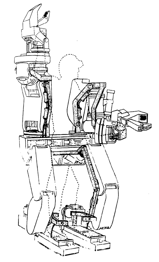
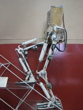
In 1972, a team from Yugoslavia developed the first functional exoskeleton: the kinematic walker. This device was the first of its kind; a powered robot consisting of a single hydraulic actuator (motor), which reduced its size. The kinematic walker was designed as a walking orthotic, and allowed for smooth movements to be made. Some movements the exoskeleton was able to perform included flexion and extension of the hip, knee, and ankle, in addition to abduction and adduction of the legs. Although this exoskeleton only weighed 12 kg, it required a separate power source and a computer, which were externally located from the exoskeleton.

For more information on treadmill-based robotics, refer to our articles on Body Weight Supported Treadmill Training and Functional Electrical Stimulation for more information on treadmill based robotics.

In 2001, the first exoskeleton products started being sold. The Lokomat, a robotic exoskeleton that was suspended over a treadmill, was one of the first exoskeletons used for rehabilitation. Meanwhile, the US Defense Advanced Research Projects Agency (DARPA) had announced their Human Performance Augmentation program which offered funding to develop exoskeletons for military use. From this funding, two separate groups developed exoskeletons – the Berkeley Lower Extremity Exoskeleton (BLEEX) and the Raytheon XOS suit. Both suits were created to help soldiers carry extra weight (up to 200 lb!) without feeling it.

Starting in 2010, more rehabilitation-based exoskeletons started entering the market. These include gait-assistive devices commonly seen in the media today, such as the Ekso, the ReWalk, and the Indego exoskeletons. These models are described in the section below


There are many types of exoskeletons that are currently available for use for individuals with SCI. While some exoskeletons have only been cleared for rehabilitation purposes, others have separate models for personal or home use. Below we discuss the general differences between rehabilitation and personal exoskeletons.

Rehabilitation Exoskeletons

The Ekso (left), the ReWalk (middle), and the REX (right) exoskeletons.4-6

Exoskeletons used for rehabilitation are generally heavier than personal exoskeletons and are often controlled by a computer and battery located in a backpack worn by the user. However, rehabilitation exoskeletons often come with more customizable abilities. For example, the Ekso and the ReWalk allow the clinician to modify the amount of power (i.e., support from the robot) for each leg, depending on the ability of the user. Similarly, the therapy version of the Indego allows the clinician to control how much weight is supported by the exoskeleton and how much movement assistance is provided by the exoskeleton. Moreover, how steps are triggered can be programmed in different ways: e.g., by the press of a button, by shifting your body weight, or by initiating a step using your own muscles. These bulkier clinical models are usually one-size-fits-all, allowing clinicians to adjust the dimensions of the device (i.e., leg length, hip width, etc.) to treat their clients of varying body sizes.

The REX differs from all of the aforementioned exoskeletons in that it is controlled with a joystick and is able to self-balance, making it essentially “hands-free”. This feature allows individuals to perform exercises while standing, including squats, lunges, and upper body exercises using both hands.

The Indego (left) and the ReWalk (right).

Personal Exoskeletons

Exoskeletons that have been designed for home use are generally lighter and provide limited support to the torso. Personal exoskeletons have a battery pack that is connected to the waist, but otherwise operates similarly to the rehabilitation models in that a weight shift initiates a step. Furthermore, some exoskeleton companies have developed apps that accompany the exoskeleton, allowing the user to independently access performance data. These personal exoskeletons are usually custom-developed to fit the user’s unique body dimensions.

While there are many purported benefits of using robotic exoskeletons among individuals with SCI, strong research evidence is lacking. Exoskeletons are largely used to enhance mobility in SCI, where they can be used for walk (gait) training during rehabilitation or in the community (at home) to perform simple daily activities. Although further research is required, some research has suggested that walking with an exoskeleton may have additional health benefits:

Spasticity

There are mixed findings regarding the effects of exoskeletal walking on spasticity. Many (weak evidence) studies have found that using exoskeletons can result in decreased spasticity. Despite these noted benefits, one weak evidence study reported mixed findings on spasticity as 26.7% of their study sample saw a decrease in spasticity, while 62.2% of participants saw no change and 11.1% saw an increase in spasticity. The impact of exoskeletal walking on spasticity may be related to the user’s baseline level of spasticity. In a weak evidence study, users who had low levels of spasticity prior to using an exoskeleton experienced an increase in spasticity; however, this increase in spasticity ultimately decreased over 12 weeks back to near zero. Individuals who already had high levels of spasticity prior to walking in an exoskeleton saw no changes in spasticity.

Bowel function

There is some weak research evidence that suggests walking with an exoskeleton can help with various bowel functions, including improved regularity of bowel movements, less time required for bowel management, and decreased enema dose. However, two (weak evidence) studies have found no effects on bowel function.


Bone Health

After SCI, bone mineral density in the legs declines at rapid rates due to inactivity and weight bearing activities have the potential to help restore bone mineral density. One study (weak evidence) found that walking in an exoskeleton may increase bone mineral density up to 14% with 6 weeks of training.

Fitness

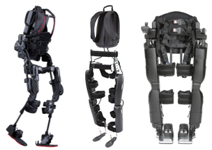
Some research (weak evidence) suggests that walking in an exoskeleton can provide good exercise for the heart and upper and lower limb strength in those with incomplete SCI. More details about the effect of exoskeletons on fitness are discussed below in the section: “Can I get exercise benefits from walking in an exoskeleton?”

Pain

A few studies (weak evidence) report a decrease in pain with exoskeleton use, with one noting a reduction in pain, but not enough to significantly impact everyday life (i.e., clinical significance). On the other hand, some weak evidence studies have found no effect of exoskeleton walking on pain.

Pressure Sores

There is some weak research evidence that walking in an exoskeleton may help avoid the negative effects of prolonged standing or sitting  (e.g., pressure ulcers).


Refer to our articles on Pressure Sores, Pain and Spasticity for more information!

While a number of benefits are associated with exoskeleton use, there are also factors to consider prior to using the device in therapy or for the long term.

Risks of using an exoskeleton

Robotic exoskeletons are generally safe when used with discretion. However, there are some risks associated with its use. Mild adverse effects that have been reported in research include: skin redness, small abrasions (i.e., scrapes), mild joint swelling, and mild bruising. Additionally, like other simple orthotics and braces, falls and fractures have been identified as a risk. It is suggested (weak evidence) that family and friends of exoskeleton users should be trained to deal with emergency situations, such as falls or the exoskeleton shutting off unexpectedly.

Considerations of using an exoskeleton in the community

While exoskeleton home use may seem promising, there are some limitations. These include:

  • Slow walking speeds, which may not be ideal for everyday activities.
  • High costs to purchase the device.
  • Lack of availability of community-based exoskeletons.
  • Limited capacity or poor efficiency in moving on uneven surfaces (e.g., hills, steps) or complex movements (turning, side-stepping, backwards walking).
  • Being prone to water damage (they are not waterproof).

There are certain situations where extra attention is needed to determine whether robotic exoskeletons are appropriate and safe. Consult a qualified health provider for further safety information. Robotic exoskeletons are not recommended for individuals:

Extreme contractures are a contraindication for using exoskeletons.

  • Who are unable to tolerate standing, even with an assistive device (walking frame, bracing), due to pain or other complications (autonomic dysreflexia, orthostatic hypotension).
  • With severe neurological injuries (apart from SCI).
  • With severe or uncontrolled spasticity.
  • With osteoporosis.
  • With fractures.
  • With severe contractures (deformities that cause joint and muscle stiffness and limit normal or functional movement of the limbs).

Using Functional Electrical Stimulation (FES) to counter spasticity

Researchers built a novel device that integrates FES into an exoskeleton to address the issue of severe spasticity affecting exoskeleton use. In this FES-exoskeleton hybrid, FES complemented the exoskeleton by stimulating tight extensor muscles to facilitate walking. The authors found that spasticity was temporarily reduced when FES was used when walking with an exoskeleton. Furthermore, it was found that the knee was more easily extended when moving from a sit-to-stand position, and the forces applied on the knee during sit-to-stand were reduced. While this (weak evidence) study provides some promise for individuals with severe spasticity to also use exoskeletons, further research is required.

Using an exoskeleton requires practice. Although walking in an exoskeleton may seem daunting at first, research suggests that walking proficiency improves over time. In both newly injured (i.e., less than 6 months since injury) and chronically injured individuals with SCI, weak evidence shows that walking in the exoskeleton improves over time.

You get faster

Among newly injured individuals, weak evidence from one study suggests that walking speed in an exoskeleton becomes 3.2x faster after 25 1-hour training sessions. Moreover, these individuals were able to walk further in an exoskeleton after their training sessions. Among chronically injured individuals, similar trends are seen with increases in exoskeleton walking speed in two weak evidence studies. In another weak evidence study, it was found that 21 sessions were required to achieve the near-maximal walking speed at the end of a 12-week period, while 62 sessions were required to achieve near-maximal walking distance.

Less effort is Required

There is both weak and moderate evidence suggesting that the amount of effort it takes to walk using an exoskeleton decreases over time. This has been evaluated both subjectively (i.e., people feel like walking in an exoskeleton is not as hard over time) and physiologically (i.e., less demand on your body). This suggests that individuals are able to walk longer distances with lower effort after training to use an exoskeleton.

Is using an exoskeleton different for those with acute injuries versus chronic injuries?

Researchers have noted differences in exoskeleton use among newly injured (e.g., in patients) and chronically injured individuals with respect to adverse effects and benefits received. Among people who have recently sustained an SCI, weak evidence indicates that the most common adverse effect was orthostatic hypotension (a sudden drop in blood pressure). One study found that orthostatic hypotension commonly occurred after the first stand or after pauses (e.g., to take vitals, or to turn). However, the frequency of orthostatic hypotension episodes tapered off after a couple of sessions. Furthermore, another study (weak evidence) suggests that newly injured individuals may see improvements in their independence and quality of life, whereas those with chronic injuries do not. More research is required to determine the significance of differences between acutely injured and chronically injured individuals who use exoskeletons.

What factors influence walking speed?

Among individuals with SCI who use current exoskeleton devices, the average walking speed is 0.26 meters per second. This speed is fairly slow, and is lower than the average speed required to walk proficiently in the community (0.8 meters per second) and to cross the street safely (1.06 meters per second). However, walking speed in an exoskeleton is subject to improvement, depending on various factors.

Some factors that influence walking speed include age, level and type of injury, and the amount of training one receives. There is some (weak) evidence suggesting that those with incomplete, lower level SCI are more likely to exhibit faster walking speeds. In particular, one (weak evidence) study found that those with lower level paraplegia (i.e., T9-L1) were able to walk at significantly higher speeds. Moreover, there was a weak correlation between older age and faster walking speeds (i.e., older adults walk slightly faster than younger adults), though this could be related to the age-related difference in injury severity (that is, older individuals had lower levels of injury). No correlation was found with a greater time since injury.

What factors influence skill acquisition?

The time it takes to acquire proficient skills to walk in an exoskeleton varies greatly, ranging from 6-23 sessions. A variety of factors influence skill acquisition, including lifestyle, age, age at injury, and body mass index (BMI). Weak evidence suggests that an active lifestyle is the most important predictor of skill performance, although being younger and having a lower BMI are also associated with higher skill level. Additionally, the authors note that while having a lower level of injury was a positive predictor of skill between 2-4 weeks of using an exoskeleton, it did not predict final skill levels.

The type of exoskeleton being used may also influence skill acquisition. For example, weak evidence notes that a device with more support to the torso may facilitate skill acquisition as it provides more stability. Although we have summarized the research on factors influencing skill acquisition, the type of exoskeleton used in each study was not accounted for. As a result, we are unable to tease apart the effects of the aforementioned factors (e.g., lifestyle, age, BMI) and of the exoskeleton type on acquiring skills. The time it takes to acquire proficient skills to walk in an exoskeleton varies greatly, ranging from 6-23 sessions. A variety of factors influence skill acquisition, including lifestyle, age, age at injury, and body mass index (BMI). Weak evidence suggests that an active lifestyle is the most important predictor of skill performance, although being younger and having a lower BMI are also associated with higher skill level. Additionally, the authors note that while having a lower level of injury was a positive predictor of skill between 2-4 weeks of using an exoskeleton, it did not predict final skill levels.

Although walking in an exoskeleton is used primarily for rehabilitation purposes, the effort required to use the device is strenuous enough to be considered exercise. For example, one (weak evidence) study found that walking in an exoskeleton requires 3.34 times more effort than pushing a wheelchair, and 1.9 times more effort compared to walking without impairment, despite walking 7.4 times slower. Not surprisingly, participants also perceive themselves to be working harder. Participants from three (weak evidence) studies exercised at a moderate intensity, which is enough to get cardiovascular benefits, while walking in a robotic exoskeleton. Although some participants from a (weak evidence) study reported working at a low intensity, the authors noted that based on their heart rate and oxygen consumption, they were actually working at a moderate intensity. This suggests that some people may actually be working their bodies harder than they feel they are!

So why is walking in an exoskeleton so much work? Research suggests that using an exoskeleton requires a lot of work from the arms and torso to support an upright posture and to shift weight to initiate stepping. However, relative to other walking orthotics (e.g., robotic gait orthoses, hip-knee-ankle-foot orthoses), FES, and bracing, walking in an exoskeleton is considerably less effort. So then why would you use an exoskeleton for exercise if using other walking orthotics is harder work? The important consideration is stamina. You might work harder walking with rigid braces, but you may tire out quickly. With an exoskeleton, the understanding is that the assisted walking could allow you to exercise at moderate intensity for much longer.

Currently, there are only two exoskeleton models that have been approved for community use in North America. As such, there is limited evidence for using an exoskeleton in the community. In order to use an exoskeleton independently, individuals should be able to put on and take off the exoskeleton without professional help. Weak evidence has shown that individuals with paraplegia are able to independently put on and take off these devices, although those with tetraplegia are not. Additionally, weak evidence has shown that the time it takes to put on and take off an exoskeleton can be reduced with practice. Regarding walking speed in indoor versus outdoor environments, one weak evidence study has found that there are no significant differences in speed.

Some studies have looked at home- and community-based skills that can be completed using an exoskeleton. One weak evidence study found that the majority of participants could walk independently without a trainer in an exoskeleton, and perform tasks such as reaching high cupboards, using a stove, and using a sink, but were not able to walk on carpet and ramps. However, the authors note that some tasks were more difficult to complete in an exoskeleton, including reaching low cupboards and opening a fridge and getting items. Other (weak evidence) studies have found that a small proportion of people were able to do more advanced community-based tasks, such as entering/exiting elevators, operating automatic doors, navigating revolving doors, and ordering at a café. More research is required to determine how quickly individuals can pick up these skills.

There are currently many models of robotic exoskeletons continuing to be developed and refined. Exoskeletons are primarily used for rehabilitation purposes, although some models are available for community use. Using an exoskeleton has been shown to be relatively safe and easy to learn. Many benefits have been reported, including being able to ambulate, improvements in bone health, heart health, spasticity, bowel functioning, fitness, and pressure sores. While there are a lot of positive findings for robotic exoskeletons, this is an emerging field and stronger research is required to support these beneficial claims. A strong consideration is to weigh these benefits against cost, and also compare how these benefits compare to other to achieve similar gains.

For a review of how we assess evidence at SCIRE Community and advice on making decisions, please see SCIRE Community Evidence.

Parts of this page have been adapted from the SCIRE Project (Professional) “Lower Limb” Chapter:

Lam T, Wolfe DL, Domingo A, Eng JJ, Sproule S (2014). Lower Limb Rehabilitation Following Spinal Cord Injury. In: Eng JJ, Teasell RW, Miller WC, Wolfe DL, Townson AF, Hsieh JTC, Connolly SJ, Noonan VK, Loh E, McIntyre A, editors. Spinal Cord Injury Rehabilitation Evidence. Version 5.0. Vancouver: p 1-74.

Available from: scireproject.com/evidence/rehabilitation-evidence/lower-limb/ 

Evidence for “What are robotic exoskeletons?” is based on the following studies:

Khan, A. S., Livingstone, D. C., Hurd, C. L., Duchcherer, J., Misiaszek, J. E., Gorassini, M. A., … Yang, J. F. (2019). Retraining walking over ground in a powered exoskeleton after spinal cord injury: a prospective cohort study to examine functional gains and neuroplasticity, 1–17. Retrieved from https://doi.org/10.1186/s12984-019-0585-x

Ali, H. (2014). Bionic Exoskeleton: History, Development and the Future. IOSR Journal of Mechanical and Civil Engineering (IOSR-JMCE), 2014, 58–62. Retrieved from http://iosrjournals.org/iosr-jmce/papers/ICAET-2014/me/volume-5/12.pdf?id=7622

Gardner, A. D., Potgieter, J., & Noble, F. K. (2017). A review of commercially available exoskeletons’ capabilities. 2017 24th International Conference on Mechatronics and Machine Vision in Practice, M2VIP 2017, 2017Decem, 1–5.

He, Y., Eguren, D., Luu, T. P., & Contreras-Vidal, J. L. (2017). Risk management and regulations for lower limb medical exoskeletons: A review. Medical Devices: Evidence and Research, 10, 89–107.

Evidence for “What is the history behind robotic exoskeletons?” is based on the following studies:

Yang, C. J., Zhang, J. F., Chen, Y., Dong, Y. M., & Zhang, Y. (2008). A review of exoskeleton-type systems and their key technologies. Proceedings of the Institution of Mechanical Engineers, Part C: Journal of Mechanical Engineering Science, 222(8), 1599–1612.

Ali, H. (2014). Bionic Exoskeleton: History, Development and the Future. IOSR Journal of Mechanical and Civil Engineering (IOSR-JMCE), 2014, 58–62. Retrieved from http://iosrjournals.org/iosr-jmce/papers/ICAET-2014/me/volume-5/12.pdf?id=7622

Evidence for “What are exoskeletons used for?” is based on the following studies:

Esquenazi, A., Talaty, M., Packel, A., & Saulino, M. (2012). The Rewalk powered exoskeleton to restore ambulatory function to individuals with thoracic-level motor-complete spinal cord injury. American Journal of Physical Medicine and Rehabilitation, 91(11), 911–921.

Kolakowsky-Hayner, S. A. (2013). Safety and Feasibility of using the EksoTM Bionic Exoskeleton to Aid Ambulation after Spinal Cord Injury. Journal of Spine.

Kozlowski, A. J., Bryce, T. N., & Dijkers, M. P. (2015). Time and effort required by persons with spinal cord injury to learn to use a powered exoskeleton for assisted walking. Topics in Spinal Cord Injury Rehabilitation, 21(2), 110–121.

Kressler, J., Thomas, C. K., Field-Fote, E. C., Sanchez, J., Widerström-Noga, E., Cilien, D. C., … Nash, M. S. (2014). Understanding therapeutic benefits of overground bionic ambulation: Exploratory case series in persons with chronic, complete spinal cord injury. Archives of Physical Medicine and Rehabilitation, 95(10), 1878-1887.e4.

Zeilig, G., Weingarden, H., Zwecker, M., Dudkiewicz, I., Bloch, A., & Esquenazi, A. (2012). Safety and tolerance of the ReWalkTM exoskeleton suit for ambulation by people with complete spinal cord injury: A pilot study. Journal of Spinal Cord Medicine, 35(2), 96–101.

Juszczak, M., Gallo, E., & Bushnik, T. (2018). Examining the effects of a powered exoskeleton on quality of life and secondary impairments in people living with spinal cord injury. Topics in Spinal Cord Injury Rehabilitation, 24(4), 336–342.

Khan, A. S., Livingstone, D. C., Hurd, C. L., Duchcherer, J., Misiaszek, J. E., Gorassini, M. A., … Yang, J. F. (2019). Retraining walking over ground in a powered exoskeleton after spinal cord injury: a prospective cohort study to examine functional gains and neuroplasticity, 1–17. Retrieved from https://doi.org/10.1186/s12984-019-0585-x

Karelis, A. D., Carvalho, L. P., Castillo, M. J. E., Gagnon, D. H., & Aubertin-Leheudre, M. (2017). Effect on body composition and bone mineral density of walking with a robotic exoskeleton in adults with chronic spinal cord injury. Journal of Rehabilitation Medicine, 49(1), 84–87.

Escalona, M. J., Brosseau, R., Vermette, M., Comtois, A. S., Duclos, C., Aubertin-Leheudre, M., & Gagnon, D. H. (2018). Cardiorespiratory demand and rate of perceived exertion during overground walking with a robotic exoskeleton in long-term manual wheelchair users with chronic spinal cord injury: A cross-sectional study. Annals of Physical and Rehabilitation Medicine, 61(4), 215–223.

Mcintosh, K., Charbonneau, R., Bensaada, Y., Bhatiya, U., & Ho, C. (2019). The Safety and Feasibility of Exoskeletal-Assisted Walking in Acute Rehabilitation After Spinal Cord Injury. Archives of Physical Medicine and Rehabilitation. Retrieved from https://doi.org/10.1016/j.apmr.2019.09.005

Stampacchia, G., Rustici, A., Bigazzi, S., Gerini, A., Tombini, T., & Mazzoleni, S. (2016). Walking with a powered robotic exoskeleton: Subjective experience, spasticity and pain in spinal cord injured persons. NeuroRehabilitation, 39(2), 277–283.

Baunsgaard, C. B., Nissen, U. V., Brust, A. K., Frotzler, A., Ribeill, C., Kalke, Y. B., … Benito Penalva, J. (2018). Exoskeleton gait training after spinal cord injury: An exploratory study on secondary health conditions. Journal of Rehabilitation Medicine, 50(9), 806–813.

Evidence for “What are the risks and considerations for using an exoskeleton?” is based on the following studies:

Tefertiller, C., Hays, K., Jones, J., Jayaraman, A., Hartigan, C., Bushnik, T., & Forrest, G. F. (2018). Initial outcomes from a multicenter study utilizing the indego powered exoskeleton in spinal cord injury. Topics in Spinal Cord Injury Rehabilitation, 24(1), 78–85.

Mcintosh, K., Charbonneau, R., Bensaada, Y., Bhatiya, U., & Ho, C. (2019). The Safety and Feasibility of Exoskeletal-Assisted Walking in Acute Rehabilitation After Spinal Cord Injury. Archives of Physical Medicine and Rehabilitation. Retrieved from https://doi.org/10.1016/j.apmr.2019.09.005

Mekki, M., Delgado, A. D., Fry, A., Putrino, D., & Huang, V. (2018). Robotic Rehabilitation and Spinal Cord Injury: a Narrative Review. Neurotherapeutics, 15(3), 604–617.

Miller, L. E., Zimmermann, A. K., & Herbert, W. G. (2016). [Miller, 2016] Clinical effectiveness and safety of powered exoskeleton-assisted walking on SCI patients, 455–466.

Van Herpen, F. H. M., Van Dijsseldonk, • R B, Rijken, • H, Keijsers, • N L W, Louwerens, J. W. K., & Van Nes, • I J W. (2019). Spinal Cord Series and Cases Case Report: Description of two fractures during the use of a powered exoskeleton. Retrieved from https://doi.org/10.1038/s41394-019-0244-2

Kandilakis, C., & Sasso-Lance, E. (n.d.). Exoskeletons for Personal Use After Spinal Cord Injury. Retrieved from https://doi.org/10.1016/j.apmr.2019.05.028

Evidence for “Are there restrictions or precautions for using robotic exoskeletons?” is based on the following studies:

Miller, L. E., Zimmermann, A. K., & Herbert, W. G. (2016). [Miller, 2016] Clinical effectiveness and safety of powered exoskeleton-assisted walking on SCI patients, 455–466.

Murray, S. A., Farris, R. J., Golfarb, M., Hartigan, C., Kandilakis, C., & Truex, D. (2018). FES Coupled with A Powered Exoskeleton for Cooperative Muscle Contribution in Persons with Paraplegia. Proceedings of the Annual International Conference of the IEEE Engineering in Medicine and Biology Society, EMBS, 2018July, 2788–2792.

Ekelem, A., & Goldfarb, M. (2018). Supplemental stimulation improves swing phase kinematics during exoskeleton assisted gait of SCI subjects with severe muscle spasticity. Frontiers in Neuroscience, 12(JUN).

Evidence for “How does walking change over time?” is based on the following studies:

Mcintosh, K., Charbonneau, R., Bensaada, Y., Bhatiya, U., & Ho, C. (2019). The Safety and Feasibility of Exoskeletal-Assisted Walking in Acute Rehabilitation After Spinal Cord Injury. Archives of Physical Medicine and Rehabilitation. Retrieved from https://doi.org/10.1016/j.apmr.2019.09.005

Ramanujam, A., Momeni, K., Husain, S. R., Augustine, J., Garbarini, E., Barrance, P., … Forrest, G. F. (2018). Mechanisms for improving walking speed after longitudinal powered robotic exoskeleton training for individuals with spinal cord injury. Proceedings of the Annual International Conference of the IEEE Engineering in Medicine and Biology Society, EMBS, 2018July, 2805–2808.

Tefertiller, C., Hays, K., Jones, J., Jayaraman, A., Hartigan, C., Bushnik, T., & Forrest, G. F. (2018). Initial outcomes from a multicenter study utilizing the indego powered exoskeleton in spinal cord injury. Topics in Spinal Cord Injury Rehabilitation, 24(1), 78–85.

Khan, A. S., Livingstone, D. C., Hurd, C. L., Duchcherer, J., Misiaszek, J. E., Gorassini, M. A., … Yang, J. F. (2019). Retraining walking over ground in a powered exoskeleton after spinal cord injury: a prospective cohort study to examine functional gains and neuroplasticity, 1–17. Retrieved from https://doi.org/10.1186/s12984-019-0585-x

Escalona, M. J., Brosseau, R., Vermette, M., Comtois, A. S., Duclos, C., Aubertin-Leheudre, M., & Gagnon, D. H. (2018). Cardiorespiratory demand and rate of perceived exertion during overground walking with a robotic exoskeleton in long-term manual wheelchair users with chronic spinal cord injury: A cross-sectional study. Annals of Physical and Rehabilitation Medicine, 61(4), 215–223.

Delgado, A. D., Escalon, M. X., Bryce, T. N., Weinrauch, W., Suarez, S. J., & Kozlowski, A. J. (2019). Safety and feasibility of exoskeleton-assisted walking during acute/sub-acute SCI in an inpatient rehabilitation facility: A single-group preliminary study. Journal of Spinal Cord Medicine. Retrieved from https://www.tandfonline.com/action/journalInformation?journalCode=yscm20

Baunsgaard, C. B., Nissen, U. V., Brust, A. K., Frotzler, A., Ribeill, C., Kalke, Y. B., … Benito Penalva, J. (2018). Exoskeleton gait training after spinal cord injury: An exploratory study on secondary health conditions. Journal of Rehabilitation Medicine, 50(9), 806–813.

Evidence for “What influences walking speed and skill acquisition when using an exoskeleton?” is based on the following studies:

Louie, D. R., Eng, J. J., & Lam, T. (2015). Gait speed using powered robotic exoskeletons after spinal cord injury: A systematic review and correlational study. Journal of NeuroEngineering and Rehabilitation, 12(1), 1–10. Retrieved from http://dx.doi.org/10.1186/s12984-015-0074-9

Hartigan, C., Kandilakis, C., Dalley, S., Clausen, M., Wilson, E., Morrison, S., … Farris, R. (2015). Mobility outcomes following five training sessions with a powered exoskeleton. Topics in Spinal Cord Injury Rehabilitation, 21(2), 93–99.

van Dijsseldonk, R. B., Rijken, H., W van Nes, I. J., van de Meent, H., W Keijsers, N. L., & W Keijsers,  el L. (2019). Predictors of exoskeleton motor learning in spinal cord injured patients. Disability and Rehabilitation, 1-7.

Khan, A. S., Livingstone, D. C., Hurd, C. L., Duchcherer, J., Misiaszek, J. E., Gorassini, M. A., … Yang, J. F. (2019). Retraining walking over ground in a powered exoskeleton after spinal cord injury: a prospective cohort study to examine functional gains and neuroplasticity, 1–17. Retrieved from https://doi.org/10.1186/s12984-019-0585-x

Evidence for “Can I get exercise benefits from walking in an exoskeleton?” is based on the following studies:

Khan, A. S., Livingstone, D. C., Hurd, C. L., Duchcherer, J., Misiaszek, J. E., Gorassini, M. A., … Yang, J. F. (2019). Retraining walking over ground in a powered exoskeleton after spinal cord injury: a prospective cohort study to examine functional gains and neuroplasticity, 1–17. Retrieved from https://doi.org/10.1186/s12984-019-0585-x

Escalona, M. J., Brosseau, R., Vermette, M., Comtois, A. S., Duclos, C., Aubertin-Leheudre, M., & Gagnon, D. H. (2018). Cardiorespiratory demand and rate of perceived exertion during overground walking with a robotic exoskeleton in long-term manual wheelchair users with chronic spinal cord injury: A cross-sectional study. Annals of Physical and Rehabilitation Medicine, 61(4), 215–223.

Kozlowski, A. J., Bryce, T. N., & Dijkers, M. P. (2015). Time and effort required by persons with spinal cord injury to learn to use a powered exoskeleton for assisted walking. Topics in Spinal Cord Injury Rehabilitation, 21(2), 110–121.

Mcintosh, K., Charbonneau, R., Bensaada, Y., Bhatiya, U., & Ho, C. (2019). The Safety and Feasibility of Exoskeletal-Assisted Walking in Acute Rehabilitation After Spinal Cord Injury. Archives of Physical Medicine and Rehabilitation. Retrieved from https://doi.org/10.1016/j.apmr.2019.09.005

Evidence for “What evidence is there for using an exoskeleton in the community?” is based on the following studies:

Tefertiller, C., Hays, K., Jones, J., Jayaraman, A., Hartigan, C., Bushnik, T., & Forrest, G. F. (2018). Initial outcomes from a multicenter study utilizing the indego powered exoskeleton in spinal cord injury. Topics in Spinal Cord Injury Rehabilitation, 24(1), 78–85.

Hartigan, C., Kandilakis, C., Dalley, S., Clausen, M., Wilson, E., Morrison, S., … Farris, R. (2015). Mobility outcomes following five training sessions with a powered exoskeleton. Topics in Spinal Cord Injury Rehabilitation, 21(2), 93–99.

Khan, A. S., Livingstone, D. C., Hurd, C. L., Duchcherer, J., Misiaszek, J. E., Gorassini, M. A., … Yang, J. F. (2019). Retraining walking over ground in a powered exoskeleton after spinal cord injury: a prospective cohort study to examine functional gains and neuroplasticity, 1–17. Retrieved from https://doi.org/10.1186/s12984-019-0585-x

Spungen, A. M., Asselin, P. K., Fineberg, D. B., Kornfeld, S. D., & Harel, N. Y. (2012). Exoskeletal-Assisted Walking for Persons with Motor-Complete Paraplegia. VA Rehabilitation Research and Development National Center of Excellence for the Medical Consequences of Spinal Cord Injury. Retreived from: http://www.ryzur.com.cn/uploadfile/2016/0830/20160830115519272.pdf

Miller, L. E., Zimmermann, A. K., & Herbert, W. G. (2016). Clinical effectiveness and safety of powered exoskeleton-assisted walking on SCI patients. Medical Devices: Evidence and Research, 9:455–466.

Image credits

  1. Walking with a Clinician ©The SCIRE Community Team
  2. Hardiman I ©Bruce R. Fick and John B. Makinson, General Elerctric Co., Public Domain
  3. Active Suit ©Robotics Laboratory, Mihailo Pupin Institute
  4. Ekso Exoskeleton ©Ekso Bionics 2020
  5. ReWalk Exoskeleton ©ReWalk Robotics 2020
  6. REX Exoskeleton ©REX Bionics Ltd 2020
  7. Indego Exoskeleton ©Parker Hannifin Corp 2020
  8. ReWalk Exoskeleton ©ReWalk Robotics 2020
  9. Spasticity ©The SCIRE Community Team
  10. Colon ©Servier Medical Art, CC BY 3.0
  11. Femur ©Servier Medical Art, CC BY 3.0
  12. Modified from: Beating heart ©Lillit Kalachyan, CC BY 3.0
  13. Lightning ©FLPLF, CC BY 3.0
  14. ModifiedfromSpasticity ©The SCIRE Community Team
  15. Ankle sprain ©Servier Medical Art, CC BY 3.0
  16. Exoskeleton Icon ©The SCIRE Community Team
  17. Downtown New York City streets ©Free-photos, Pixabay License
  18. Candles ©The SCIRE Community Team
  19. Modified from: Weight Scale ©Sandra, CC BY 3.0
  20. Wheelchair Tennis ©Gan Khoon Lay, CC BY 3.0
  21. Personal user photo courtesy of Parker Hannifin Corporation, USA


Disclaimer: This document does not provide medical advice. This information is provided for educational purposes only. Consult a qualified health professional for further information or specific medical advice. The SCIRE Project, its partners and collaborators disclaim any liability to any party for any loss or damage by errors or omissions in this publication.

Wheelchair Propulsion Assist Devices

By | | No Comments

Author: Sharon Jang | Reviewer: Jaimie Borisoff | Published: 15 May 2020 | Updated: ~

Wheelchair propulsion assist devices are pieces of equipment that can facilitate the use of a manual wheelchair. This page provides an overview of the different options available. SCIRE Community is not affiliated with and does not endorse any of the specific products mentioned on this page.

Key Points

  • Propulsion assist devices are technologies that attach to your manual wheelchair to facilitate propulsion
  • There is limited research on the effectiveness of these devices, but they are clinically well-accepted with perceived benefits associated with their use
  • There are options for both powered and non-powered propulsion assist devices, depending on your needs
  • Propulsion assist devices can be either front frame-mounted, replace your rear wheels, or rear frame-mounted
  • Propulsion assist devices are rapidly developing and changing; specific products in this article may no longer be available or unavailable in your area

An example of a front-mounted propulsion assist device.1

Propulsion assist devices are pieces of equipment that can be attached to a manual wheelchair to facilitate the ease of using a wheelchair, or expand the capabilities of a manual wheelchair. These devices generally work by reducing the amount of effort required by the user to move their wheelchair. Use of these devices may help individuals go up hills, reduce the amount of force required to start moving the wheelchair, or travel over difficult terrain such as grass. Propulsion assist devices are normally added onto the front or back of the chair frame, but also sometimes replace the rear wheels. These devices can be further split up into two categories: powered and non-powered.

As technology is rapidly progressing, novel devices are being frequently released. The development of multiple devices to fill a niche market has resulted in some products becoming unavailable in certain areas, or leaving the market altogether. The quick changes and developments in this area make conducting research on propulsion assist devices difficult. Currently, there is limited research on the use of propulsion assist devices for manual wheelchair users and most of the research is focussed on the devices that replace the rear wheels.

Using a propulsion assist device can be beneficial to manual wheelchair users. The man on the left is using a powered add-on.2

Power assist devices may be purchased by individuals with or without a prescription. Some indications for propulsion assist device use are limited strength in the upper body, or for those that are at a higher risk of an injury or pain to the arms and/or shoulders. These devices may improve participation in the community by enabling increased travel distance and access to more difficult environments.

In two studies (weak evidence), users of propulsion assist devices have reported benefits including:

  • Less strain on the cardiovascular and respiratory systems; this results in reduced overall feelings of fatigue.
  • Being able to go further with less effort (improved propulsion efficiency). This may help prevent overuse injuries.
  • Increased distance you are able to travel before feeling tired.
  • Travelling up and down hills.
  • Improved ability to travel on non-paved roads (e.g., grass, gravel, dirt), which increases the variety of spaces a wheelchair user may access.

Although there is limited research on propulsion assist devices, Pushrim Activated Power-Assist Wheelchairs (PAPAW) are the most researched device. Evidence has suggested that using PAPAWs have multiple benefits. One study provided moderate evidence for the use of PAPAWs to help individuals with SCI who have shoulder pain to propel their wheelchair further distances. The participants in this study also required less energy to wheel, and perceived that wheeling was easier with the use of PAPAWs. Other (weak) evidence has also shown that using a PAPAW may reduce the frequency of arm/shoulder injuries and the amount of energy required. Additional (weak) evidence suggests that individuals with tetraplegia using PAPAWs have improved ability to manoeuvre through a variety of textured surfaces and to complete activities of daily living.

Although propulsion assist devices can facilitate the use of a manual wheelchair, there are also some risks associated with their use. While this is not an exhaustive list, risks include:

Increased risk of wheelchair damage

Most manual wheelchair frames may not have been designed to withstand the different forces applied by propulsion assist devices. This may lead to premature wear and tear, or possible breakage, on some parts of the wheelchair, such as the footplate. Sudden breakage during use may lead to injury of the wheelchair user.

Using a propulsion assist device may increase the risk of tipping when going up or down hills.3

Decreased stability

The addition of a propulsion assist device either in front or behind the wheelchair may alter the center of gravity on the wheelchair. This may result in greater instability, increasing the risks of tipping backwards when going up and down steep hills. Forward stability may be compromised as well and induce tipping (e.g. when moving down at the bottom of a curb cut).

Greater impact forces when encountering an obstacle

Propulsion assist devices, especially motorized ones, allow wheelchair users to travel at a greater speed. However, hitting obstacles such as a rock or a curb in your wheelchair at a higher speed can result in greater forces, which may lead to a decrease in stability and increase the risk of falls.

Potential lateral stability issues

Propulsion assist devices that attach to the front pose a risk of tipping sideways. Particularly when travelling at higher speeds, sudden turns can result in tipping over.

Prior to obtaining a propulsion assist device, you should always consult your health professional. That said, some factors to consider when deciding whether to invest in an add-on include the following factors:

Upper limb function

Upper limb function is required to use these devices. For example, the individual may be responsible for attaching/detaching the device to the wheelchair, and some degree of hand function is needed for operation. Some products do have adapted controls for use by individuals with limited hand function.

Terrain

The device should facilitate the transportation of the user over surfaces that they frequently travel on, or wish to travel on. Some terrain considerations include indoor travel, outdoor travel, uneven surfaces, soft terrains e.g. grass, and hills.

Transfer ability

It is important to take into consideration how a device may impact transferring in and out of a wheelchair. Will it make transfers more difficult?

Transport

If a car is used for transportation, consideration of the weight of the propulsion device should be considered. Will the weight of the propulsion assist device and the wheelchair be manageable to lift into a car? Will the propulsion assist device fit into the car?

Weight

Is the device light enough for the user to remove by themselves? Will the weight make manual propulsion more difficult when not in use (e.g. if battery is drained before end of travel)?

Interaction with wheelchair

Propulsion assist devices require a certain part of a wheelchair to connect to, which may vary between types of wheelchair (e.g., rigid vs folding wheelchairs) and types of add-on. How the device mounts to a wheelchair should be considered, including the user’s capability for performing the attachment/detachment. Additional consideration may be needed if the device is going to be used between different wheelchairs.

Addressing casters

Casters help provide stability to a wheelchair, but become a limiting factor when a wheelchair user wishes to travel off a smooth even surface. There are some devices that connect to the front of the wheelchair via the foot plate and lift up the front caster wheels. This allows the wheelchair user to travel over uneven surfaces easier, such as dirt paths, sand, and gravel. An example of this kind of device is a FreeWheel.

Mechanical advantage

There are some devices that facilitate pushing by amplifying your efforts (a mechanical advantage) using gears or levers. Using a device with a mechanical advantage will allow you to go further with a given push. One example of a device that provides mechanical advantage is a lever-propelled wheelchair. There are some lever devices that can be connected to your wheel (others require replacement of the rear wheels) that allow the user to propel by pushing the lever forward. Using a lever has two purported benefits: 1) using a longer lever to propel a wheelchair requires less force, and 2) using a lever enables a more favorable movement pattern of the hands, wrists, and shoulder. Together these may reduce the risk of injury. An example of this device is the NuDrive.

Pushrim-Activated Power-Assist Wheelchairs (PAPAWs)

A PAPAW is a manual wheelchair with motorized rear wheels. The wheels are powered by a battery, which is attached to the back of the wheelchair. This type of wheelchair is controlled by normal pushing movements using the pushrims. PAPAWs assist individuals with limited strength or arm function by amplifying the force applied to the wheels. Each push on the pushrim by the user is sensed and proportionally amplified to increase the force to continue the forward movement. This also occurs for braking and turning (e.g., the wheels would detect a backwards force and apply a stronger braking force). This allows for users to go further with a given push, or to brake more efficiently with less force. When desired, the power assist function can be turned off. One limitation of using PAPAWs is the significantly increased weight of the wheelchair, which is especially noticeable when turned off. Another limitation is that the pushrims are damaged more easily, as this is where the sensors are located. Some examples of PAPAWs include the Alber E-motion and the Quickie Xtender.

Front mounted systems

These systems attach to the front of the wheelchair frame and typically lift up the front casters. Non–powered versions exist which are propelled via cranks and chains similar to handcycles. Most often powered, these devices are able to turn manual wheelchairs into a three-wheeled “scooter”. Front mounted systems are usually controlled with a throttle controller, which removes the need to manually propel the wheelchair. They are also able to reach significantly higher speeds in comparison to other power add-ons. A disadvantage to front mounted systems is that they add length to the wheelchair, which may impact the ability to move around indoors. In addition, the devices can be fairly heavy (e.g., the typical Batec system is 25 kg), and little to no customizations can be made. Examples of front mounted systems include the Rio Mobility Firefly, Triride, Klaxon, and Batec.

Rear mounted systems

Rear mounted propulsion device generally connect to the bar underneath the wheelchair seat (i.e., the camber tube). These devices can power the wheelchair so that no manual propulsion is required, although turning and braking still require constant user control. Currently, there are two models commercially available that are light, compact, and easily attached: the SmartDrive and the Smoov. The SmartDrive is controlled with a wristband worn by the user. A double tap of the wristband (or similar accelerometer-sensed motion of the hand/arm when tapping the wheel or other surface) will activate the motor and begin accelerating the wheelchair. Another tap sets the speed, while the next double tap will stop the motor. Gripping on the handrim is usually still needed to slow down and stop the chair. Users are not required to propel the wheelchair once it is in motion; they are only required to steer. Good reaction time is required to operate the device; a learning curve is involved when first using the device. The Smoov operates via a frame mounted control knob to set the desired speed. Starting and stopping the motor is accomplished through tapping the knob. Turning, slowing, and stopping are similar to the SmartDrive operation.

Other rear-mounted systems, such as the Spinergy ZX-1, convert manual wheelchairs into a power wheelchair. The user backs up into the device, which connects to the wheelchair with a push of the button. This add-on lifts up the rear wheels of the wheelchair, converting it into a power wheelchair that is controlled with a joystick.

The use of propulsion assist devices has the potential to protect your arms and shoulders from pain and overuse injuries, and allow you to travel further with less energy and more various terrain. Many different kinds of technologies are being rapidly developed and introduced to the market to facilitate manual wheelchair use. Thus, the world of powered propulsion assist devices is ever changing. Due to this factor, the devices discussed in this article may change, or may no longer be available for purchase.

Any reference to a specific product does not constitute or imply an endorsement by SCIRE Community. Professional advice should be sought before making any health care and treatment decisions.

For a review of how we assess evidence at SCIRE Community and advice on making decisions, please see SCIRE Community Evidence.

SCIRE Community. “Powered Mobility Devices”. Available from:  https://community.scireproject.com/topic/powered-mobility/

SCIRE Community. “Manual Wheelchairs”. Available from: https://community.scireproject.com/topic/manual-wheelchairs/

SCIRE Community. “Wheeled mobility video series”. Available from: https://community.scireproject.com/videos/wheeled-mobility/

SCIRE Community. “Wheelchair Add-ons”. Available from: https://community.scireproject.com/resources/products-and-devices/

Parts of this page has been adapted from SCIRE Project (Professional) “Wheeled Mobility and Seating Equipment Following Spinal Cord Injury” Chapter:

Titus L, Moir S, Casalino A, McIntyre A, Connolly S, Mortenson B, Guilbalt L, Miles S, Trenholm K, Benton B, Regan M. (2016). Wheeled Mobility and Seating Equipment Following Spinal Cord Injury. In Eng JJ, Teasell RW, Miller WC, Wolfe DL, Townson AF, Hsieh JTC, Connolly SJ, Loh E, McIntyre A, editors. Spinal Cord Injury Rehabilitation Evidence. Version 6.0: p 1-178.

Available from: https://scireproject.com/evidence/rehabilitation-evidence/wheeled-mobility-and-seating-equipment 

Evidence for “What are wheelchair propulsion devices” is based on:

Choukou, M.-A., Best, K. L., Potvin-Gilbert, M., Routhier, F., Lettre, J., Gamache, S., … Gagnon, D. (2019). Scoping review of propelling aids for manual wheelchairs. Assistive Technology, 0(00), 1–15. Retrieved from https://doi.org/10.1080/10400435.2019.1595789

Evidence for “What are the benefits of using a propulsion assist device” is based on:

Morgado Ramirez, D. Z., & Holloway, C. (2017). “But, I Don’t Want/Need a Power Wheelchair.” Proceedings of the 19th International ACM SIGACCESS Conference on Computers and Accessibility  – ASSETS ’17, 120–129. Retrieved from http://dl.acm.org/citation.cfm?doid=3132525.3132529

Evidence for “What are the risks of using a propulsion assist device” is based on:

Medicines and healthcare products regulator agency (2004). Guidance on the Stability of Wheelchairs, (March). Retrieved from http://www.unece.org/fileadmin/DAM/trans/doc/2004/wp29grsg/GRSG-ig-access-03-10.pdf

Ogilvie, C. (2019). Finite Element Analysis of a Wheelchair when Used with a Front-Attached Mobility Add-On by (November).

Evidence for “What should I consider when contemplating propulsion assist device” is based on:

Agency for clinical innovation (2019). Prescribing manual wheelchair with propulsion assist devices. Retrieved from: https://www.aci.health.nsw.gov.au/networks/spinal-cord-injury/spinal-seating/module-9/prescribing-manual-wheelchair-with-propulsion-assist-devices

Evidence for “What are the options for powered propulsion assist device” is based on:

Choukou, M.-A., Best, K. L., Potvin-Gilbert, M., Routhier, F., Lettre, J., Gamache, S., … Gagnon, D. (2019). Scoping review of propelling aids for manual wheelchairs. Assistive Technology, 0(00), 1–15. Retrieved from https://doi.org/10.1080/10400435.2019.1595789

Corfman TA, Cooper RA, Boninger ML, Koontz AM, Fitzgerald SG. Range of motion and stroke frequency differences between manual wheelchair propulsion and pushrim-activated power-assisted wheelchair propulsion. J Spinal Cord Med 2003;26(2):135-40

Nash MS, Koppens D, van Haaren M, Sherman AL, Lippiatt JP, Lewis JE. Power-assisted wheels ease energy costs and perceptual responses to wheelchair propulsion in persons with shoulder pain and spinal cord injury. Arch Phys Med Rehabil 2008;89:2080-5.

Image credits
  1. Batec Handbike. © SCIRE Community Team
  2. Moving in the community. © SCIRE Community Team
  3. Men in wheelchairs at heather lake, Mt. Baker Snoqualmie National Forest. © U.S. Forest Service – Pacific Northwest Region.
  4. Hand © Sandra. CC BY 3.0
  5. Mountain © iconcheese. CC BY 3.0
  6. Transfer © romzicon. CC BY 3.0
  7. Car © Priyanka, IN. CC BY 3.0
  8. Scales © Made, AU. CC BY 3.0
  9. Wheelchair © Satawat Anukul, TH. CC BY 3.0
  10. FreeWheel Wheelchair Attachment © FreeWheel 2020
  11. Stanley handling NuDrive Air © NuDrive Air 2020
  12. Alber E-Motion M25 © Manston mobility 2018
  13. Firefly Electric Attachable Handcycle for Wheelchair © Rio Mobility 2020
  14. Smart Drive © MAX Mobility, LLC 2019
  15. Spinergy ZX-1 © Spinergy 2020


Disclaimer: This document does not provide medical advice. This information is provided for educational purposes only. Consult a qualified health professional for further information or specific medical advice. The SCIRE Project, its partners and collaborators disclaim any liability to any party for any loss or damage by errors or omissions in this publication.

Wheelchair Provision

By | | No Comments

Author: Sharon Jang | Reviewer: Emma M. Smith | Published: 25 March 2020 | Updated: ~

Wheeled mobility devices like wheelchairs and scooters are an important part of independent living after spinal cord injury (SCI). This page provides an overview of the basics of the provision process and choosing a wheeled mobility device after SCI.

Key Points

  • Wheeled mobility devices such as wheelchairs and scooters are used to enhance mobility and independence for people with paralysis, weakness, or sensory loss resulting from an SCI.
  • There are many factors that play into device selection and set-up including the goal of the user, personal factors, and environmental factors.
  • The World Health Organization has outlined an 8-step wheelchair provision process to help ensure a smooth and successful transition into a using a new wheelchair.

Wheeled mobility devices are assistive technologies that are used to enhance mobility and independence following SCI. This may include a range of different wheeled devices, including manual wheelchairs, power wheelchairs and scooters.

Wheeled mobility devices are an important part of daily life for many people after SCI. They are used primarily to assist people with reduced mobility caused by weakness, paralysis, or loss of sensation, which can make it more difficult to move around independently. These devices assist individuals to move around and access their environment and community, which is an important part of being able to participate fully in life, including tasks of everyday living, work, social life, and recreation.

The main types of wheeled mobility devices typically used by people with SCI are manual wheelchairs and power wheelchairs; though there are also other options. The type of device is selected based on your physical abilities, needs, preferences, and available funding.

Manual wheelchairs

Manual wheelchairs are propelled by the user or pushed by another person. They do not typically have a battery or other power source. Manual wheelchairs are usually used by people who have enough muscle control and strength in the arms to propel the wheelchair forward on their own.

Refer to our article on Manual Wheelchairs for more information!

Power wheelchairs

Power wheelchairs are electrically powered devices that can be controlled by the user or another person. Depending on the level of movement control, they can be controlled by the hand, head, breath, or other specialized controls. Power wheelchairs are an alternative to manual wheelchairs and may be used when someone has limited arm control or strength, concerns with fatigue, pain, or injuries limiting their ability to propel a manual wheelchair, or a preference for powered mobility.

Refer to our article on Powered Mobility Devices for more information!

Other Devices

In addition to the many different types of wheelchairs that are available, there are several other power wheeled mobility devices such as mobility scooters and even Segways, which may be used in other circumstances, such as for people who are able to walk to some extent, but not for long periods of time.

Selecting a wheeled mobility device usually involves working together with your health team to find a device that fits best with your body, physical abilities, preferences, and lifestyle. Your source of funding is also an important factor, because mobility devices are often very expensive.

Your health team

Choosing a mobility device may involve working with several different health providers, which may include: occupational therapists, physiotherapists, equipment/assistive technology specialists, physicians, and sometimes equipment vendors like Durable Medical Equipment (DME) providers (people who sell medical equipment).

Assessment

A health history, physical examination, and interview will be completed to determine your functional abilities, wheelchair fit, and which device is most suitable for you. This will typically involve asking you questions about your health history, and lifestyle. In addition, your physical abilities and posture and alignment may be evaluated to determine which equipment will best suit your needs. Assessments may also involve special technologies for seating assessment, such as pressure mapping.

Refer to our article on Pressure Mapping for more information!

Factors to be considered

A wide range of factors must be considered when selecting a wheeled mobility device to ensure that it is safe and meets your needs. These factors may include:

Physical considerations

Physical considerations include your functional abilities (e.g., amount of arm/trunk control, strength, range of motion), body measurements (e.g., weight, height, and joint positioning).

Time since injury

Your potential for recovery, such as how long it has been since the injury and whether you are continuing to see improvements in function.

Medical considerations

The activities you wish to do in your wheelchair influence the selection of your wheeled mobility device. 6

Other medical conditions that may affect your movement, positioning, and wheelchair use, including pressure injuries, spasticity, shoulder injuries, and pain.

Lifestyle considerations

Lifestyle considerations include how much time you will be spending in the chair each day, what activities you will be doing in your chair, whether you will need to get the chair in or out of your car, or if you will be using the chair for sport.

Environmental considerations

Your home and work environment need to be considered in regards to what kind of mobility device is best for you. Some considerations include whether the location is carpeted, obstacles and barriers in the built environment, and the amount of space available for you to maneuver in.

Caregiver considerations

Will other people in your life who may provide assistance to you, such as paid caregivers, family members, or health providers be helping you push the mobility device? Will they help with folding it up?

Funding considerations

Your funding, such as whether you have medical coverage and insurance to reimburse the costs of equipment.

Your personal preference

It is also important to consider your preferences in device selection!

Your mobility needs may change over time

It is important to consider that your mobility needs may change over time. For example, changes that affect your functional status, the development of new medical conditions or the development of new technologies may result in a need to reassess your changing needs over time. Regular check-ins with your health team are an important part to take into consideration how your needs may change over time.

Wheelchair prescription and set-up is an important part of fitting a wheelchair appropriately. This requires consideration of several different factors. The configuration of the wheelchair will greatly affect the overall performance of the wheelchair in the community, as well as how a person functions in the wheelchair. Set-up is also important because complications may arise when inappropriate adjustments/selections are made. Choosing the right device is an important part of making sure that your day-to-day mobility is safe and meets your needs.

It impacts your safety and the prevention of health problems

The characteristics of your mobility device can impact your health in several ways. Ill-fitting or inappropriate equipment can contribute to health problems, such as:

  • Pressure injuries from rubbing, friction or areas of high pressure
  • Overuse injuries from poor positioning or too much resistance
  • Arm, shoulder, or back pain
  • Joint contractures or spasticity from poor positioning
  • Muscle imbalances or spinal deformities
  • Postural changes
  • Discomfort

Your device and how it is set-up can also affect your safety. For example, if a chair is set-up in a manner that makes it easy to tip over, it may lead to falls.

It impacts your mobility and everyday function

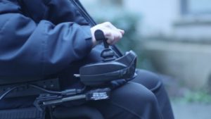
The characteristics of your mobility device can also affect how you function in day-to-day life. For example, small tweaks to your wheelchair set-up can make it easier or more difficult to maneuver and propel yourself. The characteristics of your device may also affect the environments and situations that you can use it in, such as whether it can be used outside, during sports, or can be put in and out of a car independently.

It affects your participation in a number of different activities and environments

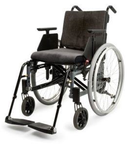
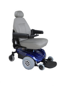
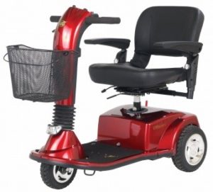
There are a variety of styles of wheelchairs and scooters, each with their own benefits and drawbacks. Properties of various mobility devices, such as the turning radius, the length and width, and the stability of the device may impact the types of activities that a wheelchair user can participate in. For example, a wheelchair that has a wide turning radius may not be able to maneuver in smaller stores with narrow aisles. Additionally, some devices perform better in inclement weather than others. This may be a consideration for individuals living in areas where it is often rains or snows.

The process of selection is a complicated process that typically involves collaboration among people with spinal cord injury, their caregivers, device prescribers (Occupational Therapists or Physical Therapists), and vendors (sometimes referred to as DME Provider or Durable Medical Equipment Provider). The World Health Organization (WHO) identifies and breaks down this complicated process into 8 important steps. These include:

1. Referral and appointment

In the first step, you will be referred to a knowledgeable health provider. The referral process varies based on the services provided by each country. Some countries may have a self-referral process, while others may require a referral to a wheelchair/mobility device service.

2. Assessment

In this step, you will be accurately assessed to determine the most appropriate wheelchair and wheelchair components for you. Factors that are evaluated during an assessment include: physical functioning, posture, lifestyle/the environment it will be used in, and the tasks that are to be performed using the wheelchair.

3. Prescription

The prescription of a device that best matches the needs of the user then occurs. A vendor or therapist will work with you to choose the right wheelchair, cushion, and parts, and note down the many details and measurements of your wheelchair. In this step, you are encouraged to try various devices and seating set ups with a clinician or vendor to determine what is the most optimal set up for you.

4. Funding and Ordering

Once a detailed prescription has been created, it is then possible to determine an accurate quote for the cost of the wheelchair. A request for funding to the appropriate source with a letter of justification is provided by the prescribing therapist and physician. Determining a source of funding should be in place prior to ordering the wheelchair. Once that has been processed and approved the vendor/DME provider can order the equipment.

5. Product preparation or initial set-up

When the equipment arrives, it will be set-up and put together for the initial fitting as prescribed by the clinician. In addition, the wheelchair is inspected to ensure that it is safe and ready for use. This step is typically completed by the vendor/DME provider.

6. Fitting and adjusting

Getting your wheelchair fitted is important to make sure your chair is suited to your needs. 10

The therapist and/or the vendor will make an appointment to check on the wheelchair to ensure that it has been properly assembled. During this time, they will also make final adjustments so that it fits properly. Some things that are checked during this step include:

  • Making sure the wheelchair is the right size
  • Ensuring that the wheelchair is properly adjusted to your needs so that secondary complications are prevented
  • Confirming that any modifications made are fitting correctly.

7. User training

In this step, you will receive instructions on how to care for your wheelchair and training on wheelchair skills. This step allows you to use your device to its greatest potential, and to receive the most benefits from the device. Some key areas that should be covered in training include:

  • How to transfer in and out of your wheelchair
  • How to handle the wheelchair
  • Basic skills to use your wheelchair
  • How to prevent and watch for pressure sores
  • How to care for the wheelchair and seating components (e.g., cushion, backrest, etc.)
  • What to do if there is a problem.

8. Follow-up, maintenance and repairs

Follow-up is often required to fine tune the set up once the equipment has been used in a variety of environments. During follow-up sessions, the effectiveness of the wheelchair will be evaluated to maximize functioning, comfort, stability, and to make sure that the equipment has been properly maintained. There is no general guideline for how often a follow-up should occur; however, follow-ups and maintenance are beneficial and can contribute to the safe use of your device.

Read our article on Wheelchair Maintenance for more information.

There are many types of wheeled mobility devices that can help you get around after a spinal cord injury. Determining which device will best suit your needs is dependent on a variety of factors. These factors are often considered in partnership with a device prescriber (such as an occupational or physical therapist), who will also help ensure your device properly fits your body and needs.

For a review of how we assess evidence at SCIRE Community and advice on making decisions, please see SCIRE Community Evidence.

Parts of this page has been adapted from SCIRE Project (Professional) “Wheeled Mobility and Seating Equipment” Chapter:

Titus L, Moir S, Casalino A, McIntyre A, Connolly S, Mortenson B, Guilbalt L, Miles S, Trenholm K, Benton B, Regan M. (2016). Wheeled Mobility and Seating Equipment Following Spinal Cord Injury. In Eng JJ, Teasell RW, Miller WC, Wolfe DL, Townson AF, Hsieh JTC, Connolly SJ, Loh E, McIntyre A, Querée M, editors. Spinal Cord Injury Rehabilitation Evidence. Version 6.0: p 1-178.

Available from: https://scireproject.com/evidence/rehabilitation-evidence/wheeled-mobility-and-seating-equipment/

Armstrong, W., Borg, J., Krizack, M., Lindsley, A., Mines, K., Pearlman, J., … Sheldon, S. (2008). Guidelines on the provision of manual wheelchairs in less resourced settings. World Health Organization. https://doi.org/10.1007/978-1-349-13869-2_70

Frost, S., Mines, K., Noon, J., Scheffler, E., & Jackson-Stoeckle, R. (2012). Wheelchair service training package: Reference manual for participants. World Health Organization.

Mortenson, W. Ben, & Miller, W. C. (2008). The Wheelchair Procurement Process: Perspectives of Clients and Prescribers. Canadian Journap of Occupational Therapy, 75(3), 167–175.

Rehabilitation Engineering and Assistive Technology Society of North America (RESNA). (2011). Wheelchair service provision guide. Retreived from: https://resna.org/sites/default/files/legacy/resources/position-papers/RESNAWheelchairServiceProvisionGuide.pdf

Image credits:

  1. Image by the SCIRE Community Team
  2. Wheelchair etac cross ©Etac Sverige AB, CC BY-SA 3.0
  3. Pride Jazzy Select power chair ©Stephen B Calvert Clariosophic, CC BY-SA 3.0
  4. Welland Transportation ©Zdlpwebb, CC BY-SA 4.0
  5. Rehabilitation equipment ©satynek, Pixabay License
  6. Modifed from: wheelchair ©Saeful Muslim, CC BY 3.0 US
  7. Toillet ©Hadi, CC BY 3.0 US
  8. praying ©Hadi, CC BY 3.0 US
  9. Image by the SCIRE Community Team
  10. Image by the SCIRE Community Team


Disclaimer: This document does not provide medical advice. This information is provided for educational purposes only. Consult a qualified health professional for further information or specific medical advice. The SCIRE Project, its partners and collaborators disclaim any liability to any party for any loss or damage by errors or omissions in this publication.

Powered Mobility Devices

By | | No Comments

Author: Sharon Jang | Reviewer: Emma M. Smith | Published: 4 March 2020 | Updated: ~

Powered wheelchairs and scooters are an important part of independent living after spinal cord injury (SCI). This page provides an overview of the basics of choosing a powered wheeled mobility device after SCI. For more general information on wheelchair provision and wheeled mobility devices, please see our page on the wheelchair provision process. Although this page mentions specific products, SCIRE Community is not affiliated with and does not endorse any of these products.

Key Points

  • Power wheelchairs may be used by people who need greater support and assistance for mobility.
  • Power wheelchairs are normally classified by their drive (rear-wheel drive, mid-wheel drive, front-wheel drive).
  • There are many adjustable parts on a power wheelchair to ensure your safety and comfort.
  • Other powered mobility options other than wheelchairs are available (i.e., scooters, Segways); however, they are not often used by individuals with SCI.

A typical power wheelchair includes a metal frame containing a motor and battery, a seat with a backrest, footrests, small caster wheels in the front and/or back, and two large rear wheels at the back. It is propelled and maneuvered using a joystick or other control function.1

Power wheelchairs are mobility devices powered by a battery and motor, and operated by the user or by another person, such as a caregiver. Power wheelchairs may be controlled in a number of different ways: by hand using a joystick, by head movements through head array systems, by the breath through sip-and-puff controllers, and by other specialty controls. This allows power wheelchairs to be used by individuals with high cervical SCI.

Power wheelchairs are typically used by those with limited control of the muscles at the elbow (complete injuries above C5) or other reasons which make it difficult to propel a manual wheelchair. Additional factors that may encourage the use of a power wheelchair over a manual wheelchair include:

  • Improved independence (if the individual is unable to effectively propel a manual wheelchair)
  • Avoidance of overuse injuries
  • Increased speed (if the individual has weak upper body strength)
  • Lengthening the amount of time that a person can spend in the wheelchair
  • Improves the ability to participate in important activities more easily

Types of power wheelchairs are described by the base. The base is the bottom portion of a power wheelchair that houses the motors, batteries, drive wheels, casters and electronics. The seating system sits on top of this base. The base is classified according to the drive wheel location relative to the system’s center of gravity. The three classifications of power wheelchairs are rear-wheel drive, mid-wheel drive, or front-wheel drive, each with their own benefits and drawbacks.

Rear-Wheel Drive.2

Rear-wheel drive

Rear-wheel drive wheelchairs have drive wheels located behind the user’s center of gravity, with casters in front. Benefits of rear-wheel drive wheelchairs include increased stability when travelling at higher speeds. However, rear-wheel drive wheelchairs are the most likely to tip backwards when going uphill, and have a large turning radius which may make it difficult to maneuver in indoor or tight spaces.

Mid-wheel drive

Mid-wheel drive wheelchairs have drive wheels located right below the user’s center of gravity, with caster wheels both in front and behind the drive wheels (Image 3). Benefits of a mid-wheel drive wheelchairs include:

Mid-wheel drive power wheelchair with tilt-in space capability.3

  • Having the smallest turning radius, thus making them most effective for indoor mobility
  • Being highly sensitive to change in direction
  • Being the most stable when going up hills and uneven ground
  • Given that mid-wheel drive wheelchairs have a total of 6 wheels as opposed to 4, the user may experience a bumpier ride; however, this can be offset with good quality suspensions. Additionally, mid-wheel drive wheelchairs are not the most efficient at going over uneven ground or soft terrain – users may get stuck when going over these surfaces.

Front-wheel drive

Front-wheel drive power wheelchair with recline capability.4

Front-wheel drive wheelchairs have the drive wheels located in the front of the chair, with caster wheels behind (Figure 4). Some benefits of this type of wheelchair include:

  • Being the most stable type of wheelchair when going on uneven terrain and up hills
  • Being the best at going over obstacles
  • Having the ability to turn around tight corners well
  • Allowing the user to pull up close to surfaces as the footplate is closer to the wheelchair since there are no casters in the way

Some drawbacks to front-wheel drive wheelchair include increased difficulty turning in small spaces due to a long back end, less stability when going at high speeds (may tend to fishtail, making it hard to drive in a straight line).

Drive controls

Drive controls for power wheelchairs come in a variety of forms and they are chosen depending on the physical capabilities of the user. Joysticks are used if you have enough arm movement to drive the wheelchair. There are different styles of joysticks available for use, depending on your hand function. This may include ball shaped controls, knob shaped both large and small, stick shaped, as well as U-shaped to name a few. Selecting an appropriate joystick according to your abilities will provide control and precision when driving. For people without the strength or endurance to drive with their arms they may choose to drive with their head, chin or use their mouth with a sip and puff control that is controlled by breath.

Various types of joystick controllers include U-shaped (A), stick (B), and ball (C).5-7


Novel drive systems

With the advancement of technology, more unique drive systems are becoming available to accommodate for a range of abilities. Recently, there has been emerging evidence for:

  • Tongue drive systems: This new system allows individuals to drive a power wheelchair with their tongues. For this to work, an individual will need to have their tongue pierced and have a titanium magnetic barbell fitted. Once in place, the user will wear a headset with magnetic sensors. This allows for individuals to move their tongues to certain teeth/spots in their mouth to drive their chair.
  • Eye gaze systems: This system uses an eye tracking device, which allows the user to drive a powered wheelchair with their eyes. To maneuver, the user simply looks left to go left, looks right to go right, and blinks for 1 second to start or stop the wheelchair. Weak evidence suggests that users are satisfied with the system and that it may be feasible to accurately drive a wheelchair with the eyes.
  • Facial drive systems: New programs allow individuals to drive their wheelchair with various facial movements/emotions as they choose. This includes movements including raised eyebrows, head movement to the left/right, or head movement up/down.

Ultimately, we must note that the tongue drive system is very new, and more research will need to be conducted before its use in practice.


Positioning functions

Power wheelchairs can have the added function of repositioning the SCI user. Repositioning helps shift weight to other parts of the body. This is helpful in providing comfort, supporting posture, and lessening pressure on certain parts of the body.

Tilting

Tilting a power wheelchair maintains the hip and knee angles by shifting both the seat and backrest together. This reduces the chances of rubbing on the skin when moving between positions. Generally, tilting with a power positioning device to a minimum angle of 30° is needed before the beneficial effects of weight shifting can be obtained, while a 55° tilt has been recommended. Additional benefits of tilting include assisting with respiratory function, providing a more comfortable position to rest in without having to transfer to a bed, and increasing stability and balance when travelling on uneven surfaces (e.g., hills).

Reclining

Reclining a power wheelchair consists of the backrest moving backwards/downwards while the seat remains stable. This increases the angle between the seat and the backrest. Some benefits of reclining include stretching out the hips, facilitating toileting and changing catheters, and facilitating transfers for caregivers. There is some weak research evidence that suggests that reclining to 120º may help reduce the pressure applied by the buttock, but reclining back this far also increases the amount of rubbing, which can lead to sores.

Repositioning and pressure sores

One recommended strategy to prevent the development of pressure sores includes weight shifting. Relieving weight off the buttock may help prevent pressure sores by allowing the tissues under pressure to regain blood flow. This is normally done using strategies such as leaning forward or lifting off the seat. If you have a higher level of injury, you may not be able to relieve pressure off your buttock independently. This is where the tilt function of a power wheelchair can come in handy – tilting backwards may help reduce the pressure applied on your bottom and allow for proper blood flow to occur. For more information on pressure relief and sores, check out our page on pressure sores.


Standing

Some wheelchairs can support standing in people with SCI. Not only does standing decrease pressure at the sit bones and coccyx, it also provides many other physiological advantages such as maintaining bone mass density, improving circulation, and enhancing functional tasks such as reaching for items on a shelf. It can also be helpful in managing spasticity and for social interaction or certain job functions.

Elevation

The elevation function of a power wheelchair raises the height of the seat. This is an option that can be added to the chair to enable individuals to reach and access things independently or without shoulder strain. It can also improve social interaction enabling the wheelchair to move to a height where they do not have to look up or be blocked in a room full of people.

Arm rests

Arm rests are multifunctional pieces that are located on the sides of the wheelchair. First, arm rests act as a support for the arm and the shoulder. When in motion, the arm rest help with balance during sudden stops, going up/down hills, and with balance in general. In addition, swelling in the arms may be reduced when the arms are kept elevated. Resting arms on the arm rest may also help maintain upper body posture as the weight of the arms is supported (versus hanging and pulling the upper body downward).

Individuals with cervical level injuries may require a specialized shaped armrest to provide support. 12

Secondly, arm rests can act as a source of stability when weight shifting to relief pressure. Individuals with a SCI can use their arm rest to push themselves up when shifting, or use the arm rest to act as a stabilizer when shifting their weight. For someone who adjusts their position often, gel padded arm rests may be used to increase comfort and support.

Arm rests can be highly adjustable based on various needs.

  • Having an arm rest that is too high up may push up the shoulders and may be uncomfortable
  • The use of adjustable arm rests may be helpful if the individual wants to be able to pull up to a table
  • Some arm rests come with a side panel built in to provide thigh support
  • Some arm rests have the function to be flipped up, while other types of arm rests may be completely removed from the wheelchair
  • A removable arm rest may be used to help facilitate transfers

Additionally, arm rests for individuals with injuries C4 or above may require special adjustments. Arm troughs (or a more formed arm rest) may be used to provide additional support to the arm to prevent it from moving. In addition, elbow blocks may be used to help support an upright position when tilting. In the case that an individual experiences spams in the arm, sheepskin or gel pads may be used to protect the skin when the arm rests in the trough.

Foot rests: footplates and leg rests

A footplate is generally a single plate (left), while foot rests consist of individual pieces (right). 13,14

Depending on the drive mode, power wheelchairs may have a footplate or leg rests. A foot plate is often connected to the base of the power wheelchair, and is one piece of metal that can be flipped up and down and supports both of the feet. In contrast, leg rests are two separate supports for each foot.

Where the foot rest sits is dependent on the drive base and the physical characteristics of the person. A rear wheel drive wheelchair has footplates the furthest away from the chair as there are caster wheels located on the front. This results in added length to the wheelchair, which may make it harder to turn. The use of a single centre-mounted footplate may help reduce the length of the wheelchair. Front wheel drive wheelchairs do not have front casters that interfere with the foot rests, as the caster wheels are located in the rear. As a result, the foot rests on front wheel drive wheelchairs do not interfere.

Some power wheelchairs may have an option for powered footrests, allowing the user to adjust the angle of the footplates through a motor. To prevent the legs from dangling when using the tilt or recline function, some power wheelchairs may have a foot rest elevation function. This function helps to increase the height and angle of the foot rest to elevate the legs when reclining, and lowers back into the default position when returning to an upright position.

The batteries on a power wheelchair are designed to run for generally 3-9 hours of continuous use, and are made to last for up to 5 years. Some factors that influence how far you can drive on one charge of your battery can be categorized into fixed and varying factors. Many of these factors impact each other, and changing one aspect may influence another.

Fixed factors

Fixed factors influence how long your battery lasts, but cannot be changed. Examples include:

  • Weight and size of the battery – a larger battery will allow for a greater driving distance, but adds to the overall weight of the wheelchair
  • The drive type of the wheelchair – rear wheel drive wheelchairs tend to use more power than a front or mid wheel drive
  • Weight of the wheelchair – a lighter chair will use less battery
  • Weight distribution – more weight applied on the caster wheels will use more battery power during manoeuvering

Varying factors

Varying factors are considerations that can influence your battery life in which you can have some control over to a certain extent. These factors include:

Using a powered wheelchair on smooth even ground in moderate temperatures can help prolong the battery life.15

  • The payload of the wheelchair – this is a combination of the user’s weight in addition to any items they may be carrying (e.g., respiratory products, groceries, battery charger, etc.). Most often the maximum payload the battery can handle is much greater than the average weight the power wheelchair carries.
  • Properties of the driving surface – driving on uneven, rough, slippery, or soft (e.g., gravel, grass) will use more battery power.
  • Temperature – using your power wheelchair in extreme weather (hot or cold) can impact how long the battery lasts for. Using your wheelchair in over 30 degrees or under 0 degrees Celsius will reduce the battery’s normal capacity.
  • Driving behaviour – performing a lot of stop and go actions, going up hills, and climbing curbs consume more battery
  • Low air pressures in the tires – if you are using pneumatic tires and they are underinflated, this will use up battery faster
  • Powered options – using the tilt/reclining functions or powered leg rest functions may reduce the driving range of your wheelchair

If you are using your power wheelchair every day, you should be charging your wheelchair daily. When charging your battery, it is better to charge it for longer amounts of time than in multiple short charges (i.e., less than 2 hours). A minimum charge time of 12 hours or longer has been recommended; power wheelchair batteries will not overcharge! If you are using a new power wheelchair, note that it may take 10-20 charges prior to the batteries reaching their fullest capacity.

While the majority of people who use wheeled mobility devices after SCI will use a manual or power wheelchair, other devices are also sometimes used.

Scooters

Scooters are powered devices that are typically used for getting around in the community. They come in a variety of sizes and in 3 wheeled and 4 wheeled designs. They are often used by individuals whose functional abilities do not require a full-time wheelchair, but may need support when moving around in the community. This support may help a person move greater distances, conserve energy, or get around safely if they have impaired balance, pain, or fatigue. Scooters are typically larger devices compared to wheelchairs, and are controlled by hand controls similar to a bike or motorcycle. They are usually cheaper than power wheelchairs but do not have as small a turning radius and also cannot facilitate complex seating systems. Due to their longer length, they are also more difficult to accommodate in wheelchair taxis or vans.

Additionally, there are tiller-like devices that can be added on to powered wheelchairs. This allows powered wheelchair users to operate their device similarly to a scooter. An example of this device is the JoyBar.

Segways

Segways are powered, self-balancing two-wheeled devices with arm bars that a person can stand on to move around in the community. While Segways are not intended specifically to be a rehabilitation device, some people with higher levels of functional abilities (people who are able to stand and balance effectively) may use these devices to assist with getting around in the community in a similar way to scooters. They are considered to be smaller, faster, and more manoeuvrable than power wheelchairs. Early research has suggested that Segways may be a potential mobility option for people with SCI who have limited walking abilities, although there may be some difficulties getting on or off the device.

In more recent times, several seated Segway-like devices have started entering the market. These devices (such as the Nino), allow individuals with upper trunk function to drive the wheelchair by leaning forward and to brake the wheelchair by leaning back. More research is required to determine how useful and practical it would be for individuals with spinal cord injury.

As described in this article, many factors play into the selection and set up of a powered mobility device. This article provides you with information on the various parts of a power wheelchair that can be adjusted to suit your needs. If you think that a feature needs to be adjusted or added, consult with your health care or wheelchair provider.

Any reference to a specific product does not constitute or imply an endorsement by SCIRE Community. Professional advice should be sought before making any health care and treatment decisions.

For a review of how we assess evidence at SCIRE Community and advice on making decisions, please see SCIRE Community Evidence.

Parts of this page has been adapted from SCIRE Professional “Wheeled Mobility and Seating Equipment” Module:

Titus L, Moir S, Casalino A, McIntyre A, Connolly S, Mortenson B, Guilbalt L, Miles S, Trenholm K, Benton B, Regan M. (2016). Wheeled Mobility and Seating Equipment Following Spinal Cord Injury. In Eng JJ, Teasell RW, Miller WC, Wolfe DL, Townson AF, Hsieh JTC, Connolly SJ, Loh E, McIntyre A, Querée M, editors. Spinal Cord Injury Rehabilitation Evidence. Version 6.0: p 1-178.
Available from: https://scireproject.com/evidence/wheeled-mobility-and-seating-equipment/ 


Evidence for “What types of power wheelchairs are there?” is based on:

Queensland Spinal Cord Injuries Services. (2019). Comparison of front, mid, and rear wheel drive power chairs. Retrieved from: https://www.health.qld.gov.au/__data/assets/pdf_file/0028/428482/pdwc-comparison.pdf

Evidence for “What parts of a power wheelchair should I know about?” is based on:

Agency for Clinical Innovation. (2020). Ke2p the big picture in mind. Retrieved from: https://www.aci.health.nsw.gov.au/networks/spinal-cord-injury/spinal-seating/module-10/keep-the-big-picture-in-mind

Boninger, M. and the Model Systems Knowledge Translation Center. (2019). The power wheelchair: what the spinal cord injury consumer needs to know. Retrieved from: https://msktc.org/sci/factsheets/wheelchairs/The-Power-Wheelchair

Giesbrecht EM, Ethans KD, & Staley D. Measuring the effect of incremental angles of wheelchair tilt on interface pressure among individuals with SCI. Spinal Cord 2011;49:827- 31.

Kim J, Park H, Bruce J, Sutton E, Rowles D, Pucci D, et al. The tongue enables computer and wheelchair control for people with spinal cord injury. Science translational medicine 2013;5(213):213ra166.

Kim J, Park H, Bruce J, Rowles D, Holbrook J, Nardone B, et al. Qualitative assessment of tongue drive system by people with high-level spinal cord injury. Journal of rehabilitation research and development 2014;51(3):451-466

Kim J, Park H, Bruce J, Rowles D, Holbrook J, Nardone B, et al. Assessment of the tongue- drive system using a computer, a smartphone, and a powered-wheelchair by people with tetraplegia. IEEE Transactions on Neural Systems and Rehabilitation Engineering 2016;24(1):68-78.

Laumann A, Holbrook J, Minocha J, Rowles D, Nardone B, West D, et al. Safety and efficacy of medically performed tongue piercing in people with tetraplegia for use with tongue- operated assistive technology. Topics in spinal cord injury rehabilitation 2015;21(1):61-76.

Queensland Spinal Cord Injuries Service. (2019). Powerdrive Wheelchair Features. Retrieved from: https://www.health.qld.gov.au/__data/assets/pdf_file/0024/425625/pdwc-features.pdf

Stewart, D. (2019). Wheelchair arm rests. Retrieved from: https://mobilitybasics.ca/wheelchairs/armrests

United Spinal Association (2019). Wheelchair armrests – what do they really do? Retrieved from: https://www.unitedspinal.org/wheelchair-armrests/

Evidence for “What impacts battery life” is based on

Invacare. (2020). How to charge and maintain a battery on a power wheelchair. Retrieved from: https://www.passionatepeople.invacare.eu.com/charge-maintain-battery-power-wheelchair/

Karman. (2020). Power wheelchair batteries: understanding how the battery works. Retrieved from: https://www.karmanhealthcare.com/power-wheelchair-batteries-understanding-how-the-battery-works/

Scootaround. (2019). How long do wheelchair batteries last? (and battery life tips!). Retrieved from: https://scootaround.com/en/how-long-do-wheelchair-batteries-last-and-battery-life-tips

Image credits:

  1. Modified from: Wheel isolated ©MBGX2, Pixabay License
  2. Mid wheel drive Image ©Br Yonten Phuntsok, Pixabay License
  3. Wheel isolated ©MBGX2, Pixabay License
  4. Front wheel drive ©Stephen B Calvert Clariosophic, CC BY-SA 3.0
  5. U-shaped joystick handle ©Bodypoint
  6. Large ball ©Permobil
  7. Joystick ©Wheelchair and Scooter Repair
  8. Midwheel drive power wheelchair with tilt-in-space capability ©Model systems knowledge translation center (MSKTC)
  9. Front wheel drive power wheelchair with recline capability ©Model systems knowledge translation center (MSKTC)
  10. Power elevating seat ©Model systems knowledge translation center (MSKTC)
  11. 2019 F5 Corpus VS ©Permobil
  12. Molded wheelchair arm rest ©Comfort Company
  13. Intrepid mid-wheel power wheelchair ©Intrepid
  14. Conventional footrest ©David Stewart
  15. Motorized wheelchair wheelchair elderly man ©Kevin Philips, Pixabay License
  16. Welland Transportation ©Zdlpwebb, CC BY-SA 4.0
  17. JoyBar © Joybar
  18. Segway ©Ivva,CC BY-SA 2.0 
  19. Nino Segway Wheelchair ©Gyronova


Disclaimer: This document does not provide medical advice. This information is provided for educational purposes only. Consult a qualified health professional for further information or specific medical advice. The SCIRE Project, its partners and collaborators disclaim any liability to any party for any loss or damage by errors or omissions in this publication.

Manual Wheelchairs

By | | No Comments

Author: Sharon Jang | Reviewer: Emma M. Smith | Published: 3 March 2020 | Updated: ~

Manual wheelchairs are a type of wheeled mobility device that are an important part of independent living after spinal cord injury (SCI). This page provides an overview of manual wheelchairs. SCIRE Community is not affiliated with and does not endorse any of the specific products mentioned on this page.

Key Points

  • Manual wheelchairs are typically used by people with enough movement control and strength in the arms to propel the wheelchair independently
  • A manual wheelchair consists of many different parts, most of which can be altered to suit your needs
  • Manual wheelchairs can be adjusted to prevent injury and to promote comfort
  • Multiple factors play into injuries related to manual wheelchair use, including the way you push your wheelchair, your wheelchair set up, and the surfaces you are wheeling on

Manual wheelchairs are wheelchairs that are propelled by the user or pushed by another person. They do not have a battery or other power source.

For the most part, manual wheelchairs are used by people who have enough muscle control and strength in their arms to propel the wheelchair forward on their own. For people with SCI, this typically means that a person needs to at least have function of the biceps (the muscle that bends the elbow), which is intact for people with complete injuries at C5 and below. However, it can be difficult to propel a wheelchair for individuals with C5 and C6 injuries, so only some individuals with these types of injuries will be able to do so. Use of a manual wheelchair is more common in people with control of the triceps (the muscle that straightens the elbow), which is intact in people with complete injuries at C7 and below. In some cases, a manual wheelchair can be pushed by another person or propelled using the legs.

As a bridge between the more common manual and power wheelchairs, power-assist add-ons combine features of manual and power wheelchairs to provide greater assistance for those who need it. Additionally, non-powered propulsion assist add-ons may also be used to facilitate wheeling. Propulsion assist options are available for manual wheelchairs and come in several different styles. The use of powered add-ons has been reported to improve efficiency and to reduce strain on the cardiovascular system. The following article will focus on manual wheelchairs that are propelled with the arms.

Refer to our chapter on Propulsion Assist Devices for more information!

A typical manual wheelchair contains a frame, seat with a backrest, footrests or footplate, two small caster wheels in the front, and two large rear wheels at the back. The rear wheels contain hand rims which are used to propel and maneuver the wheelchair. Wheel locks on the rear wheels help prevent the wheelchair from moving when it is not in use or when getting in or out of it.

There are many different styles of manual wheelchairs, most of which allow for the ability to adjust and change aspects of the set up. This can be important in the first few years that a person uses a wheelchair as a person’s skills and priorities may change. Nonetheless, it is important to try to get as lightweight and compact a wheelchair as possible while also ensuring it is safe, durable and flexible.

There are many different components of manual wheelchairs that can be customized and changed to find the best set-up for an individual. Below, we list some of the common options and changes that may be possible when setting up a manual wheelchair. In general, changes to the set-up of the manual wheelchair are a series of tradeoffs. Although changes are usually intended to achieve a desirable improvement (such as greater ease of wheeling), sometimes they need to be balanced with reduced stability and an increased risk of tipping over. Please speak to your health providers before making any major changes to your wheelchair set-up.

Frame design

Manual wheelchairs come in two basic frame designs, folding frames or rigid frames. Folding frames have a crossbar under the seat that allows the frame to fold in half. Rigid frames may also fold but in a different way, where the backrest folds down. In many manual wheelchairs, wheels can removed if they are being put in a vehicle.

Folding manual wheelchairs

Folding wheelchairs are designed to be folded vertically and take up minimal storage space. This allows for easy portability (such as fitting the wheelchair into a car). However, these wheelchairs also have many moving parts that may break down or loosen over time, and are heavier than rigid wheelchairs. Folding wheelchair often have flip up, swing away or swing in footrests so they may be used by individuals that do not use the wheelchair full time, can stand or take some steps, or by those who foot propel.

Rigid manual wheelchairs

Rigid wheelchairs tend to be lighter and more durable than folding wheelchairs, which makes them the common choice for people with complete SCI injuries. The rear tires often have to be removed to fit into a vehicle because they cannot fold inward. Some rigid manual wheelchairs can still be ordered with adjustability, which can be important if seating components change.


Frame materials

The majority of manual wheelchair frames are made of either aluminum or stainless steel. Ultralight and sports wheelchairs are often constructed from high performance aluminum, carbon fiber, chromium-molybdenum, nickel alloy steel, or titanium. Titanium and composites can be considerably more expensive but are more lightweight.

Seat width, depth and slope

It is important that the width, depth and slope (also known as dump) of the seat from front to back are well-fitted to the overall function and characteristics of the user. A wheelchair that is too narrow in width may cause skin problems due to rubbing, while a wheelchair that is too wide may hinder propulsion, and will interfere with the accessibility and maneuverability of the chair. A wheelchair that is too short in depth may provide insufficient support to the hips and stability to the bottom, while a wheelchair that is too long in depth may cause knee pain or a slouching posture.  In addition, an increased seat slope (i.e., one that is higher in the front than the back) makes transfers more difficult as one would need to lift up at an incline, but are better for people with a higher level of injury because this slope helps them find balance and stability to wheel.

Footrest angle and frame taper

The footplate is angled away from the frame of the chair and the post of the casters.6

When considering the footrest angle between the seat and the footplate hanger, the knee flexion range of movement and hamstring length needs to be considered as well as the visual field as the client may not be able to see their feet. For example, a greater frame angle improves wheelchair access by making the chair shorter but may make the chair more unstable in the forward direction. Some wheelchairs have a frame taper, which is the narrowing of the frame towards the front and continues down to the footplates. This improves access from the front of the wheelchair, making toilet transfers easier. However, the taper may not be compatible with the cushion and may impact the ability of the wheelchair to fold.

Rear Wheels

Spoked wheels (left) versus mag wheels (right).7,8

The rear wheels are set up with hand rims on them so that self-propelling is possible. Wheel material and the amount of air in the wheels affect how effective the wheels are in minimizing vibration or shock to prevent triggering of spasticity and to increase comfort. Wheel rims are made from a variety of materials including aircraft grade aluminum, plastic, fibreglass, reinforced nylon, titanium, carbon graphite, or steel. Types of rear wheels include: spoked wheels (made of metal, normally has more than 30 spokes), mag wheels (made out of synthetic material, less than 10 spokes), carbon-graphite mag wheels, and power assist wheels. The more spokes, the better the vibration absorption. The most common wheel size for adults is 60cm (24”) and for children it is 50cm (20”).

Tires

Airless (solid) tires tend to provide bumpier ride.9

Wheelchairs may have solid tires, foam-filled tires, or air (pneumatic) tires. Solid tires do not run the risk of popping, however, they often result in a bumpier ride. On the contrary, air tires provide a softer ride, but require regular maintenance of air pressure. A lack of air in the tires (less than 50% of the recommended PSI) makes propelling a wheelchair more difficult. It is recommended that tires be re-inflated every two months to keep the pressure above 50%. Some type of tires include: standard pneumatic tires, high pressure pneumatic tires, outdoor pneumatic tires, puncture proof tires, and solid plastic tires.

Hand rims/Push rims

Hand rims are attached to the rear wheel rim on wheelchairs that are intended for self-propulsion. This part is optional, and some users may opt not to use them. Hand rims are commonly made out of aluminum, and may be anodized or vinyl coated. Some downsides to using aluminum hand rims are that they may turn your hand black, and can become very sharp if they are damaged. Other models of hand rims have a rubber like material and assist individuals without full handgrip with their push stroke. In addition, flexible hand rims have been developed to adapt to the shape of the hand in order to reduce strain resulting from the bending action of the wrist and fingers.

Front caster wheels

The small front wheels are called caster wheels, and they help maneuver and steer the position of the wheelchair. Caster diameter, material, and position are important factors to consider that affect wheelchair balance and performance. Sizes available include 75mm (3”), 125mm (5”), 150mm (6”), 180mm (7”), 200mm (8”), and 250mm (10”). Bigger caster wheels are more stable, have a bigger turning radius, and easier to wheel over thresholds but are less maneuverable and responsive. A point of caution is that when caster wheels are trailing, a person’s weight will be more forward and may be in front of the caster wheel, which can cause tipping and is a frequent cause of falls in manual wheelchairs (see image below). Caster stem angle must always lie at right angles to the floor to ensure casters track correctly, or they may “flutter” when the wheelchair is pushed or can cause the front of the wheelchair to rise and fall during turning.

Forward casters (left) versus trailing casters (right).11

When choosing the type of caster, one should consider the most common type of surface they will be propelling on (e.g. outdoors or indoors), the desired front seat height, and the front frame angle of the wheelchair. The construction materials available for caster wheels include plastic, urethane and aluminum. Pneumatic caster tires, usually 150mm (6”)” and 200mm (8”) in diameter, have more shock absorption features making them ideal for outdoor wheeling, while 75mm (3”) and 125mm (5”) urethane caster tires are more common and are good for indoor propulsion and sport specific wheelchairs. Those who experience significant spasticity and/or discomfort when propelling wheelchairs often opt for pneumatic casters to achieve a smoother ride.

Wheel locks

Wheel locks are used on the large rear wheels for safety with transfers and when no movement is wanted. They vary in styles including push to lock, pull to lock, and scissor style locks. To decrease the force required to engage/disengage the wheel locks, extended brake levers can be applied, as they minimize the forward reach required to access the wheel locks. The type and position of the lock should be influenced by the user’s reaching ability, balance, strength, and hand function as well as the impact of wheel lock position on transfers. Some clients with good physical skills may not even require wheel locks at all.

Anti-tippers

Anti-tippers are an optional set of smaller wheels connected to a metal poles that are attached to the frame of your wheelchair 381 to 508mm (1.5 to 2″) above the ground. Anti-tippers are commonly found on rear of the chair to prevent a backwards fall, however they can also be found on the front of the wheelchair to prevent forward tipping. Anti-tippers may be especially useful for new wheelchair users, those who have recently switched to a new wheelchair, or if one’s health is declining. While they are considered a safety item, they also have drawbacks. Firstly, anti-tippers may interfere with wheelchair skills requiring you to lean backwards, such as climbing a curb or doing a wheelie. Secondly, if the anti-tippers are set too low, it may impede on going over obstacles or up hills.

There are many necessary considerations for setting up a manual wheelchair properly for mobility and function. It is important to work with a knowledgeable health care professional and vendor to ensure the correct decisions are made.

These considerations may include:

Various factors can influence your ideal wheelchair set up. In this photo, some considerations include having a dog and wheeling on grass.13

  • Activities of Daily Living: It is important to think about where you need to take your chair, how close you can get to or under surfaces, reach for things, and how easily you can remove components and put them back on.
  • Mobility: You need to consider what surfaces you will wheel on (carpet, flooring, snow, rain). How the chair will move over inclines and transitions as well as in confined spaces. Is it a primary chair or just an indoor chair or a transport chair?
  • Positioning: It is important to consider your posture, comfort and interacting with people and your environment.
  • Psychosocial: You want to consider how you look and feel and how you will interact with people and your environment.
  • Transfers: You need to look at the ability to get in and out of the wheelchair safely so the height, stability, ease of moving and weight are some of the things to consider.

The three most important adjustments to have in a manual wheelchair are back angle, axle position and seat to floor height. These adjustments are important as they can have an impact on pain, wounds, or postural issues. The wheelchair set-up is essential to allow for safety and balance of the chair.

Axle Position (vertical and horizontal)

The horizontal position of the rear wheel axle can affect how much energy is required to move the wheelchair. Studies have shown that placing the rear wheel axle closer to the front of the wheelchair can make it easier to propel. However, it can also affect the stability of the wheelchair, and make the wheelchair more likely to tip backwards. This is because there is more weight behind the axle. Decisions on where to place the axle will depend on the person’s wheelchair skills. The distance between the shoulder and the axle of the rear wheel can also affect efficiency. One study showed that greater distances may result in greater energy requirements. Adding adjustable axles to manual wheelchairs can help individuals customize their wheelchairs to improve propulsion. This can lead to reduced risks of upper body injuries specifically shoulder injuries.

Backrest height and angle

The appropriate backrest height is largely a trade-off between posture, comfort and freedom of movement. A backrest is usually 406mm (16″) tall, and often does not come up any higher than your shoulder blades – any higher and it will impede your ability to push your wheelchair. Most manual wheelchairs have the ability to modify the height of the backrest (so it can be modified as function increases or decreases). A higher backrest is seen to be more comfortable, and is often used by individuals with limited trunk functioning as it provides more support. On the contrary, a shorter back rest allows for more movement of the trunk, which can be functionally useful (e.g., it will allow you to reach for objects sideways and behind you).

Using a wheelchair with backrest height that is not optimal may have negative consequences. If the backrest is too low, there will be a lack of support which may lead to postural instability. However, most often individuals are using backrests that are too high. This may result in a limited reach (and thus reduction in functional activity), pain, and a slouching posture as a result of compensating for sliding forward.

Other Adjustments

Rear wheel lateral position (space from frame) and camber

The lateral position of the rear wheel is the distance the wheel is from the wheelchair frame. This distance can affect the overall width of the wheelchair and how accessible the wheel is.

Camber relates to the rear wheels being set on an angle from their axle position, where the distance between the top of the wheels is less than at the bottom of the wheels. The angle can range from 0º up to an extreme 12º, although the average camber for day wheelchairs varies from 0º to 4º. Some wheelchair models offer adjustability in camber angles within the same chair. Stability, wheeling efficiency, and turning maneuverability can be enhanced with wheel camber, especially when moving over side slopes. In addition, the hands are better protected against trauma because the wheels touch the floor spanning a wider area than the hands have in contact with the hand rims. Too much camber can make the overall width of the wheelchair wider so assessment of the spaces and doorways needed to access is essential when deciding on camber.

Camber and Sports Wheelchairs

A large camber is often seen on sport wheelchairs, measuring up to 15º. A greater camber is of beneficial use to wheelchair sports, as it provides more lateral support to prevent tipping over to the side when turning quickly and sharply. It also helps to create turns that are smoother and sharper when in motion.


Footrest height and length

Proper support of the feet and legs is important to the comfort and safety of the wheelchair user. If the foot and thigh is not properly supported it may result in the leg or foot moving excessively and may result in instability, pain, and spasticity. In addition, it can create areas of pressure on the leg or the foot, ultimately leading to pressure injuries. Support can be adjusted through altering the height and length of the footrest.

The foot rest should be 1-2 inches off the ground to allow for optimal clearance. If it is too low, it may catch on door threshold and other objects, and may cause difficulties when trying to go up or down hills or slopes. Should the footrest unexpectedly hit an obstacle, there is a high risk of the user falling out of their wheelchair.

Refer to our article on Wheelchair Seating for more information on footrest set-ups

Using a manual wheelchair can be tough on the user’s arms and shoulders. Research has shown that 25-80% of manual wheelchair users experience injuries in their wrists, elbows, and or/shoulders. There have been multiple (weak) studies that have identified multiple factors that can be addressed in order to reduce/prevent the chances of injury:

Muscles required for propulsion

Muscles in the chest, upper back, shoulders, and arms that are used in propelling a wheelchair.17

When pushing a manual wheelchair, many muscles in the upper back, chest, shoulder, and arms are used. Pushing a manual wheelchair is not the most efficient way of getting around, as only between 2-14% of force applied by the arms goes into propelling a wheelchair, depending on the level of injury and style of propelling. The shoulder muscles are particularly strained during wheelchair propulsion as they are relatively smaller muscles that are responsible for both stabilizing the shoulder and applying force to a wheelchair to push it forward . Shoulder pain occurs in 31-73% of manual wheelchair users, and weak evidence suggests that individuals with tetraplegia experience more pain due to greater forces being applied with their arms.


Kinematics: the technique you use when propelling a wheelchair can impact your risk of shoulder pain/injury. For example, pushing a wheelchair at increasing speeds/intensities may contribute to the development of shoulder pain. Additional factors to be considered include the angle of your joints (i.e., the elbows, wrist) when pushing, and the angle at which you push your tires at.

Propulsion pattern: the pattern you push your wheelchair with (e.g., where do your hands go when you push and after you’re done pushing) may also have an influence on your risk of injury. Some weak evidence suggests that using the semi-circular and double-loop-over may reduce the risk of nerve injury and are the most optimal ways to push your wheelchair. Further evidence (weak) has indicated that arcing may be more efficient for short bouts of high intensity pushing (like when going uphill).

Body weight: Weak evidence suggests that having a higher body weight may be related to a higher risk of injury while propelling your wheelchair. This is due to the fact that moving a heavier body requires higher forces created by the shoulders. Body weight management is important in decreasing the amount of force created in your arms when pushing your wheelchair, and reduces the risk of injury.

Wheelchair set up: Having the rear wheels on a manual wheelchair placed in a forward axle position can help improve push rim biomechanics, reduce the amount of force put on your shoulders when propelling, and the frequency of propulsion (i.e., you do not have to push as much to go as far). Research has suggested the use of manual wheelchairs with adjustable axle positions so that the rear wheels can be optimally adjusted.

Wheeling on uneven surfaces: Some research suggests that wheeling across cross slopes (such as a driveway) can increase the forces on the arm and may lead to overuse injuries. In addition, more physiological effort  (i.e., heart rate, how much oxygen is being used, rating of perceived exertion) is required to go up slopes more than 2% incline, and more physiological and physical effort is required to go up slopes greater than 8%.

Manual wheelchairs are highly customizable pieces of equipment that can be tailored to your needs. In order to optimally use your wheelchair, certain adjustments to the backrest, frame, and tires can be made. Using manual wheelchairs requires upper arm function and strength. Improper set ups and techniques may lead to injuries.

It is best to discuss all treatment options with your health providers to find out which treatments are suitable for you.

For a review of how we assess evidence at SCIRE Community and advice on making decisions, please see SCIRE Community Evidence.

Parts of this page has been adapted from SCIRE Project (Professional) “Wheeled mobility and seating equipment following spinal cord injury” Chapter:

Titus L, Moir S, Casalino A, McIntyre A, Connolly S, Mortenson B, Guilbalt L, Miles S, Trenholm K, Benton B, Regan M. (2016). Wheeled Mobility and Seating Equipment Following Spinal Cord Injury. In Eng JJ, Teasell RW, Miller WC, Wolfe DL, Townson AF, Hsieh JTC, Connolly SJ, Loh E, McIntyre A, editors. Spinal Cord Injury Rehabilitation Evidence. Version 6.0: p 1-178.

Available from: https://scireproject.com/evidence/rehabilitation-evidence/wheeled-mobility-and-seating-equipment/ 


Evidence for “What are the components of a manual wheelchair?” is based on:

Boninger, M., and the Model Systems Knowledge Translation Center. (2019). The manual wheelchair: What the spinal cord injury consumer needs to know. Retrieved from: https://msktc.org/sci/factsheets/wheelchairs/The-Manual-Wheelchair

Spinal outreach team and The University of Queensland School of health and rehabilitation sciences. (n.d.). Manual wheelchairs: Information resource for service providers. Retrieved from: https://www.health.qld.gov.au/__data/assets/pdf_file/0026/429911/manual-wheelchairs.pdf

Evidence for “What adjustments can I make to my manual wheelchair?” is based on:

Sprigle, S. (2014). Measure it: proper wheelchair fit is key to ensuring function while protecting skin integrity. Advanced Skin Wound Care, 27(12), 561-72.

Evidence for “What are health concerns related to manual wheelchair use?” is based on:

Arva, J., Fitzgerald, S. G., Cooper, R. A., & Boninger, M. L. (2001). Mechanical efficiency and user power requirement with a pushrim activated power assisted wheelchair. Medical Engineering and Physics, 23(10), 699–705. https://doi.org/10.1016/S1350-4533(01)00054-6

Boninger, M. L., Souza, A. L., Cooper, R. A., Fitzgerald, S. G., Koontz, A. M., & Fay, B. T. (2002). Propulsion patterns and pushrim biomechanics in manual wheelchair propulsion. Archives of Physical Medicine and Rehabilitation, 83(5), 718–723. https://doi.org/10.1053/apmr.2002.32455

Curtis, K. A., Drysdale, G. A., Lanza, R. D., Kolber, M., Vitolo, R. S., & West, R. (1999). Shoulder pain in wheelchair users with tetraplegia and paraplegia. Archives of Physical Medicine and Rehabilitation, 80(4), 453–457. https://doi.org/10.1016/S0003-9993(99)90285-X

Gil-Agudo, A., Del Ama-Espinosa, A., Pérez-Rizo, E., Pérez-Nombela, S., & Pablo Rodríguez-Rodríguez, L. (2010). Upper limb joint kinetics during manual wheelchair propulsion in patients with different levels of spinal cord injury. Journal of Biomechanics, 43(13), 2508–2515. https://doi.org/10.1016/j.jbiomech.2010.05.021

Kulig, K., Newsam, C. J., Mulroy, S. J., Rao, S., Gronley, J. K., Bontrager, E. L., & Perry, J. (2001). The effect of level of spinal cord injury on shoulder joint kinetics during manual wheelchair propulsion. Clinical Biomechanics, 16, 744–751.

Rankin, J. W., Richter, W. M., & Neptune, R. R. (2011). Individual muscle contributions to push and recovery subtasks during wheelchair propulsion. Journal of Biomechanics, 44(7), 1246–1252. https://doi.org/10.1016/j.jbiomech.2011.02.073

Richter, W. M., Rodriguez, R., Woods, K. R., & Axelson, P. W. (2007). Consequences of a Cross Slope on Wheelchair Handrim Biomechanics. Archives of Physical Medicine and Rehabilitation, 88(1), 76–80. https://doi.org/10.1016/j.apmr.2006.09.015

Image credits

  1. Kuschall wheelchair model R33 ©Tim99~commonswiki, CC BY-SA 4.0
  2. Image modified from Different kinds of handicap equipments ©brgfx, Freepik License
  3. Wheelchair ©George Hodan, CC0 1.0
  4. Wheelchair parts (main pic) ©Memasa CC BY-SA 3.0
  5. The SCIRE Community Team
  6. Modified from disabled, stroller, the disease, wheelchair, disability, wheel, transportation, medical equipment, metal, mode of transportation, CC0 1.0
  7. Wheelchair disability paraplegic injured disabled ©stevepb, Pixabay License
  8. wheelchair, old, vintage, isolated, wheel, antique, transportation, white, retro, transport, CC0 1.0
  9. The SCIRE Community Team
  10. Wheelchair disabled person with reduced mobility man ©SGENET, Pixabay License
  11. Forward versus trailing casters ©Ian Denison
  12. Modified from Black and grey wheelchair, CC0 1.0
  13. Woman, dog, pet, friend, outdoors, grass, female, person, jacket, pal, CC0 1.0
  14. Axle Position by the SCIRE Community Team
  15. Modified from Disabled people set Free Vector ©Macrovector, Freepik License
  16. Euroleague – LE Roma vs Toulouse IC-27 ©Pierre-Selim, CC BY-SA 3.0
  17. Muscles that move the humerus ©Betts et al, CC BY-SA 4.0
  18. Stylized illustration of stroke pattern classification during wheelchair propulsion ©Emily Churton and Justin WL Keogh, CC BY 2.0


Disclaimer: This document does not provide medical advice. This information is provided for educational purposes only. Consult a qualified health professional for further information or specific medical advice. The SCIRE Project, its partners and collaborators disclaim any liability to any party for any loss or damage by errors or omissions in this publication.

Supported Standing

By | | No Comments

Author: SCIRE Community Team | Reviewer: Darryl Caves | Published: 17 January 2018 | Updated: ~

Standing with supportive equipment is a therapy option after spinal cord injury (SCI). This page outlines basic information about the use of supported standing after SCI.

Key Points

  • Passive standing using supportive equipment is a therapy option for people who do not stand as part of their everyday mobility.
  • Passive standing involves using equipment such as standing frames, tilt tables, orthoses or standing wheelchairs to support an upright position for a period of time.
  • Standing involves a change in posture that challenges the circulatory system, loads the legs, and provides different sensory stimulation.
  • Research evidence suggests that standing may improve blood pressure control and spasticity management. There is conflicting evidence about whether passive standing helps with bone density or bowel and bladder health.
  • Further research is needed to better understand the benefits of standing after SCI and how long standing should be done for to achieve those benefits.
Running shoes

Standing is a therapy option following SCI.1

Standing is an important part of functional movement in humans. Standing is needed for walking, and also provides a challenge to the circulatory system, bones, and muscles in ways that cannot be achieved in sitting or lying.

Passive standing (standing with support instead of by muscle activation) may have treatment benefits after SCI, even when the recovery of walking and standing abilities is unlikely. Standing may have benefits in treating health conditions associated with SCI, such as conditions involving the musculoskeletal, circulatory, breathing, bowel and bladder systems. It remains a key treatment tool used in rehabilitation.

Supported standing involves the use of special equipment to support an upright posture. The type of equipment used depends on the person’s unique characteristics and abilities (such as the amount of muscle control in the arms, legs and trunk), the equipment that is available, and other medical concerns like joint contractures, spasticity and osteoporosis. Equipment used for standing may include a wide range of different devices such as:

Tilt tables

Illustration showing a person lying on a tilt table and secured with a band around the waist. There is an arrow showing that the tilt table is moving to an upright position.

Tilt tables can be moved from a horizontal to a vertical position.2

Tilt tables are flat surfaces that can be tilted from a horizontal position into a vertical standing position. The person is strapped securely to the table while in a horizontal lying position and the table can be tilted vertically. Tilt tables are typically the first devices that are used to work towards standing because the table can be gradually increased by degrees. This is often needed because it may take some time to tolerate being upright and maintaining a safe blood pressure. It is also a good device to test a person’s physical tolerance and safety for standing.

Standing frames

Standing frames are simple frames that have padding at the joints to support a standing position. There are many different types of standing frames. The frame needs to be fitted to the person’s unique physical abilities and body type and minimize areas of excess pressure.

Standing wheelchairs

Photograph of a person using a standing frame with cushions behind and in front of the legs to help align them. The person is resting their arms on a small desk that is attached to the frame.

Standing using a standing frame.3

Standing wheelchairs are wheelchairs that can extend from a sitting position into standing. There are many different types of standing wheelchairs, from manual devices to motorized systems. However, standing wheelchairs are expensive and not commonly available.

Body weight-supported treadmill training

Suspension body weight support systems involve a harness system that is suspended from above to support a percentage of body weight while standing. These systems are typically used while walking on a treadmill (body weight-supported treadmill training) or sometimes while walking over ground. This type of system is usually used for people with incomplete injuries that may work towards standing or stepping independently.

See our article on Body Weight Supported Treadmill Training.

Robotic exoskeletons

Robotic exoskeletons are a relatively new and emerging technology that is typically used for walking and walking training, but may also have benefits related to standing. However, this equipment is costly and not available in most settings.

Walkers, crutches or canes may be used by people with incomplete SCI and good strength in the arms who need only minimal support in standing.

Orthoses

Orthoses and braces may be used to brace the hip, knee and/or ankle joints to keep them from bending. This can help to support the person in an upright standing posture with training and rehabilitation. Orthoses and braces are typically custom-made and usually used by people with paraplegia who have good upper body strength and hip flexibility. Orthoses and braces used for standing may include:

  • Knee Ankle Foot Orthoses (KAFOs) provide support at the knee, ankle and foot.
  • Reciprocating Gait Orthoses (RGOs) are more complex orthoses that are made of a left and right KAFO that are linked together with a rigid brace at the pelvis or abdomen. The brace has hip joints that are built with an alternating stepping mechanism. When one leg is extended, the other flexes forward, providing assistance for stepping. Although normally used for walking, reciprocating gait orthoses can also be used to help support a standing position.


    Functional electrical stimulation

    A person walking between parallel bars with FES applied to their legs and two other people supporting the feet.

    FES can be applied to the leg muscles during assisted walking.4

    Functional electrical stimulation (FES) involves the use of electrical stimulation to activate muscles that are weak or paralyzed after an SCI during a purposeful activity. FES over the trunk or leg muscles may be used while standing with equipment for added benefits.


    See our article on Functional Electrical Stimulation (FES) for more information.


    Standing equipment may be expensive and sometimes requires repeated visits to healthcare facilities, which can sometimes be a barrier to regular standing. It is important for the individual to work with their health providers to find appropriate equipment that is safe and suitable.

Once appropriate equipment and strategies for standing are selected with assistance from a health provider, standing is gradually introduced over time. The amount of time spent standing, the amount of load that is taken through the legs, and the final standing position will be adjusted until a suitable standing position can be maintained. During these first several sessions, health providers will monitor for any adverse effects related to the treatment.

A person walking between parallel bars with his wheelchair behind him. A heathcare provider on the side watches him.

Standing and stepping activities may be combined to optimize therapy.5

Current research findings are unable to tell us how long or how often standing should be done to have benefits. Studies have used standing for 20 to 60 minutes, three to four times per week to study the effects of this treatment. It will be different for everyone. The standing prescription will be based on the person’s unique situation.

Depending on the treatment goals, standing may also involve:

  • Adding extra weight while standing
  • Using standing together with functional electrical stimulation (FES) to activate the muscles of the legs and/or trunk
  • Weight-shifting, balance or stepping activities

Supported standing is considered to be a relatively safe treatment for use after SCI. However, there are some situations in which standing may not be appropriate and some possible risks. This is not a complete list; please consult a health provider for further safety information.

Standing should not be used in the following situations:

  • By people with recent broken bones (fractures) or a high risk of fractures (such as people with severe osteoporosis)
    Diagram comparing bone with normal bone mineral density to bone with osteoporosis.

    People with osteoporosis have weaker bones which can pose a risk during supported standing.6

  • Where the standing equipment places excess pressure on areas where there are injuries, sores, and wounds; or areas of skin prone to pressure injuries
  • By individuals whose limbs cannot be brought into a good standing position due to other conditions like joint contractures, spasticity, or heterotopic ossification
  • By people with medical conditions where heart rate or blood pressure are uncontrolled, such as those who are unable to stay upright without a major drop in blood pressure (severe orthostatic hypotension)
  • By people with muscle or joint injuries or other conditions that may be worsened by standing

Risks of standing may include:

  • Pressure injuries if the position and equipment used for standing creates too much pressure or shear while standing – it is essential that the equipment used for standing is appropriately fitted to prevent skin damage
  • Blood pooling in the legs may lead to feelings of light-headedness, dizziness or fainting (orthostatic hypotension)
  • Broken bones (fractures) are possible in weight-bearing positions in people with osteoporosis
  • Increased spasticity or autonomic dysreflexia in some people
  • Pain in the standing position

For more information on these topics, see our articles on Pressure Injuries, Orthrostatic Hypotension, Osteoporosis, Spasticity, and Autonomic Dysreflexia

If using electrical stimulation, the safety precautions and risks associated with use of functional electrical stimulation (FES) also apply.

Bone health

It is not clear if standing helps to maintain or increase bone density in the legs after SCI. Current research evidence is inconclusive and further studies are needed.

Blood pressure and circulationA person taking a blood pressure reading of another person wearing a blood pressure cuff

Another proposed use of standing after SCI is to help with blood pressure control. One study provides weak evidence that standing with a harness and assistance from health providers helps to increase resting blood pressure and reduce drops in blood pressure when standing (orthostatic hypotension) in people with cervical SCI.

Spasticity

There is weak evidence that standing may help to reduce spasticity short term in people with SCI. There are also surveys that report that many people with SCI report that regular standing helped to reduce their spasticity.

Bowel problems

There is not enough evidence to determine whether standing can also improve bowel function. Further research in this area is needed.

Supported standing serves as a therapy option for people such as individuals with SCI as they do not normally stand as part of their everyday mobility. This therapy involves equipment such as standing frames, tilt tables, orthoses, or standing wheelchairs to support an upright position for a designated period of time. While there is research supporting the benefits of supported standing, conflicting evidence is still apparent. Further research is needed to better understand the benefits of supported standing after SCI.

For a review of how we assess evidence at SCIRE Community and advice on making decisions, please see SCIRE Community Evidence.

Parts of this page have been adapted from the SCIRE Project “Bone Health”, “Orthostatic Hypotension”, “Spasticity”, and “Bowel Dysfunction and Management”  Chapters:

Craven C, Lynch CL, Eng JJ (2014). Bone Health Following Spinal Cord Injury. In Eng JJ, Teasell RW, Miller WC, Wolfe DL, Townson AF, Hsieh JTC, Connolly SJ, Noonan VK, Loh E, McIntyre A, editors. Spinal Cord Injury Rehabilitation Evidence. Version 5.0. Vancouver: p 1- 37.

Available from: http://scireproject.com/evidence/rehabilitation-evidence/bone-health/

Krassioukov A, Wecht JM, Teasell RW, Eng JJ (2014). Orthostatic Hypotension Following Spinal Cord Injury. In: Eng JJ, Teasell RW, Miller WC, Wolfe DL, Townson AF, Hsieh JTC, Connolly SJ, Noonan VK, Loh E, McIntyre A, editors. Spinal Cord Injury Rehabilitation Evidence. Version 5.0. Vancouver: p. 1-26.

Available from: https://scireproject.com/evidence/rehabilitation-evidence/orthostatic-hypotension/

Hsieh JTC, Connolly SJ, McIntyre A, Townson AF, Short C, Mills P, Vu V, Benton B, Wolfe DL (2016). Spasticity Following Spinal Cord Injury. In Eng JJ, Teasell RW, Miller WC, Wolfe DL, Townson AF, Hsieh JTC, Connolly SJ, Curt A, Mehta S, Sakakibara BM, editors. Spinal Cord Injury Rehabilitation Evidence. Version 6.0.

Available from: http://scireproject.com/evidence/rehabilitation-evidence/spasticity/

Coggrave M, Mills P, Willms R, Eng JJ, (2014). Bowel Dysfunction and Management Following Spinal Cord Injury. In Eng JJ, Teasell RW, Miller WC, Wolfe DL, Townson AF, Hsieh JTC, Connolly SJ, Noonan VK, Loh E, McIntyre A, editors. Spinal Cord Injury Rehabilitation Evidence. Version 5.0. Vancouver: p 1- 48.

Available from: https://scireproject.com/evidence/rehabilitation-evidence/bowel-dysfunction-and-management/


Evidence for “Does standing work for treating the symptoms of SCI?” is based on the following studies:

Bone health

[1] Dudley-Javoroski S, Saha PK, Liang G, Li C, Gao Z, and Shields RK. High dose compressive loads attenuate bone mineral loss in humans with spinal cord injury. Osteoporos Int 2012; 23:2335-2346.

[2] Goktepe A, Tugco I, Alaca, R, Gunduz S, Nikent M. Does standing protect bone density in patients with chronic spinal cord injury? JSCM 2008;31:197-201.

[3] Needham-Shropshire BM, Broton JG, Klose KJ, Lebwohl N, Guest RS, Jacobs PL. Evaluation of a training program for persons with SCI paraplegia using the Parastep 1 ambulation system: part 3. Lack of effect on bone mineral density.  Arch Phys Med Rehabil 1997;78:799-803.

[4] Kunkel CF, Scremin AM, Eisenberg B, Garcia JF, Roberts S, Martinez S. Effect of “standing” on spasticity, contracture, and osteoporosis in paralyzed males. Arch Phys Med Rehabil 1993;74:73-78.

[5] Kaplan PE, Roden W, Gilbert E, Richards L, Goldschmidt JW. Reduction of hypercalciuria in tetraplegia after weight-bearing and strengthening exercises. Paraplegia 1981;19:289-293.

[6] Ben M, Harvey L, Denis S, et al.  Does 12 weeks of regular standing prevent loss of ankle mobility and bone mineral density in people with recent spinal cord injuries? Aust J Physiother. 2005;51:251-256.

[7] de Bruin ED, Frey-Rindova P, Herzog RE, Dietz V, Dambacher MA, Stussi E. Changes of tibia bone properties after spinal cord injury: effects of early intervention. Arch Phys Med Rehabil 1999;80:214-220.

[8] Dudley-Javoroski S, and Shields RK. Active-resisted stance modulates regional bone mineral density in humans with spinal cord injury. Journal of Spinal Cord Medicine 2013; 36: 191-199.

Blood Pressure and Circulation

[1] Harkema SJ, Ferreira CK, van den Brand RJ, Krassioukov AV. Improvements in orthostatic instability with stand locomotor training in individuals with spinal cord injury. J Neurotrauma 2008;25:1467-1475.

Spasticity

[1] Odeen I, Knutsson E. Evaluation of the effects of muscle stretch and weight load in patients with spastic paraplegia. Scand J Rehabil Med 1981;13:117-21.

[2] Bohannon R. Tilt table standing for reducing spasticity after spinal cord injury. Arch Phys Med Rehabil 1993;74:1121-2.

[3] Kunkel C, Scremin A, Eisenberg B, Garcia J, Roberts S, Martinez S. Effect of “standing” on spasticity, contracture, and osteoporosis in paralyzed males. Arch Phys Med Rehabil 1993;74:73-8.

[4] Dunn R, Walter J, Lucero Y, Weaver F, Langbein E, Fehr L, et al. Follow-up assessment of standing mobility device users. Assist Technol 1998;10:84-93.

[5] Eng JJ, Levins S, Townson A, Mah-Jones D, Bremner J, Huston G. Use of prolonged standing for individuals with spinal cord injuries. Phys Ther 2001;81:1392-9.

[6] Shields R & Dudley-Javoroski S. Monitoring standing wheelchair use after spinal cord injury: a case report. Disabil Rehabil 2005;27:142-6.

Bowel problems

[1] Hoenig H, Murphy T, Galbraith J, Zolkewitz M. Case study to evaluate a standing table for managing constipation. SCI Nursing 2001;18:74-7.

Other references

Dunn RB, Walter JS, Lucero Y, Weaver F, Langbein E, Fehr L, Johnson P, Riedy L. Follow-up assessment of standing mobility device users. Assist Technol. 1998;10(2):84-93.

Glickman LB, Geigle PR, Paleg GS. A systematic review of supported standing programs. J Pediatr Rehabil Med. 2010; 3(3),197-213.

Sadeghi M, McLvor J, Finlayson H, Sawatzky B. Static standing, dynamic standing and spasticity in individuals with spinal cord injury. Spinal Cord 2016;54:376-82.

Spinal Cord Injury Centre Physiotherapy Lead Clinicians. Clinical guideline for standing adults following spinal cord injury. 2013 Apr. Available from: https://www.mascip.co.uk/wp-content/uploads/2015/05/Clinical-Guidelines-for-Standing-Adults-Following-Spinal-Cord-Injury.pdf. Accessed Dec 1, 2017.

Image credits

  1. 58/365 ©John Lustig, CC BY 2.0
  2. Image by SCIRE Community Team
  3. Standing frame ©Memasa, CC BY-SA 3.0
  4. Functional Electrical Stimulation Therapy for walking ©MilosRPopovic, CC BY-SA 4.0
  5. Image by SCIRE Community Team
  6. Modified from: oesteoporosis_eng ©go elsewhere…, CC BY-NC 2.0
  7. KRT LIFE HEALTH-BLOOD-PRESSURE PG ©Fort George G. Meade Public Affairs Office, CC BY 2.0


Disclaimer: This document does not provide medical advice. This information is provided for educational purposes only. Consult a qualified health professional for further information or specific medical advice. The SCIRE Project, its partners and collaborators disclaim any liability to any party for any loss or damage by errors or omissions in this publication.

Body Weight Supported Treadmill Training

By | | No Comments

Author: SCIRE Community Team | Reviewers: Tania LamShannon Sproule | Published: 29 November 2017 | Updated: ~

Body weight supported treadmill training is a therapy that can be used to support walking training after spinal cord injury (SCI). This page outlines basic information about the use of body weight supported treadmill training after SCI.

Key Points

  • Body weight supported treadmill training is a therapy modality in which part of a person’s body weight is supported while walking on a treadmill.
  • It is usually used to work on walking ability, walking speed, and fitness in people with some control of movement in their legs after SCI (usually people with incomplete SCI).
  • Research evidence supports that body weight supported treadmill training is effective to help improve walking in people with incomplete SCI. It may also have benefits for fitness, reducing spasticity, and overall wellness.
  • The relationship between body weight supported treadmill training and stepping movements after complete SCI is not well understood. Further research is needed to understand whether body weight supported treadmill training has potential treatment benefits on walking (locomotor) function for people with complete injuries.

Body weight supported treadmill training is a therapy modality in which part of a person’s body weight is supported while walking on a treadmill. It is usually done using an overhead suspension system attached to a harness that supports part of a person’s body weight over a treadmill. While supported, the person walks with or without assistance from health providers on a treadmill.

Man supported with harness walks on a treadmill with a female clinician standing beside him

Man engages in body weight supported treadmill training.

Body weight supported treadmill training is usually used to work on walking in people with some control of movement in their legs after SCI (usually people with incomplete SCI).

The goals of treatment with body weight supported treadmill training may include:

To practice walking and standing

Body weight supported treadmill training is usually used to work on walking and standing skills after incomplete SCI. Because the body weight is partially supported, walking can be practiced even when a person cannot stand or walk independently. This may also allow for walking training to begin earlier after injury.

To work on walking quality and speed

Body weight supported treadmill training may be used to practice better walking patterns and prevent unwanted movement compensations that can happen during unsupported walking. It may also allow a person to safely practice walking at faster speeds. This may provide important feedback to the nervous system to help with learning.

To train fitness and health

Standing upright and walking may have benefits for cardiovascular fitness and overall health. It may also have other benefits, such as improving spasticity and feelings of wellness.

Body weight supported treadmill training usually involves the use of an overhead harness and suspension system that supports the person in standing over a treadmill. There are other forms of body weight support training, such as underwater treadmills, anti-gravity treadmills and robotic assisted systems, although these are less common in standard clinical settings.

The amount of body weight that is supported will be different for each person depending on the characteristics of their SCI (such as the level of injury), the level of support provided by the health providers, and the person’s experience with the training.

Equipment

Photograph of a treadmill with handrails and equipment at the back of the treadmill supporting a harness that is placed above the treadmill.

Equipment for a suspension type system includes a treadmill, overhead suspension system, and harness1

Body weight supported treadmill training may involve the use of several different pieces of equipment, depending on the type of support provided. The most common type of harness and suspension system may involve a variety of pieces of equipment such as:

  • A harness
  • Groin and abdominal straps and padding
  • An overhead suspension system
  • A treadmill with adjustable speeds
  • A ramp up to the treadmill
  • Additional tubing or strapping
  • Parallel bars
  • Braces and orthoses

Some body weight supported treadmill training systems may also involve the use of computer systems which control the training and/or robotic systems which guide movement of the legs.

Procedures

The exact procedures depend on the type of equipment used and the person’s physical abilities. General procedures for use of a standard harness and overhead suspension system may include the following steps:

Female clinician adjusting the harness on a man sitting up out of his wheelchair

Female clinician adjusting the harness, preparing a man for the treadmill.

  • In order to ensure this treatment is safe for you, your health providers will measure your heart rate, blood pressure, and assess your risk of fractures before beginning this treatment.
  • Your health providers with help you put the harness and groin straps on in a lying or standing position. The harness is then tightened so it does not slide up when weight is supported.
  • The harness is then securely connected to the overhead suspension over the treadmill and you are lifted up using a mechanical lift to support some weight. There are usually bars on the side to hold on to for balance.
  • Once you are standing upright, your health provider will then turn on the treadmill and gradually increase the speed of the treadmill. Depending on your needs and abilities, hands-on assistance or braces may be used to help move the legs or control the trunk and pelvis.

Training usually begins with maximum body weight support at a slow speed. The amount of support is usually between 35% and 50% of body weight, but depends on your ability to stand on one leg without it buckling. As you get used to the training, the amount of support provided is reduced and the speed or time spent on the treadmill can be increased. It is important to maintain a good quality walking pattern to practice normal movement patterns.

Illustration showing a person walking on a treadmill supported by a harness around their pelvis and waist attached to an overhead suspension system. Suspension system is behind the treadmill and on the other side has an off-weighting system and a height adjustable winch. There is a healthcare provider supporting the movement of the person's legs while seated next to the treadmill.

Body weight supported treadmill training is usually done using an overhead suspension system and harness that supports the body over a treadmill. Hands-on assistance or braces may be used to help move the legs or control the trunk and pelvis.

Amount of training

Your health provider will determine how long the training will last, depending on you and your training goals, as well as the availability of equipment and staff. Body weight supported treadmill training is often done for 15 to 30 minutes two to five times per week. However, we do not know what the optimal amount of training is.


Additional therapies

Body weight supported treadmill training is just one of many different walking therapies for people with SCI. It is often accompanied by other forms of walking training such as:

  • Walking overground (off the treadmill) with or without an assistive device, such as a walker. This may be used to help reinforce walking after treadmill training in a form that is more realistic to everyday movement.
    A leg with two electrodes on the thigh for functional electrical stimulation execise therapy

    FES training can also strengthen muscles used for walking

  • Functional electrical stimulation (FES) can be applied to the muscles of the legs and trunk during treadmill training to stimulate muscle activity. This may help to create stronger muscle contractions in weakened muscles when walking. Special FES systems (such as foot control systems that raise the toes up with each step) may be used to help with coordination when stepping.

It is important to speak with a health provider about body weight supported treadmill training to make sure it is safe and suitable for you and to learn how to use the equipment correctly.

Refer to our article on Functional Electrical Stimulation (FES) for more information!

There are some situations in which body weight supported treadmill training may be unsafe to use. This not a complete list, speak to a health provider about whether this treatment is safe and appropriate for you.

Body weight supported treadmill training should not be used in the following situations:

  • By people with medical conditions where heart rate, blood pressure, or seizures are uncontrolled
  • By individuals who are unable to stay upright for 5-10 minutes without a major drop in blood pressure
  • By people at risk of broken bones (fractures), such as people with severe osteoporosis or recent fractures
  • By people with joint limitations (such as contractures) which limit walking, weight-bearing, or standing
  • In areas where the harness may put pressure on open wounds or areas at risk of pressure sores
  • By people using mechanical ventilation

Body weight supported treadmill training should be used with caution in the following situations:

  • When there are tubes or lines attached to the body, such as a feeding tube or indwelling catheter
  • By people with severe and uncontrolled spasticity
  • By people with blood clots or a history of blood clots
  • By people with other major medical conditions or injuries
  • By people prone to autonomic dysreflexia

There are some risks and side effects that should be discussed before participating in body weight supported treadmill training. This is not a complete list; ask your health providers for more detail.

Risks and side effects of body weight supported treadmill training may include:

  • Groin discomfort or pain around the harness
  • Skin irritation near where skin or clothes are shearing against the harness
  • Abdominal discomfort or difficulty breathing if the harness is too tight
  • Broken bones (fractures)
  • Muscle strain, soreness, or injuries
  • Worsening of muscle spasms
  • Autonomic dysreflexia
  • Changes in blood pressure that may cause light-headedness and dizziness

In addition to the risks and side effects of body weight supported treadmill training, there are also practical limitations its use, including:

  • It is challenging to use and sometimes requires assistance from up to four people
  • The equipment and staff time needed for body weight supported treadmill training can be very costly
  • Many facilities do not have the staff or equipment to use body weight supported treadmill training in their day to day programs

Walking

Research studies have found that body weight supported treadmill training may help to:

  • Improve walking ability in people with chronic incomplete SCI (weak evidence)
  • Improve walking to a similar degree as walking off the treadmill at a similar intensity in people with recent incomplete SCI (moderate evidence)
  • Improve functional walking in people with incomplete SCI when used together with functional electrical stimulation (FES) of the leg muscles (moderate evidence)

Man walking while holding parallel bars

However, the benefits for walking do not appear to be unique to this type of training. Most walking strategies which involve weight-bearing (including walking overground, treadmill walking, and walking with FES) appear to be equally effective at improving walking after incomplete SCI.

Cardiovascular fitness

Several studies have looked at the effects of body weight supported treadmill training on different aspects of cardiovascular fitness after SCI. Taken altogether, these studies provide early evidence that body weight supported treadmill training helps to improve many aspects of cardiovascular fitness and health in people with complete and incomplete tetraplegia and paraplegia.

Other effects

In addition to benefits for walking and fitness, body weight supported treadmill training may also have other effects after SCI.

  • Body weight supported treadmill training may help to improve spasticity (weak evidence)
  • Body weight supported treadmill training may lead to greater life satisfaction and well-being (weak evidence)
  • Body weight supported treadmill training has been thought to improve bone density after SCI, however, early research suggests that it may not help to prevent bone loss after SCI (weak evidence).

Although we tend to think about walking as being entirely voluntary, the ability to step and walk is actually related to both conscious and unconscious (automatic) processes. Some of the automatic walking processes are thought to be controlled within the spinal cord by networks of nerve cells known as central pattern generators or CPGs.

What are central pattern generators (CPGs)?

Central pattern generators (CPGs or spinal pattern generators) are networks of nerve cells in the spinal cord that generate rhythmic movement patterns. These networks do not require signals from the brain or sensation to keep going once they are activated.

CPGs were discovered when researchers found that animals with complete SCI demonstrated stepping movements when they were supported over a treadmill. These animals could not start the movement themselves, but once it was triggered (typically by electrical stimulation, application of certain drugs, or sensory stimulation to an area between the pubic bone and sacrum called the perineum), the stepping movements continued in a rhythmic pattern which resembled walking.

These networks of nerve connections are thought to be located within the spinal cord itself and exist to allow repetitive movements to continue without the need to think about each step.

Evidence for central pattern generators in humans with complete SCI

A diagram showing how the pathway of locomotor CPG is cut short by a complete SCI

Generation of rhythmic movement through the CPG is debatable in people with SCI2

Researchers are still unsure about whether central pattern generators can be activated after complete SCI in humans. Researchers have suggested several observations that may show evidence of central pattern generators after complete SCI in humans, including:

  • Spontaneous rhythmic movements below the level of injury;
  • Stepping-like movements when electrical stimulation is applied through an electrode implanted over the spinal cord (epidural stimulation); and
  • Rhythmic muscle contractions that can be induced through treatment with certain drugs.

However, there is debate among researchers about whether these findings really show evidence of central pattern generators or not. It is also not clear if central pattern generators are activated during body weight supported treadmill training after SCI.

Refer to our article on Epidural Stimulation for more information!

Automatic stepping is not walking

It is also important to consider that automatic stepping is not walking. Walking is much more complex, involving many other components, such as strength to support the body weight, balance to stay upright and shift weight, and sensation and voluntary control to adapt to the environment and situation. For these reasons, even if central pattern generators are activated after complete SCI, we do not know whether this will help a person regain walking ability or have any other benefits for functional walking.

Further research is needed to better understand central pattern generators after complete SCI. At this time, body weight supported treadmill training continues to be used clinically as a treatment for people with incomplete SCI who retain some movement in the legs.

Overall, the research evidence suggests that body weight supported treadmill training has positive effects on walking after incomplete SCI that are similar to other forms of walking training. It may also have benefits for fitness, spasticity, and wellness after SCI, although more high quality research is needed to confirm.

Body weight supported treadmill training appears to be relatively safe when used appropriately, however the equipment and support needed for this treatment may not be commonly available for regular use. If you are interested in this treatment, discuss your options with your health providers to find out if it is suitable to you.

For a review of how we assess evidence at SCIRE Community and advice on making decisions, please see SCIRE Community Evidence.


Parts of this page have been adapted from the SCIRE Professional “Lower Limb and Walking”, “Cardiovascular Health and Exercise”, “Bone Health”, “Mental Health” and “Spasticity” modules:


Lam T, Wolfe DL, Domingo A, Eng JJ, Sproule S (2014). Lower Limb Rehabilitation Following Spinal Cord Injury. In: Eng JJ, Teasell RW, Miller WC, Wolfe DL, Townson AF, Hsieh JTC, Connolly SJ, Noonan VK, Loh E, McIntyre A, editors. Spinal Cord Injury Rehabilitation Evidence. Version 5.0. Vancouver: p 1-74.
Available from: https://scireproject.com/evidence/lower-limb-and-walking/

Warburton DER, Krassioukov A, Sproule S, Eng JJ (2014). Cardiovascular Health and Exercise Following Spinal Cord Injury. In Eng JJ, Teasell RW, Miller WC, Wolfe DL, Townson AF, Hsieh JTC, Connolly SJ, Noonan VK, Loh E, McIntyre A, editors. Spinal Cord Injury Rehabilitation Evidence. Version 5.0. Vancouver: p 1-48.
Available from: https://scireproject.com/evidence/cardiovascular-health-and-exercise/

Craven C, Lynch CL, Eng JJ (2014). Bone Health Following Spinal Cord Injury, In Eng JJ, Teasell RW, Miller WC, Wolfe DL, Townson AF, Hsieh JTC, Connolly SJ, Noonan VK, Loh E, McIntyre A, editors. Spinal Cord Injury Rehabilitation Evidence. Version 5.0. Vancouver: p 1-37.
Available from: https://scireproject.com/evidence/bone-health/

Orenczuk S, Mehta S, Slivinski J, Teasell RW (20140). Depression Following Spinal Cord Injury. In Eng JJ, Teasell RW, Miller WC, Wolfe DL, Townson AF, Hsieh JTC, Connolly SJ, Noonan VK, Loh E, McIntyre A, editors. Spinal Cord Injury Rehabilitation Evidence. Version 5.0. Vancouver: p 1-35.
Available from: https://scireproject.com/evidence/mental-health/depression/

Hsieh JTC, Connolly SJ, McIntyre A, Townson AF, Short C, Mills P, Vu V, Benton B, Wolfe DL (2016). Spasticity Following Spinal Cord Injury. In Eng JJ, Teasell RW, Miller WC, Wolfe DL, Townson AF, Hsieh JTC, Connolly SJ, Curt A, Mehta S, Sakakibara BM, editors. Spinal Cord Injury Rehabilitation Evidence. Version 6.0.
Available from: https://scireproject.com/evidence/spasticity/

Evidence for “Does body weight supported treadmill training improve walking after incomplete SCI?” is based on the following studies:

Walking

[1] Behrman AL, Ardolino E, VanHiel LR, Kern M, Atkinson D, Lorenz DJ, Harkema SJ. Assessment of functional improvement without compensation reduces variability of outcome measures after human spinal cord injury. Arch Phys Med Rehabil 2012;93:1518-29.

[2] Buehner JJ, Forrest GF, Schmidt-Read M, White S, Tansey K, Basso DM. Relationship between ASIA examination and functional outcomes in the NeuroRecovery Network Locomotor Training Program. Arch Phys Med Rehabil 2012;93:1530-40.

[3] Lorenz DJ, Datta S, and Harkema SJ. Longitudinal patterns of functional recovery in patients with incomplete spinal cord injury receiving activity-based rehabilitation. Arch Phys Med Rehabil 2012;93:1541-52.

[4] Winchester P, Smith P, Foreman N, Mosby J, Pacheco F, Querry R, and Tansey K. A prediction model for determining over ground walking speed after locomotor training in persons with motor incomplete spinal cord injury. J of Spinal Cord Med 2009;32:63-71.

[5] Hicks AL, Adams MM, Martin Ginis K, Giangregorio L, Latimer A, Phillips SM, and McCartney N. Long-term body-weight-supported treadmill training and subsequent follow-up in persons with chronic SCI: effects on functional walking ability and measures of subjective well-being. Spinal Cord 2005;43:291-298.

[6] Wirz M, Zemon DH, Rupp R, Scheel A, Colombo G, Dietz V, and Hornby TG. Effectiveness of automated locomotor training in patients with chronic incomplete spinal cord injury: A multicenter trial. Arch Phys Med Rehabil 2005;86:672-680.

[7] Thomas SL, Gorassini MA. Increases in corticospinal tract function by treadmill training after incomplete spinal cord injury. J Neurophysiol 2005;94:2844-2855.

[8] Protas EJ, Holmes SA, Qureshy H, Johnson A, Lee D, and Sherwood AM. Supported treadmill ambulation training after spinal cord injury: a pilot study. Arch Phys Med Rehabil 2001;82:825-831.

[9] Wernig A, Nanassy A, and Muller S. Maintenance of locomotor abilities following Laufband (treadmill) therapy in para- and tetraplegic persons: follow-up studies. Spinal Cord 1998;36:744-749.

[10] Field-Fote EC, Roach KE. Influence of a locomotor training approach on walking speed and distance in people with chronic spinal cord injury: A randomized clinical trial. Physical Therapy 2011;91:48-60.

[11] Dobkin B, Apple D, Barbeau H, Basso M, Behrman A, Deforge D, Ditunno J, Dudley G, Elashoff R, Fugate L, Harkema S, Saulino M, and Scott M. Weight-supported treadmill vs over-ground training for walking after acute incomplete SCI. Neurology 2006;66:484-493.

[12] Hitzig SL, Craven BC, Panjwani A, Kapadia N, Giangregorio LM, Richards K, Masani K, and Popovic MR. Randomized trial of functional electrical stimulation therapy for walking in incomplete spinal cord injury: effects on quality of life and community participation. Top Spinal Cord Inj Rehabil 2013;19(4):245-58.

[13] Field-Fote EC, Lindley SD, and Sherman AL. Locomotor training approaches for individuals with spinal cord injury: a preliminary report of walking-related outcomes. J Neurol Phys Ther 2005;29:127-137.

[14] Field-Fote EC, Tepavac D. Improved intralimb coordination in people with incomplete spinal cord injury following training with body weight support and electrical stimulation. Phys Ther 2002;82:707-715.

[15] Field-Fote EC. Combined use of body weight support, functional electric stimulation, and treadmill training to improve walking ability in individuals with chronic incomplete spinal cord injury. Arch Phys Med Rehabil 2001;82:818-824.

[16] Hesse S, Werner C, and Bardeleben A. Electromechanical gait training with functional electrical stimulation: case studies in spinal cord injury. Spinal Cord 2004;42:346-352.

Cardiovascular fitness

[1] Miller PJ, Rakobowchuk M, Adams MM, Hicks AL, McCartney N, Macdonald MJ. Effects of short-term training on heart rate dynamics in individuals with spinal cord injury. Auton Neurosci 2009;150(1-2):116-21.

[2] Jack LP, Allan DB, Hunt KJ. Cardiopulmonary exercise testing during body weight supported treadmill exercise in incomplete spinal cord injury: a feasibility study. Technol Health Care 2009; 17(1):13-23.

[3] Soyupek F, Savas S, Ozturk O, Ilgun E, Bircan A, Akkaya A.E ffects of body weight supported treadmill training on cardiac and pulmonary functions in the patients with incomplete spinal cord injury. J Back Musculoskelet Rehabil 2009; 22(4):213-8.

[4] Ditor DS, Macdonald MJ, Kamath MV, Bugaresti J, Adams M, McCartney N, et al. The effects of body-weight supported treadmill training on cardiovascular regulation in individuals with motor-complete SCI. Spinal Cord 2005b;43(11):664-73.

Other effects

[1] Giangregorio LM, Hicks AL, Webber CE, Phillips SM, Craven BC, Bugaresti JM, et al. Body weight supported treadmill training in acute spinal cord injury: impact on muscle and bone. Spinal Cord 2005;43:649-657.

[2] Hicks AL, Adams MM, Martin GK, Giangregorio L, Latimer A, Phillips SM, McCartney N. Long-term body-weight-supported treadmill training and subsequent follow-up in persons with chronic SCI: Effects on functional walking ability and measures of subjective well-being. Spinal Cord 2005;43:291-8.

[3] Adams M, Hicks A. Comparison of the effects of body-weight-supported treadmill training and tilt-table standing on spasticity in individuals with chronic spinal cord injury. J Spinal Cord Med 2011;34:488-94.

Evidence for “Can body weight supported treadmill training cause stepping after complete SCI?” is based on the following studies:

[1] Minassian K, Hofstoetter US, Dzeladini F, Guertin PA, Ijspeert A. The Human Central Pattern Generator for Locomotion. Neuroscientist. 2017 Mar 1:1073858417699790. doi: 10.1177/1073858417699790. [Epub ahead of print]
[2] Kern H, McKay WB, Dimitrijevic MM, Dimitrijevic MR. Motor control in the human spinal cord and the repair of cord function. Curr Pharm Des. 2005;11:1429-1439.

[3] Dietz V, Muller R, Colombo G. Locomotor activity in spinal man: significance of afferent input from joint and load receptors. Brain. 2002;125:2626-2634.

[4] Guertin PA. Central pattern generator for locomotion: anatomical, physiological, and pathophysiological considerations. Front Neurol. 2013 Feb 8;3:183.

[5] Yang JF, Gorassini M. Spinal and brain control of human walking: implications for retraining of walking. Neuroscientist. 2006 Oct;12(5):379-89.

[6] Illis LS. Is there a central pattern generator in man? Paraplegia. 1995 May;33(5):239-40.

Other references:

Body Weight Support Treadmill Training. Summary by: Lisa Taipalus BScPT, NEO Regional Stroke Best Practice Consultant for Physiotherapy, December 2009 (updated June 2011).

Duncan PW, Sullivan KJ, Behrman AL, Azen SP, Wu SS, Nadeau SE, et al. Body-weight-supported treadmill rehabilitation after stroke. N Engl J Med 2011;364(21):2026-2036.

Senthilvelkumar T, Magimairaj H, Fletcher J, Tharion G, George J. Comparison of body weight-supported treadmill training versus body weight-supported overground training in people with incomplete tetraplegia: a pilot randomized trial. Clin Rehabil 2015; 29(1):42-49.

Morawietz C, Moffat F. Effects of locomotor training after incomplete spinal cord injury: a systematic review. Arch Phys Med Rehabil 2013;94(11):2297-2308.

Hicks AL, Ginis KA. Treadmill training after spinal cord injury: it’s not just about the walking. J Rehabil Res Dev 2008; 45(2):241-218.

Image credits:

  1. Image by SCIRE Community Team
  2. ‘Figure 1. Device for body weight support (LINAK, Silkeborg, Denmark) and treadmill (FITEX T-5050; Fitex, Gwangju, Korea) and treadmill’ from: Joon Lee B, Lee HJ, Lee, WH. The effects of intensive gait training with body weight support treadmill training on gait and balance in stroke disability patients: a randomized controlled trial. Phys Ther Rehabil Sci. 2013;2(2),104-110.
  3. Image by SCIRE Community Team
  4. Image by SCIRE Community Team
  5. Image by SCIRE Community Team
  6. Image by SCIRE Community Team
  7. ‘Figure 3 `Central’ tonic input, external train of electrical stimulation, delivered by SCS can induce stepping movements’ from: Pinter, M. M., & Dimitrijevic, M. R. (1999). Gait after spinal cord injury and the central pattern generator for locomotion. Spinal cord, 37(8), 531.


Disclaimer: This document does not provide medical advice. This information is provided for educational purposes only. Consult a qualified health professional for further information or specific medical advice. The SCIRE Project, its partners and collaborators disclaim any liability to any party for any loss or damage by errors or omissions in this publication.

Functional Electrical Stimulation (FES)

By | | No Comments

Author: SCIRE Community Team | Reviewer: Shannon Sproule | Published: 10 October 2017 | Updated: ~

Functional electrical stimulation is a treatment that activates muscles below the spinal cord injury (SCI) during exercise and activity. This page outlines basic information about functional electrical stimulation and its use for movement and strength after SCI.

Key Points

  • Electrical stimulation can be used to activate muscles that are weak or paralyzed after an SCI.
  • Functional electrical stimulation (FES) involves stimulating the muscles during an activity like exercise or movement.
  • FES is relatively safe and widely available treatment option for improving muscle strength and fitness after SCI.
  • Overall, the research evidence suggests that FES is most likely effective for improving muscle strength after SCI. It may also improve fitness, walking skills, bone density and other symptoms, although more high quality research is needed to confirm.

Functional electrical stimulation (FES) is a type of neuromodulation where electrical stimulation is applied to the nerves located outside the spinal cord and brain. This stimulation causes the muscles to contract and can assist with purposeful or functional movement in weak or paralyzed muscles.

FES is delivered using a variety of handheld or specialized commercial electrical therapy machines connected to electrodes that are placed on the skin’s surface. Systems are also available with implanted electrodes in the muscles, although this is very specialized and not widely available.

FES electrodes are placed on the leg muscles to be used during stationary cycling.1

Muscle stimulation is used for several reasons after SCI:

To promote movement and strength in weak or paralyzed muscles: Muscle stimulation is used early in rehabilitation to promote movement in muscles that are not moving or only producing a flicker of movement. It may promote recovery of movement function by assisting with normal movements and with repetition of movements.

To improve fitness and health: When FES is used as part of a rhythmic exercise like cycling, walking, or rowing, it can help to maintain health of the heart, lungs, and circulation. It may also help to maintain healthy bones.

To assist with functional movement activities like stepping, getting up to standing, and grasping: FES can be used to assist with purposeful movements by improving muscle contractions (for weakened muscles), mobility or range of movement as well as possibly decreasing spasticity.

To maintain muscle mass below the SCI: Regular use of FES may help to prevent muscle loss that happens when the muscles that are paralyzed are not used. Unless neurological return occurs this improvement will stop if the FES is discontinued.

To control the muscles of breathing and bladder function: This includes the use of surgically implanted diaphragmatic pacers (FES systems that create muscle contractions in the diaphragm to stimulate regular breaths) and bladder control systems (FES systems that stimulate the muscles of urination). However, this page will focus on FES used for movement and strength after SCI.

Watch our YouTube video about FES!

Other names for FES of muscles

You may hear other names for FES such as “neuromuscular electrical stimulation” (NMES) or simply “electrical stimulation” (ES). These terms are often used to refer to electrical stimulation of the muscles during more passive activities (like lying down or sitting), while ”FES” usually describes stimulation during purposeful activities like cycling or walking. However, in practice these terms are often used interchangeably to describe similar or related treatments and the goals of all are to promote strength, movement and function and decrease pain and spasticity.

There are a number of other neuromodulation techniques that are used for various purposes in SCI, including transcutaneous electrical nerve stimulation, sacral nerve stimulation, and intrathecal Baclofen, described in other SCIRE Community articles.

It is important to speak with a health provider about using FES to make sure it is safe and suitable for you and to learn how to use the equipment correctly.

FES is usually applied through electrodes that are placed on the surface of the skin, although electrodes can also be implanted into the muscles. Electrodes are placed over nerves or part of the muscles below the SCI that respond well to electrical stimulation. The electrodes are then attached to an adjustable machine that generates the stimulation. Your health provider will determine the settings that are used for the treatment and how long it will last.

The electrical stimulation is then gradually turned up until the muscles begin to tense or contract. Depending on your sensation, as the machine is turned up, you may feel pins and needles or other unusual sensations, which may take some time to get used to. The aim is to create a forceful but tolerable muscle contraction.

FES is applied through electrodes on the leg muscles during assisted walking.2

If the electrical stimulation goes well, it is then combined with a movement task. This may be as simple as lifting a wrist or ankle or more complex such as cycling on a stationary bike, rowing on a rowing machine, grasping, or stepping in parallel bars or a body weight support treadmill system.

The length of each session will vary depending on the goals of the treatment. Time may be required to enable your muscles to tolerate longer sessions as the muscles may fatigue quickly. Sessions are usually done several times per week for several weeks to gain training benefits.

Your health provider will monitor your response to the treatment and inspect the skin for any redness or irritation after the treatment has ended. Once you have learned to use FES safely, you may be able to use it on your own.

Our bodies naturally use electrical signals as part of the nervous system. When we move, the brain generates and sends electrical impulses along the spinal cord and nerves to tell the muscles to move.

Spinal cord injury can interrupt this pathway, preventing electrical impulses from passing through the spinal cord to reach the muscles. However, if the nerves and muscles below the injury are not damaged, they can still respond to electrical signals.

A leg with two electrodes on the thigh for functional electrical stimulation execise therapy

FES electrodes are placed over nerves or over electrically-sensitive parts of the muscles below the SCI. The specific type of electrical stimulation used with FES can trigger the nerve cells of movement (motor neurons) to send signals that cause muscle movement. An intact peripheral nerve and healthy muscle tissue are required to enable the external source of electricity to facilitate muscle contraction.

FES does not work for nerve injuries outside the spinal cord

FES can only be used for muscle weakness or paralysis caused by injuries to the spinal cord, but not injuries to the conus medullaris, cauda equina, or the nerves outside of the spinal cord. The nerve cells in these structures (called lower motor neurons) must to be intact for FES to work.

Like exercise, regular treatment with FES is usually needed to maintain the effects of the treatment. For people with complete injuries, when FES treatments are stopped, the treatment effects will usually go away over time. For people with incomplete injuries, the goal is for some carryover of strength and movement be retained after the treatment is stopped.

A cartoon pen and clipboard with check marks and x marksThere are some situations in which FES may be unsafe to use. This not a complete list, speak to a health provider about your health history and whether FES is safe for you.

FES should not be used in the following situations:

  • Near implanted medical devices like heart pacemakers
  • On areas of active cancer, or by people with bleeding disorders or other major medical conditions
  • On areas with blood clots, bleeding, damaged skin, infection, or poor circulation
  • By pregnant women
  • Electrodes should not be placed over the eyes, through the head, through the chest or abdomen, or on the front of the neck or genitals
  • By people with recent broken bones
  • By people with damage to the nerves or muscles near the area where FES is used

FES should be used with caution in the following situations:

FES is often used with the following conditions after SCI but should be monitored closely. Speak to your health provider for more information.

FES is generally well tolerated by people who can use it safely (see above for when FES may be unsafe). Serious medical complications from FES are rare. However, there are risks and side effects that should be discussed with a health provider before using FES.

More common risks and side effects of FES include:

Other less common risks and side effects of FES include:

  • Mild electrical burns near the electrodes
  • Skin breakdown near the electrodes
  • Fainting
  • Worsening of muscle spasms (spasticity)
  • Muscle and joint injuries, such as joint swelling or muscle strains
  • Broken bones
  • Mild electrical shocks (from improper use or faulty equipment)

In some cases, risks and side effects may be caused by improper use of the equipment. It is essential to learn to use the equipment from a health provider and to only use FES according to their direction and with the settings that they recommend.

For some people, side effects of FES may be stronger at first, but as their body gets used to FES with repeated treatments, their physical reactions may reduce over time.

Several studies have shown that FES helps to improve strength and fitness after SCI.

Strength

Cartoon of a flexed armStudies have shown that both FES arm exercise and FES cycling helps to maintain or improve strength after SCI. However, FES cycling may be more effective for maintaining strength after injury than improving strength that has already been lost. This is supported by moderate evidence from five studies.

Cardiovascular fitness

Heart with a barbellFifteen studies have looked at FES for improving many different aspects of fitness after SCI. Taken altogether, these studies provide weak evidence that FES training done at least 3 days per week for 2 months helps to improve many aspects of cardiovascular fitness after SCI.

Walking

A person walking between parallel bars with his wheelchair behind him. A heathcare provider on the side watches him.
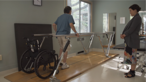
Studies show that FES improves walking speed and distance in people with both incomplete and complete SCI. Some of these studies also showed that regular use of FES carried over to improve walking even without FES. This is supported by weak evidence from eight studies.

The effects of FES treatment may also help to prevent complications of SCI like pressure sores, bone loss, spasticity, and orthostatic hypotension. These benefits may accompany gains in strength or fitness related to FES treatment.

Pressure sores

A woman lifting up her buttocks off her wheelchair by straightening her arms
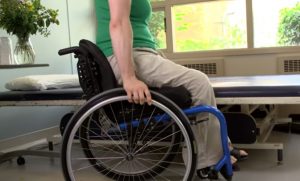
Although it is commonly thought that increased muscle bulk from FES will reduce the risk of pressure sores, there are not very many studies which have looked at whether this actually happens. One study provides weak evidence that FES cycling for 2 years reduced the number of pressure ulcers that occurred after SCI. Another study showed that regular FES cycling showed a trend toward reducing seat pressures.

Bone health

Silhouette of a fractured boneResearch studies show that FES cycling does not prevent bone loss after SCI (moderate evidence from two studies). However, it may help to increase bone density that has already been lost, although the evidence for this is conflicting (based on six studies). It is not clear whether any gains in bone density last long-term or if continued FES treatment is needed for them to be maintained.

For more information on bone density, read our article about Osteoporosis.

Spasticity

It is not clear what effects FES has on spasticity after SCI. There is conflicting evidence from three studies about whether FES cycling can help to reduce spasticity after SCI.

Click here for our article on Spasticity.

Orthostatic hypotension

Three studies provide moderate evidence that FES of the legs during a single change in position reduced orthostatic hypotension. However, this only shows that FES prevents orthostatic hypotension while it is applied, and further research is needed to look at what benefits this could have to people living with SCI.

Click here for our article on Orthostatic Hypotension.

Overall, the research evidence suggests that FES is most likely effective for improving muscle strength after SCI. It may also have effects on fitness, walking skills, bone density, skin health, spasticity, and orthostatic hypotension, although more high quality research is needed to confirm. FES appears to be safe when used appropriately and is widely available in most rehabilitation settings. Discuss this treatment with your health providers to find out if it is a suitable treatment option for you.

For a review of how we assess evidence at SCIRE Community and advice on making decisions, please see SCIRE Community Evidence.

Parts of this page have been adapted from the SCIRE Professional “Lower Limb”, “Upper Limb”, “Bone Health”, “Cardiovascular Health and Exercise”, “Orthostatic Hypotension”, “Pressure Ulcers”, and “Spasticity” chapters:

Lam T, Wolfe DL, Domingo A, Eng JJ, Sproule S (2014). Lower Limb Rehabilitation Following Spinal Cord Injury. In: Eng JJ, Teasell RW, Miller WC, Wolfe DL, Townson AF, Hsieh JTC, Connolly SJ, Noonan VK, Loh E, McIntyre A, editors. Spinal Cord Injury Rehabilitation Evidence. Version 5.0. Vancouver: p 1-74.
Available from: http://scireproject.com/evidence/lower-limb/

Connolly SJ, McIntyre A, Mehta, S, Foulon BL, Teasell RW. (2014). Upper Limb Rehabilitation Following Spinal Cord Injury. In Eng JJ, Teasell RW, Miller WC, Wolfe DL, Townson AF, Hsieh JTC, Connolly SJ, Noonan VK, Loh E, McIntyre A, editors. Spinal Cord Injury Rehabilitation Evidence. Version 5.0: p 1-77.
Available from: http://scireproject.com/evidence/upper-limb/

Craven C, Lynch CL, Eng JJ (2014). Bone Health Following Spinal Cord Injury. In Eng JJ, Teasell RW, Miller WC, Wolfe DL, Townson AF, Hsieh JTC, Connolly SJ, Noonan VK, Loh E, McIntyre A, editors. Spinal Cord Injury Rehabilitation Evidence. Version 5.0. Vancouver: p 1- 37.
Available from: https://scireproject.com/evidence/bone-health/

Warburton DER, Krassioukov A, Sproule S, Eng JJ (2014). Cardiovascular Health and Exercise Following Spinal Cord Injury. In Eng JJ, Teasell RW, Miller WC, Wolfe DL, Townson AF, Hsieh JTC, Connolly SJ, Noonan VK, Loh E, McIntyre A, editors. Spinal Cord Injury Rehabilitation Evidence. Version 5.0. Vancouver: p. 1-48.
Available from: https://scireproject.com/evidence/cardiovascular-health-and-exercise/

Krassioukov A, Wecht JM, Teasell RW, Eng JJ (2014). Orthostatic Hypotension Following Spinal Cord Injury. In: Eng JJ, Teasell RW, Miller WC, Wolfe DL, Townson AF, Hsieh JTC, Connolly SJ, Noonan VK, Loh E, McIntyre A, editors. Spinal Cord Injury Rehabilitation Evidence. Version 5.0. Vancouver: p. 1-26.
Available from: https://scireproject.com/evidence/orthostatic-hypotension/

Hsieh J, McIntyre A, Wolfe D, Lala D, Titus L, Campbell K, Teasell R. (2014). Pressure Ulcers Following Spinal Cord Injury. In Eng JJ, Teasell RW, Miller WC, Wolfe DL, Townson AF, Hsieh JTC, Connolly SJ, Noonan VK, Loh E, McIntyre A, editors. Spinal Cord Injury Rehabilitation Evidence. Version 5.0. 1-90.
Available from: https://scireproject.com/evidence/skin-integrity-and-pressure-injuries/

Hsieh JTC, Connolly SJ, McIntyre A, Townson AF, Short C, Mills P, Vu V, Benton B, Wolfe DL (2016). Spasticity Following Spinal Cord Injury. In Eng JJ, Teasell RW, Miller WC, Wolfe DL, Townson AF, Hsieh JTC, Connolly SJ, Curt A, Mehta S, Sakakibara BM, editors. Spinal Cord Injury Rehabilitation Evidence. Version 6.0.
Available from: https://scireproject.com/evidence/spasticity/


Evidence for “Strength” is based on the following studies:

[1] Baldi JC, Jackson RD, Moraille R, and Mysiw WJ. Muscle atrophy is prevented in patients with acute spinal cord injury using functional electrical stimulation. Spinal Cord 1998;36:463-469.

[2] Scremin AM, Kurta L, Gentili A, Wiseman B, Perell K, Kunkel C, and Scremin OU. Increasing muscle mass in spinal cord injured persons with a functional electrical stimulation exercise program. Arch Phys Med Rehabil 1999;80:1531-1536.

[3] Crameri RM, Weston A, Climstein M, Davis GM, and Sutton JR. Effects of electrical stimulation-induced leg training on skeletal muscle adaptability in spinal cord injury. Scand J Med Sci Sports 2002;12:316-322.

[4] Gerrits HL, de Haan A, Sargeant AJ, Dallmeijer A, and Hopman MT. Altered contractile properties of the quadriceps muscle in people with spinal cord injury following functional electrical stimulated cycle training. Spinal Cord 2000;38:214-223.

[5] Needham-Shropshire BM, Broton JG, Cameron TL, Klose J. Improved motor function in tetraplegics following neuromuscular stimulation-assisted arm ergometry. J Spinal Cord Med 1997;20:49-55.

[6] Cameron T, Broton JG, Needham-Shropshire B, Klose KJ. An upper body exercise system incorporating resistive exercise and neuromuscular electrical stimulation (nms). J Spinal Cord Med 1998;21:1-6.

Evidence for “Cardiovascular Fitness” based on:

[1] Berry HR, Kakebeeke TH, Donaldson N, Perret C, Hunt KJ. Energetics of paraplegic cycling: adaptation to 12 months of high volume training. Technology and Health Care 2012; 20: 73-84.

[2] Griffin L, Decker MJ, Hwang JY, Wang B, Kitchen K, Ding Z, et al. Functional electrical stimulation cycling improves body composition, metabolic and neural factors in persons with spinal cord injury. J Electromyogr Kinesiol 2009;19(4):614-22.

[3] Zbogar D, Eng JJ, Krassioukov AV, Scott JM, Esch BT, Warburton DE. The effects of functional electrical stimulation leg cycle ergometry training on arterial compliance in individuals with spinal cord injury. Spinal Cord 2008;46(11):722-6.

[4] Crameri RM, Cooper P, Sinclair PJ, Bryant G, Weston A. Effect of load during electrical stimulation training in spinal cord injury. Muscle Nerve 2004;29(1):104-11.

[5] Hjeltnes N, Aksnes AK, Birkeland KI, Johansen J, Lannem A, Wallberg-Henriksson H. Improved body composition after 8 wk of electrically stimulated leg cycling in tetraplegic patients. Am J Physiol 1997;273(3 Pt 2):R1072-9.

[6] Mohr T, Andersen JL, Biering-Sorensen F, Galbo H, Bangsbo J, Wagner A, et al. Long-term adaptation to electrically induced cycle training in severe spinal cord injured individuals. Spinal Cord 1997;35(1):1-16.

[7] Barstow TJ, Scremin AM, Mutton DL, Kunkel CF, Cagle TG, Whipp BJ. Changes in gas exchange kinetics with training in patients with spinal cord injury. Med Sci Sports Exerc 1996;28(10):1221-8.

[8] Faghri PD, Glaser RM, Figoni SF. Functional electrical stimulation leg cycle ergometer exercise: training effects on cardiorespiratory responses of spinal cord injured subjects at rest and during submaximal exercise. Arch Phys Med Rehabil 1992;73(11):1085-93.

[9] Hooker SP, Figoni SF, Rodgers MM, Glaser RM, Mathews T, Suryaprasad AG, et al. Physiologic effects of electrical stimulation leg cycle exercise training in spinal cord injured persons. Arch Phys Med Rehabil 1992;73(5):470-6.

[10] Gerrits HL, de Haan A, Sargeant AJ, van Langen H, Hopman MT. Peripheral vascular changes after electrically stimulated cycle training in people with spinal cord injury. Arch Phys Med Rehabil 2001;82(6):832-9.

[11] Ragnarsson KT, Pollack S, O’Daniel W, Jr., Edgar R, Petrofsky J, Nash MS. Clinical evaluation of computerized functional electrical stimulation after spinal cord injury: a multicenter pilot study. Arch Phys Med Rehabil 1988;69(9):672-7.

[12] Taylor JA, Picard G, Widrick JJ. Aerobic capacity with hybrid FES rowing in spinal cord injury: comparison with arms-only exercise and preliminary findings with regular training. PM R 2011;3(9):817-24.

[13] Kahn NN, Feldman SP, Bauman WA. Lower-extremity functional electrical stimulation decreases platelet aggregation and blood coagulation in persons with chronic spinal cord injury: a pilot study. J Spinal Cord Med 2010;33(2): 150-8.

[14] Hakansson NA, Hull ML. Can the efficacy of electrically stimulating pedaling using a commercially available ergometer be improved by minimizing the muscle stress-time integral? Muscle Nerve 2012; 45:393-402.

Evidence for “Walking” is based on the following studies:

[1] Thrasher TA, Flett HM, and Popovic MR. Gait training regimen for incomplete spinal cord injury using functional electrical stimulation. Spinal Cord 2006;44:357-361.

[2] Ladouceur M, and Barbeau H. Functional electrical stimulation-assisted walking for persons with incomplete spinal injuries: changes in the kinematics and physiological cost of overground walking. Scand J Rehabil Med 2000a;32:72-79.

[3] Ladouceur M, and Barbeau H. Functional electrical stimulation-assisted walking for persons with incomplete spinal injuries: longitudinal changes in maximal overground walking speed. Scand J Rehabil Med 2000b;32:28-36.

[4] Wieler M, Stein RB, Ladouceur M, Whittaker M, Smith AW, Naaman S, Barbeau H, Bugaresti J, and Aimone E. Multicenter evaluation of electrical stimulation systems for walking. Arch Phys Med Rehabil 1999;80:495-500.

[5] Klose KJ, Jacobs PL, Broton JG, Guest RS, Needham-Shropshire BM, Lebwohl N, Nash MS, and Green BA. Evaluation of a training program for persons with SCI paraplegia using the Parastep 1 ambulation system: part 1. Ambulation performance and anthropometric measures. Arch Phys Med Rehabil 1997;78:789-793.

[6] Granat MH, Ferguson AC, Andrews BJ, and Delargy M. The role of functional electrical stimulation in the rehabilitation of patients with incomplete spinal cord injury–observed benefits during gait studies. Paraplegia 1993;31:207-215.

[7] Stein RB, Belanger M, Wheeler G, Wieler M, Popovic DB, Prochazka A, and Davis LA. Electrical systems for improving locomotion after incomplete spinal cord injury: an assessment. Arch Phys Med Rehabil 1993;74:954-959.

[8] Granat M, Keating JF, Smith AC, Delargy M, and Andrews BJ. The use of functional electrical stimulation to assist gait in patients with incomplete spinal cord injury. Disabil Rehabil 1992;14:93-97.

Evidence for “Bone health” is based on the following studies:

[1] Eser P, de Bruin ED, Telley I, Lechner HE, Knecht H, Stussi E. Effect of electrical stimulation-induced cycling on bone mineral density in spinal cord-injured patients. Eur J Clin Invest 2003;33:412-419.

[2] Lai CH, Chang WHS, Chan WP, Peng CW, Shen LK, Chen JJJ, Chen SC. Effects of Functional Electrical Stimulation Cycling Exercise on Bone Mineral Density Loss in the Early Stages of Spinal Cord Injury. J Rehabil Med 2010; 42:150-154.

[3] Mohr T, Podenphant J, Biering-Sorensen F, Galbo H, Thamsborg G, Kjaer M. Increased bone mineral density after prolonged electrically induced cycle training of paralyzed limbs in spinal cord injured man. Calcif Tissue Int 1997;61:22-25.

[4] Chen SC, Lai CH, Chan WP, Huang MH, Tsai HW, Chen JJ. Increases in bone mineral density after functional electrical stimulation cycling exercises in spinal cord injured patients. Disabil Rehabil 2005;27:1337-1341.

[5] Frotzler A, Coupaud S, Perret C, Kakebeeke TH, Hunt KJ, Donaldson Nde N, Eser P. High-volume FES-cycling partially reverses bone loss in people with chronic spinal cord injury. Bone. 2008 Jul;43(1):169-76. Epub 2008 Mar 20.

[6] Pacy PJ, Hesp R, Halliday DA, Katz D, Cameron G, Reeve J. Muscle and bone in paraplegic patients, and the effect of functional electrical stimulation. Clin Sci (Lond) 1988;75:481-487.

[7]  Leeds EM, Klose J, Ganz W, Serafini A, Green BA. Bone mineral density after bicycle ergometry training. Archives of Physical Medicine and Rehabilitation 1990;71:207-9.

[8] BeDell KK, Scremin AM, Perell KL, Kunkel CF. Effects of functional electrical stimulation-induced lower extremity cycling on bone density of spinal cord-injured patients. Am J Phys Med Rehabil 1996;75:29-34.

Evidence for “Pressure Ulcers” is based on the following studies:

[1] Dolbow DR, Gorgey AS, Dolbow JD, Gater DR. Seat pressure changes after eight weeks of functional electrical stimulation cycling: a pilot study. Top Spinal Cord Inj Rehabil. 2013 Summer;19(3):222-8.

[2] Petrofsky JS. Functional electrical stimulation, a two year study. J Rehabil. 1992;58(3):29–34

Evidence for “Spasticity” is based on the following studies:

[1] Kapadia N, Masani K, Craven B, et al. A randomized trial of functional electrical stimulation for walking in incomplete spinal cord injury: Effects on walking competency. J Spinal Cord Med 2014;37:511-24.

[2] Manella K & Field-Fote E. Modulatory effects of locomotor training on extensor spasticity in individuals with motor-incomplete spinal cord injury. Restor Neurol Neurosci 2013;31:633-46.

[3] Ralston K, Harvey L, Batty J, et al. Functional electrical stimulation cycling has no clear effect on urine output, lower limb swelling, and spasticity in people with spinal cord injury: A randomised cross-over trial. J Physiother 2013;59:237-43.

[4] Kuhn D, Leichtfried V, Schobersberger W. Four weeks of functional electrical stimulated cycling after spinal cord injury: a clinical cohort study. Inter J Rehabil Res 2014;37:243-50.

[5] Mazzoleni S, Stampacchia G, Gerini A, Tombini T, Carrozza M. FES-cycling training in spinal cord injured patients. Eng Med Biol Soc 2013:5339-41.

[6] Sadowsky C, Hammond E, Strohl A, et al. Lower extremity functional electrical stimulation cycling promotes physical and functional recovery in chronic spinal cord injury. J Spinal Cord Med 2013;36:623-31.

[7] Reichenfelser W, Hackl H, Hufgard J, Kastner J, Gstaltner K, Gföhler M. Monitoring of spasticity and functional ability in individuals with incomplete spinal cord injury with a functional electrical stimulation cycling system. J Rehabil Med 2012;44:444-9.

[8] Krause P, Szecsi J, Straube A. Changes in spastic muscle tone increase in patients with spinal cord injury using functional electrical stimulation and passive leg movements. Clin Rehabil 2008;22:627-34.

[9] Mirbagheri M, Ladouceur M, Barbeau H, Kearney R. The effects of long-term FES-assisted walking on intrinsic and reflex dynamic stiffness in spastic spinal-cord-injured

[10] Granat M, Ferguson A, Andrews B, Delargy M. The role of functional electrical stimulation in the rehabilitation of patients with incomplete spinal cord injury–observed benefits during gait studies. Paraplegia 1993;31:207-15.

[11] Thoumie P, Le C, Beillot J, Dassonville J, Chevalier T, Perrouin-Verbe B et al. Restoration of functional gait in paraplegic patients with the RGO-II hybrid orthosis. A multicenter controlled study. II: Physiological evaluation. Paraplegia 1995;33:654-9.

Evidence for “Orthostatic Hypotension” is based on the following studies:

[1] Faghri PD, Yount J. Electrically induced and voluntary activation of physiologic muscle pump: a comparison between spinal cord-injured and able-bodied individuals. Clin Rehabil 2002;16:878-885.

[2] Elokda AS, Nielsen DH, Shields RK. Effect of functional neuromuscular stimulation on postural related orthostatic stress in individuals with acute spinal cord injury. J Rehabil Res Dev 2000;37:535-542.

[3] Sampson EE, Burnham RS, Andrews BJ. Functional electrical stimulation effect on orthostatic hypotension after spinal cord injury. Arch Phys Med Rehabil 2000; 81: 139-143.

Other references:

Electrophysical Agents – Contraindications And Precautions: An Evidence-Based Approach To Clinical Decision Making In Physical Therapy. Physiother Can. 2010 Fall;62(5):1-80.

Gibbons RS, Shave RE, Gall A, Andrews BJ. FES-rowing in tetraplegia: a preliminary report. Spinal Cord. 2014 Dec;52(12):880-6.

Martin R, Sadowsky C, Obst K, Meyer B, McDonald J. Functional electrical stimulation in spinal cord injury: from theory to practice. Top Spinal Cord Inj Rehabil. 2012 Winter;18(1):28-33.

Warms CA, Backus D, Rajan S, Bombardier CH, Schomer KG, Burns SP. Adverse events in cardiovascular-related training programs in people with spinal cord injury: a systematic review. J Spinal Cord Med. 2014 Nov;37(6):672-92.

Image credits

  1. E-Stim Therapy ©Rankn Jordan, CC BY-NC-SA 2.0
  2. Functional electrical stimulation ©MilosRPopovic, CC BY-SA 4.0
  3. Image by SCIRE Community Team
  4. Checklist ©lastspark,CC BY 3.0 US
  5. Muscle © Smalllike, CC BY 3.0 US
  6. cardio ©emma Mitchell, CC BY 3.0 US
  7. Image by SCIRE Team.
  8. Image by SCIRE Team.
  9. fracture ©fahmionline, CC BY 3.0 US


Disclaimer: This document does not provide medical advice. This information is provided for educational purposes only. Consult a qualified health professional for further information or specific medical advice. The SCIRE Project, its partners and collaborators disclaim any liability to any party for any loss or damage by errors or omissions in this publication.