


Author: SCIRE Community Team | Reviewer: Riley Louie | Published: 21 September 2017 | Updated: ~
This page provides an overview of the structures of the spinal cord and how the spinal cord works.
Key Points
- The spine is a long column of 33 small back bones (vertebrae) that are connected together by ligaments and discs.
- The main areas of the spine are the cervical spine (neck), thoracic spine (upper and mid back), lumbar spine (low back), sacrum (part of the pelvis), and coccyx (tailbone).
- The spinal cord is a large bundle of nerve tissues located within the center of the spine.
- The spinal cord is made up of nerve cells (neurons) that relay nerve signals between the brain and body to control movement, sensation, and other functions. The spinal cord is also the centre for reflexes.
The spine is a long column of 33 small back bones called vertebrae that are connected together by strong fibrous ligaments and shock-absorbing fibrous discs. The spine is part of the skeleton.
The spine supports the weight of the body, allows the torso to move, anchors many of the muscles, and protects the spinal cord from damage. The spine is also called the vertebral column, spinal column, or backbone.
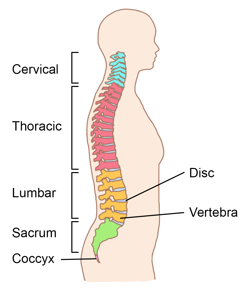
The five major areas of the spine.1
Areas of the spine:
- The cervical spine has 7 cervical vertebrae and forms the neck.
- The thoracic spine has 12 thoracic vertebrae and forms the upper and mid back.
- The lumbar spine has 5 lumbar vertebrae and forms the low back.
- The sacrum is made up of 5 sacral vertebrae that are fused together to form a wedge-shaped bone that is part of the pelvis.
- The coccyx is made up of 3 to 5 coccygeal vertebrae that are fused together to form the tailbone.
The spine has a hollow canal in its centre that runs the length of the spine from the base of the skull down to the sacrum called the spinal canal. The spinal canal contains the spinal cord.
Naming the bones of the spine
The bones of the spine are named and numbered according to:
- The area of the spine that the bone is located in (cervical, thoracic, lumbar, or sacral), which is often abbreviated simply as a letter (i.e. ‘C’ for ‘cervical spine’ or ‘C-spine’, ‘T’ for ‘thoracic spine’ or ‘T-spine’); and
- The number of the bone within that area. The numbering begins at ‘1’ for the vertebra closest to the head and increases with each vertebra down to the tailbone (counting up as you move down the spine). The numbering then begins again at ‘1’ in the next area of the spine.
For example, the bone at the top of the lumbar area of the spine (the low back) is called the ‘first lumbar vertebra’ or ‘L1’.
The spinal cord is divided into 31 segments, which each give rise to a pair of spinal nerves.3
The spinal cord is a long bundle of nerve tissue that is located in the spine. It is the main pathway for nerve signals travelling between the brain and the rest of the body. It is also the centre for the body’s reflexes.
The spinal cord is made up of millions of microscopic nerve cells. Protective layers of tissue called meninges cover the spinal cord and a special fluid called cerebrospinal fluid cushions the spinal cord within the spine.
The path of the spinal cord
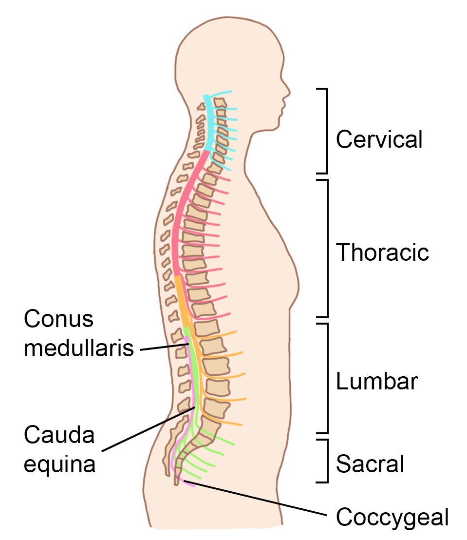
The spinal cord begins at the base of the skull, where it connects to the brain through the brainstem. The spinal cord then runs down through the spine’s hollow central spinal canal.
The spinal cord is shorter than the spine, so it does not travel the full length of the spine. It ends at a point called the conus medullaris near the first or second lumbar vertebra. From this point down, spinal nerves branching from the end of the spinal cord sit within the spinal canal in a bundle called the cauda equina.
Spinal cord segments
Spinal nerves extending from the spine.4
The spinal cord is divided into 31 nerve segments from top to bottom:
• 8 cervical segments
• 12 thoracic segments
• 5 lumbar segments
• 5 sacral segments
• 1 coccygeal segment
Each segment is a part of the spinal cord that gives rise to a pair of spinal nerves (one right and one left). The spinal nerves exit the spine through archways between the bones and continue outward to become the nerves of the arms, legs, and body.
The spinal cord is part of the nervous system. The nervous system is the body’s main communication system. It allows messages to be passed from one area of the body to another. This is possible because of special cells called neurons.
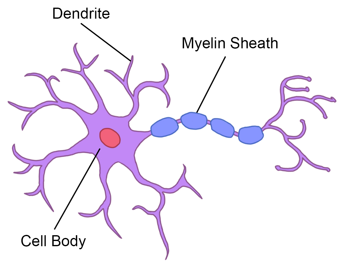
Neurons are cells that transmit nerve signals in the body.5
Neurons
Neurons are the main cells of the nervous system. Neurons generate, conduct, and pass along nerve signals within the nerves, spinal cord, and brain.
Neurons communicate with other cells (such as other neurons, muscle cells and sensory receptors) through connections called synapses. These connections allow the cells to pass electrical and chemical nerve signals to other cells.
The brain and spinal cord together make up the central nervous system. The central nervous system is the main control system of the body.
- The brain is the command center of the nervous system. It sends commands to the rest of the body which control movement, breathing, and other functions. The brain also receives signals about sensations from the whole body, which it interprets to help manage bodily functions.
- The spinal cord is the main pathway for information travelling between the brain and the rest of the body. It acts like a highway along which nerve signals can travel between distant areas of the body. This includes both commands sent from the brain to the body and sensations from the body to the brain. The spinal cord is also the center for reflexes (a reflex is a muscle response to a touch stimulus).
The main functions of the spinal cord are to pass along information related to movement, sensation, reflexes, and organ function.
Movement (motor neurons)
 The spinal cord provides a pathway for movement commands to travel from the brain to the muscles. This is called motor function. Neurons that send movement commands are called motor neurons.
Movement begins in special movement (motor) areas of the brain that plan and generate nerve signals to create movements. Upper motor neurons from this area of the brain have long nerve fibers (axons) that project down the spinal cord, where they pass their signals to lower motor neurons that travel out of the spinal cord and into the body. These lower motor neurons connect with muscle cells to pass along movement commands that tell the muscles to contract.
Sensation (sensory neurons)
The spinal cord is also a pathway for sensations traveling from the body to the brain. This is called sensory function. Neurons that send sensory information are called sensory neurons.
The body’s tissues (like the skin, muscles, and organs) contain special sensory receptors. Sensory receptors detect sensations such as touch, pressure, vibration, or temperature. When a sensation is detected, a signal is sent along the nerve fiber (axon) of a sensory neuron to the spinal cord.
The signal then travels up the spinal cord where it is passed along to other neurons in the brainstem and brain. When these signals reach the sensation centers in the brain, they are interpreted and the person feels the sensation.
Spinal reflexes
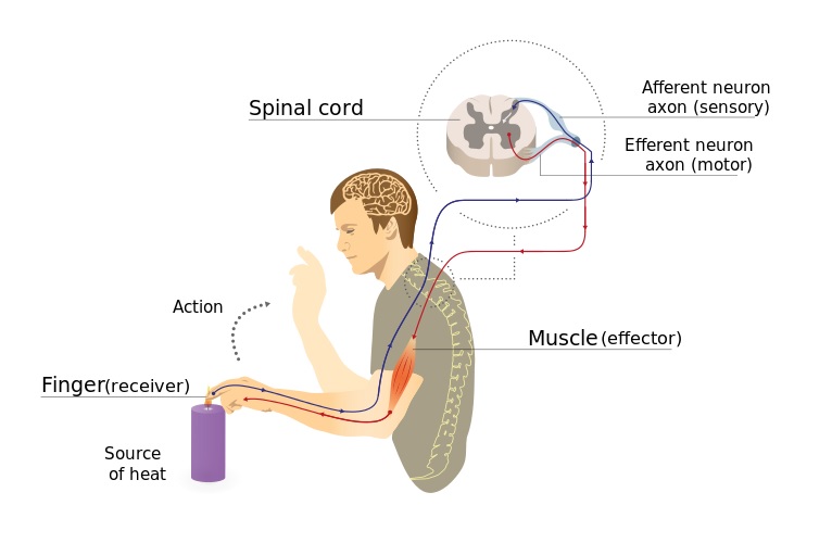
Pain signals from touching something hot travels to the spinal cord and back to the muscles without going to the brain first.7
Reflexes are automatic responses that happen in the spinal cord and do not travel to the brain first. For example, when the tendon below the knee cap is tapped, it causes the knee jerk reflex.
Spinal reflexes involve neurons for both sensation and movement. When certain sensory receptors are activated, a nerve signal is sent through sensory neurons to the spinal cord. In the spinal cord, the signal is passed on to lower motor neurons involved in the reflex movement. The motor neurons then send a signal out of the spinal cord to the muscles, causing an immediate muscular response.
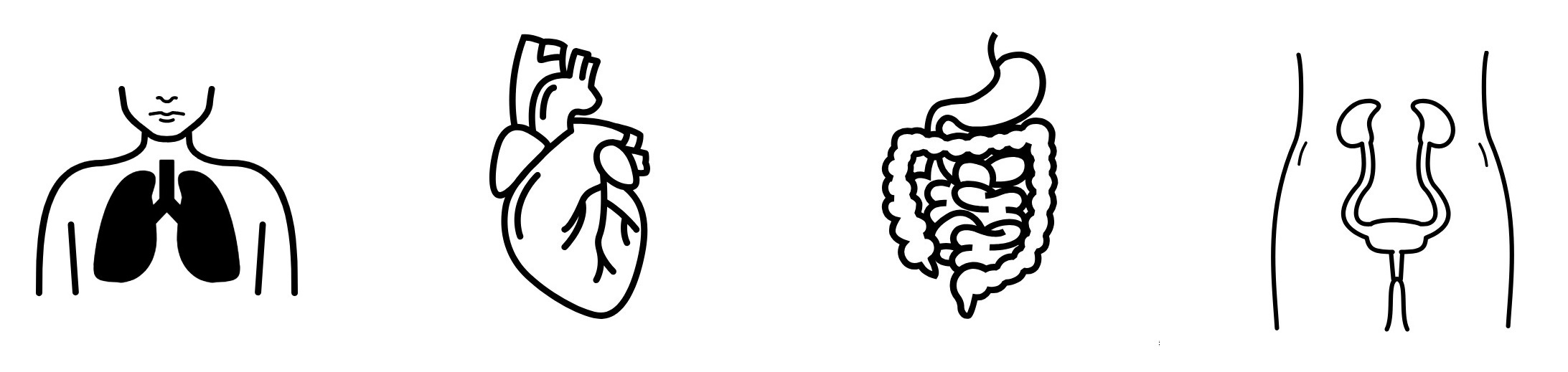
Internal organ (autonomic) function
The spinal cord also plays a role in controlling some of the functions of the internal organs through the autonomic nervous system.
The autonomic nervous system
The autonomic nervous system controls largely unconscious bodily processes such as blood pressure, heart rate, breathing rate, body temperature, digestion, bladder, bowel, and sexual function.
It has two divisions:
- The sympathetic nervous system prepares the body for stressful or emergency situations. It is often called the ‘fight or flight’ system, because it prepares the body for action. For example, it increases heart rate and slows digestion.
- The parasympathetic nervous system prepares the body for normal, non-emergency situations. It is often called the ‘rest and digest’ system, because it allows the body to restore itself. For example, it slows heart rate and increases digestion.
The sympathetic and parasympathetic systems have different (and often opposite) effects on the organs and work together to control bodily functions according to the situation.
The autonomic nervous system controls various body functions.8-11
Neurons that control the function of the autonomic nervous system begin in the brain or brainstem. Some of these neurons leave in the cranial nerves (nerves that arise directly from the brain and brainstem), and the rest travel down the spinal cord, where they branch from certain areas:
- The nerves of the sympathetic nervous system arise from the thoracic and lumbar spinal cord from the levels of T1 to L2.
- Some of the nerves of parasympathetic nervous system arise from the sacral spinal cord, from S2 to S4 (the others leave from the brainstem).
After leaving the spinal cord, the neurons connect (synapse) with other neurons in clusters of nerve cells called ganglia. From these ganglia, motor neurons project out to the organs and signal changes to their function.
Bican O, Minagar A, Pruitt AA. The spinal cord: a review of functional neuroanatomy. Neurol Clin. 2013 Feb;31(1):1-18.
Moore KL, Dalley AF, Agur AMR. Clinically Oriented Anatomy. 6th ed. Philadelphia: Lippincott Williams & Wilkins, 2010.
Image credits
- Image by SCIRE Community Team
- Spinal cord ©Vectors Market, CC BY 3.0 US
- Image by SCIRE Community Team
- BIO 120 Lab Spinal Cord 035 ©djneight, CC BY-NC-ND 2.0
- Image by SCIRE Community Team
- Lifting weights ©skeeze, CC0 1.0
- Imgnotraçat arc reflex eng ©MartaAguayo, CC BY-SA 3.0
- Lung ©mungang kim, CC BY 3.0 US
- Heart ©Laymik, CC BY 3.0 US
- Digestive System ©Design Science, CC0 1.0
- Excretory system ©Olena Panasovska, CC BY 3.0 US
Disclaimer: This document does not provide medical advice. This information is provided for educational purposes only. Consult a qualified health professional for further information or specific medical advice. The SCIRE Project, its partners and collaborators disclaim any liability to any party for any loss or damage by errors or omissions in this publication.
