COVID-19 Factsheet: Social Isolation and SCI

By | | No Comments

Author: SCIRE Professional Team | Reviewer: Rachael Neal | Published: 23 July 2020 | Updated: ~

In the time of COVID-19, physically distancing is necessary to limit virus transmission and to keep everyone as safe as we possibly can. However, social isolation can have serious effects on mental health for everyone, including those with spinal cord injury (SCI).

Key Points

  • Social isolation is the absence of relationships and interactions with one another and is linked to mental health issues like depression and anxiety.
  • People with SCI may be at increased risk for the negative effects of social isolation.
  • Consider seeking help if you experience symptoms such as marked changes in personality, difficulty coping with daily activities, prolonged sadness, or excessive worry.
  • Strategies to maintain good health while physically distancing include daily structure, reaching out to others, good diet/hydration, and regular exercise.

Download PDF

Expand All

What is social isolation?

Social isolation is described as the absence of relationships, often due to physical separation from others. This differs from loneliness, which is a state of distress caused by feeling alone or separated.

Social isolation has been linked to various mental health issues, such as higher fatigue and depression rates. People who are socially isolated often report more difficulties with access and fewer close relationships.

Am I at increased risk for these potential effects if I have an SCI?

People with SCI are not necessarily more vulnerable to the effects of social isolation. However, research shows a higher proportion of depression and anxiety in people with SCI compared to the general population. The COVID-19 pandemic may limit access to caregivers or peer support networks that people with SCI depend on which can worsen symptoms of depression and anxiety.

You may want to reach out to a professional if you experience any of the following symptoms to the extent that it is concerning to you or close others:

  • Marked changes in mood, eating or sleeping patterns
  • Excessive anxiety
  • Prolonged sadness, depression, or apathy (disinterest)
  • Thoughts or statements about hurting yourself, harming others, or suicide
  • Increased substance use or using substances in ways that could be harmful
  • Excessive anger, irritability, hostility, or violent behavior

Look after your general health

There are direct links between mental health and physiological health. So, it is especially important at a time like this to take care of your general health. Stay mentally and physically active, have proper nutrition, hydrate well, and get enough sleep. Maintaining physical health can help serve as a buffer for emotional stress as we are more likely to feel that we have the resources to cope with stress when we are rested and eating well versus when we are tired and hungry.

Manage your worry

Some degree of worrying at a time like this is normal, but it is important to note if your worry is excessive, or if worry thoughts are interfering with your ability to do other things.
Strategies you can employ if you are feeling worried include:
• Noticing and limiting your exposure to things that trigger your worry,
• Practice postponing your worry to a later time, and
• Relying on reputable news sources to ensure your information regarding the pandemic is up-to-date and accurate.

Set a routine

Setting a routine can help you maintain a sense of normalcy during the pandemic. Stick to the usual structure of your day as best as you can by eating, exercising, and sleeping at the same times that you normally would. By setting a routine, many people find that they feel have a sense of purpose and predictability that is comforting. Using structure can also encourage us to work on tasks, which can bring feelings of accomplishment, and that feeling of accomplishment is naturally rewarding.

Stay connected

In the digital age, there are many ways to stay connected virtually to your loved ones. Videochatting, calling, and messaging is possible through many different platforms. ForaHealthyMe SCIO: Home is an example of a virtual platform established for connecting people with SCI.
As restrictions to physical distancing are slowly relaxing, it is possible to physically meet some of your loved ones in small numbers and at a safe distance from each other (it is still important be cautious and maintain physical distancing).

Be knowledgeable and take appropriate precautionary measures

A study has shown that being up-to-date with accurate health information (e.g., treatment, local outbreak situation) and taking particular precautionary measures (e.g., hand hygiene, wearing a mask) was associated with a lower psychological impact of the outbreak and lower levels of stress, anxiety, and depression. Taking recommended steps to protect your health (e.g., washing your hands, mask use) can also remind you of the strategies that are currently within your control, which can also increase your self-confidence in coping with uncertainty over time.

Reach out for help if needed

Most of us are currently facing challenges that we have never faced before. It is natural and encouraged that you seek help if you require assistance in any aspect of your life. Seeking help when you need it is a sign of strength and good judgment!
There are many resources you can currently access that provide information, emotional support, and advice to people with SCI. Below are a few examples of such resources.

Resources to access

BC Government COVID-19 mental health support: Virtual Mental Health Supports During COVID-19 – Province of British Columbia

BC Mental Health Hotline: dial 310-6789, and do not add 604, 778 or 250 before the number. It’s free and available 24 hours a day.

We also recommend: keltyskey.com/, BounceBack bouncebackbc.ca/ and a “FACE COVID” e-book that can be found at actmindfully.com.au.

For good plain language information on COVID in multiple languages, visit flattenthecurve.com and for reliable data on COVID cases worldwide, visit coronavirus.jhu.edu/map.html.

You can also check out the other resources listed on our site: scireproject.com

Physical distancing is a necessary precaution that needs to be taken during the COVID-19 pandemic, but it can have adverse mental health effects if you do not take steps to stay socially connected. To maintain good mental health when isolation is inevitable, we recommend maintaining good physical health, managing your worry, setting a routine, staying connected with family and friends, and staying knowledgeable about the current situation.

Frank, M., Heinemann, A. and Wong, A., 2016. An Empirical Investigation of a Biopsychosocial Model of Social Isolation in Persons with Neurological Disorders. Archives of Physical Medicine and Rehabilitation, 97(10), p.e20.

Brooks, S., Webster, R., Smith, L., Woodland, L., Wessely, S., Greenberg, N. and Rubin, G., 2020. The Psychological Impact of Quarantine and How to Reduce It: Rapid Review of the Evidence. SSRN Electronic Journal, 395(10227).

Migliorini C, Tonge B, Taleporos G. Spinal cord injury and mental health. Aust N Z J Psychiatry. 2008;42(4):309‐314. doi:10.1080/00048670801886080

Mayoclinic.org. 2020. Mental Health: What’s Normal, What’s Not – Mayo Clinic. [online] Available at: <https://mayoclinic.org/healthy-lifestyle/adult-health/in-depth/mental-health/art-20044098?p=1> [Accessed 10 June 2020].

Ohrnberger J, Fichera E, Sutton M. The relationship between physical and mental health: A mediation analysis. Soc Sci Med. 2017;195:42-49. doi:10.1016/j.socscimed.2017.11.008

Wang C, Pan R, Wan X, et al. Immediate Psychological Responses and Associated Factors during the Initial Stage of the 2019 Coronavirus Disease (COVID-19) Epidemic among the General Population in China. Int J Environ Res Public Health. 2020;17(5):1729. Published 2020 Mar 6. doi:10.3390/ijerph17051729

Image credits

  1. Woman in House © Mohamed Hassan, CC0 1.0
  2. Image by SCIRE Team
  3. Doctor © CO. Department of Health Care Policy and Financing, CC0 1.0
  4. Image by SCIRE Team
  5. Notebook © Gentaur CC0 1.0
  6. Image by SCIRE Team
  7. Man on computer © user:cth103_t CC0 1.0
Disclaimer: This document does not provide medical advice. This information is provided for educational purposes only. Consult a qualified health professional for further information or specific medical advice. The SCIRE Project, its partners and collaborators disclaim any liability to any party for any loss or damage by errors or omissions in this publication. SCIRE receives no compensation and there are no conflicts declared with sources of information on this factsheet.

Wheelchair Seating

By | | No Comments

Author: Sharon Jang | Reviewer: Emma M. Smith | Published: 25 May 2020 | Updated: ~

Wheeled mobility devices like wheelchairs and scooters are an important part of independent living after spinal cord injury (SCI). This page provides an overview of the basics of wheelchair seating after SCI.

Key Points

  • Wheelchair seating includes parts of the wheelchair that help you maintain a proper and comfortable posture when seated. This includes the backrest, foot rests, and cushion.
  • Proper wheelchair seating is important to prevent pressure sores, maintain posture, and promote function.
  • There are many different types of seating cushion, each with its own benefits and drawbacks.
  • There are three main aspects of backrests to be considered when getting fitted to a wheelchair: the height, the shape, and the stiffness.

A wheelchair seating assessment is a complex process where healthcare professionals assess your body and fit you to a wheelchair to suit your needs. How you fit in your wheelchair can significantly impact your health and comfort while using your device. A proper seating assessment can be used to prevent conditions (for example, spine deformation) from getting worse, to correct posture, and to accommodate for other aspects that are not changeable. The seating process is done in combination with other rehabilitation interventions (such as exercises, physical therapy, and spasticity management), and should be done with specific therapeutic goals in mind.

In this document, we will focus on the components on seating. Seating components consist of backrests, cushions, and accessories (e.g., footrests and arm rests). Having appropriate seating is important to having good posture while in your wheelchair, being able to function while using the wheelchair, and maintaining good skin health.

Refer to our article on Wheelchair Provision for more information!

Pressure sores

As you may be spending the majority of your time in your wheelchair, sitting pressures are of concern to skin health. Research (moderate evidence) has suggested that the sitting postures of individuals with SCI are different than able-bodied individuals. In the SCI population, higher sitting pressures are exhibited. Up to half of pressure ulcers occur on the sit bones, and are likely to have developed when sitting. Having a proper cushion suited to the way you sit can help to prevent pressure sores by redistributing pressure.

Maintaining posture

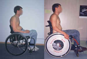
Your wheelchair setup can have a major effect on your posture while seated.1

The way your wheelchair is set up can impact your sitting posture. After an SCI, you may not be able to control the stability of your trunk, which can lead to spinal deformities and abnormal sitting positions. However, the use of a proper cushion and supports can help address sitting posture. For example, one study (weak evidence) suggests that the use of lateral supports (i.e., pads supporting the side to prevent side to side leaning) can help improve the alignment of the spine and to help improve posture while reducing the amount of effort required by muscles for postural control.

Psychosocial concerns

The way you are able to present yourself while sitting in your wheelchair may impact your psychosocial health, or your sense of confidence and self-esteem.

Quality of life

Having proper seating may impact your quality of life. Good seating can allow you to fully participate in leisure activities, and may allow you to go to school or to work. These activities depend on your sitting tolerance (how long you are able to sit in your chair comfortably) and your ability to effectively travel in your wheelchair.

There are two main purposes of wheelchair cushions: 1) to help improve function and achieve a balanced posture, and 2) to redistribute the pressure applied by the sit bones when seated. Taken together, the cushion can impact the amount of support provided, which in turn determines how long someone can sit for, how easily transfers can be done, and posture of the hips. There are many types of cushions available on the market, as no one cushion is suitable for all individuals with SCI. To determine which is best suited for your needs, various factors are considered, including:

  • The amount of pressure reduction/redistribution required
  • Temperature effects (warm temperatures may lead to sweating, which may make the skin more vulnerable to injury and infections)
  • Your level of injury
  • Your ability to relieve pressure off your sit bones
  • Your transfer techniques
  • Your lifestyle

Although new cushions may be able to provide support and redistribute pressure applied by the sit bones, the effectiveness of a cushion decreases over time. Some research studies have looked into factors that impact deterioration of a cushion. Weak evidence suggests that how a cushion is used is a bigger indicator of deterioration compared to age of the cushion. For example, factors such as how you transfer in and out of your wheelchair, frequency of curb jumps or high vibration activities, exposure to the elements (rain, snow, etc.), moisture, and exposure to extreme temperatures can negatively impact the quality of your cushion.

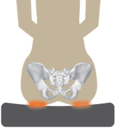
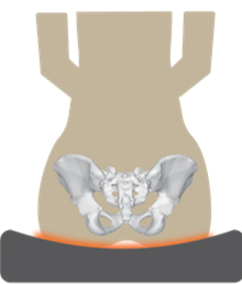
When sitting, pressures are created (orange) from the force of your sit bones on your buttock.2


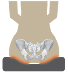
During offloading, a greater surface area of the buttock is in touch with the cushion.3

Pressure sores develop when there is a lot of pressure applied at one point. We can counter this focussed pressure through pressure redistribution, i.e. spreading out the sitting pressures. This can be done in three ways: offloading, immersion, and immersion and envelopment.

When offloading, pressure is distributed over a greater surface area. To achieve this, pressure is distributed over the majority of the buttock (i.e., more of the buttock is in contact with the cushion) rather than just on the sit bones (see top right image). To encourage this position, a piece of the cushion may be taken out so there is a grooved surface.

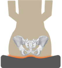
Immersion and envelopment is a combination of sinking into the cushion and having the cushion form to the shape of your buttock.5


During immersion, the buttock sinks into the cushion.4

Immersion refers to a property of the cushion material, which allows the body to “sink” into it. Given the material’s ability to be compressed, pressure is redistributed by increasing the amount of body surface area that is in contact with the cushion.

Immersion and envelopment redistributes pressure by allowing the body to sink into the material, while the material conforms to the body’s shape. This maximizes pressure distribution by increasing the amount of surface area in contact with the cushion.

There are five common types of cushion material: foam, gel, air, honeycomb urethane, and alternating pressure.

Foam cushions

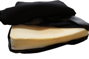
Foam cushions require protection from getting wet. The cushion above features an extra lining under the cushion cover to help keep it dry.6

Foam cushions are a cheaper option, and come in a variety of densities, ranging from soft memory foam to higher density foam. Foam is able to adapt to shapes, is low maintenance, and can provide support while spreading sitting pressures. One downside to using foam is that it wears out and loses its shape quickly. In addition, foam cushions are only able to provide a limited amount of pressure relief and comfort. Foam cushions also must be protected from getting wet. The use of foam cushions is recommended for those with basic sitting needs.

Gel cushions

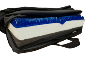
A variety of gel cushions exist. These include a gel matrix (left) and a gel cushion (right).7

There are different types of gel cushions including gel matrix and gel cushions. Gel cushions aim to provide seating comfort by using gel placed on top of a layer of supportive foam. Gel cushions are able to relieve pressure points and distribute pressure over a larger area, while providing a stable surface to support positioning. They may also help counter effects of high temperatures with its cooling properties. Some drawbacks to using a gel cushion include heavier weight and a lack of shock absorption. Gel cushions also have the potential to “bottom out” (when all the gel is pushed aside, massaging may be required to redistribute the gel), and a potential for leaks.

Air cushions

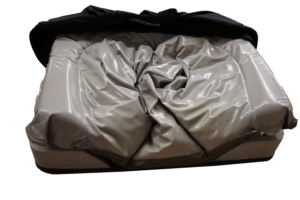
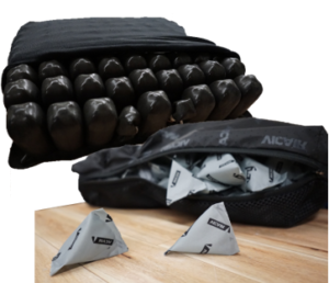
Air cushions are often comprised of a group of small air-filled cells. In some air cushions, the cells are interconnected, while other versions contain multiple separate air sacs contained within a cushion cover. These cells support the weight of the user, and spread out the pressure of sitting through shifting air to the surrounding cells. These cushions are customizable in regards to the number of cells and the amount of air each cell is able to hold. If the cells are interconnected, the amount of air in the cushions can be adjusted using a pump. Moderate evidence has suggested that the use of air cushions can reduce the risk of a pressure sore through reducing the amount of pressure produced while sitting and promotion of air flow. Furthermore, air cushions are generally waterproof. Although these cushions are great for providing pressure distribution, they are not optimal for stability and postural support. In addition, they may be considered high maintenance as the pressure of the cushion has to be checked frequently and manual pumping of air into the cushion is required.

Honeycomb urethane cushions

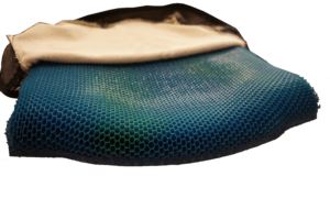
These light-weight durable cushions resemble a honeycomb in that they are composed of multiple open cells. These cells are able to distribute pressure evenly while avoiding the risk of being punctured. Air flow is also promoted throughout the open cells to prevent skin breakdown. As these cushions are made out of urethane, a material that resembles rubber, they also provide good shock absorbance. However, compared to other cushions, they provide moderate positioning capabilities and are not modifiable in shape.

Alternating pressure cushions

Alternating pressure cushions consist of multiple air bladders (similar to an air cushion) with an added battery-operated microprocessor that controls the amount of air in each part of the cushion. Every 4-6 minutes, the air in each segment of the cushion alternates (i.e., inflates or deflates) to help relieve pressure of the users’ bottom. This cushion is suitable for individuals who are unable to effectively relieve seated pressure. There has been weak evidence suggesting that alternating pressure cushions have provide good user satisfaction and comfort. Some negatives for this cushion include its high cost, heavy weight (as it requires a pump and a motor), and susceptibility to punctures.

While cushion marketing may promote a reduction in sitting pressure, more research is required to determine whether reducing the pressure on the sit bones or decreasing risk factors will prevent pressure ulcers. Pressure mapping is one technique that may be used to help determine the areas that are prone to pressure sore.

Refer to our article on Pressure Mapping for more information!

Custom Contour Cushions

These cushions are made custom to the shape of your buttock in an attempt to reduce pressure. Often, custom contour cushions are made with a combination of the aforementioned materials, accounting for the high pressure areas identified by pressure mapping. There is moderate research evidence that suggests that custom contoured cushions may create a safe sitting surface for individuals with SCI through the ability to redistribute sitting pressures. There is also weak evidence that using a custom contour cushion may help increase sitting stability and posture. If using a custom contour cushion, avoid getting the cushion wet as it may deform, and be careful to properly position yourself on the cushion as there is only one position that is optimal for comfort and pressure distribution.

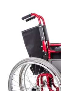
The main purposes of backrests are to provide stability and support to the trunk and the hips. Backrests are adjustable in three different ways: the height, the shape, and the stiffness.

Height

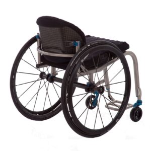
For manual wheelchair users, the height of the backrest can vary. Low backrests provide support for the lower back, and are often preferred by active users as they allow for more mobility in the upper spine. However, the use of a low backrest requires complete or partial trunk control, as it does not provide much stability. Higher backrests typically span the majority of the back, but should come up to just under the shoulder blades. A higher back rest can provide more support, but a backrest that is too high may hinder mobility.

There has been some research on the impact of various backrest heights on the range of motion required for propulsion and reaching/grasping motions. One study (weak evidence) has shown that the height of the backrest may influence the efficiency of pushing a manual wheelchair. In particular, the authors suggest that a low back rest may be more beneficial for wheelchair propulsion, as it allows for greater movement in the shoulder, a greater push rate (i.e., more pushes per minute), and a greater propulsion stroke. In addition, a lower back rest has been found to allow paraplegics to apply more force when propelling their wheelchairs. However, with regards to reaching, a (weak evidence) study found that backrest height did not have an effect.

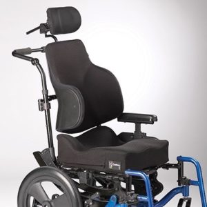
As power wheelchair users do not need to manually propel their wheelchair, the height of the backrest is typically higher. These backrests typically span the entire length of the back, and provide more support to the spine. Moreover, having a taller backrest can provide a resting position for tilt or recline functions.

Shape

Backrests generally come in three different shapes: flat, general contour, and custom contoured.

Flat backrests

Flat backrests are flat or slightly curved in shape, and often consist of a stiff flat surface (e.g., plastic, plywood) that is layered with foam and covered with material. This style of back allows for the greatest range of motion of the arms, thus creating increased freedom. Another advantage of the flat backrest is that it is very adjustable and can accommodate a large variety of support accessories, such as lateral supports, chest straps, and headrests. A drawback to this style of back is that it provides limited support. It does not accommodate for the shape of the spine, making it less suitable for individuals with lordosis (i.e., sway back) or kyphosis (i.e., a hunched back).

General contoured backrests

General contoured backrests are off-the-shelf backs that are shaped, but are not customized to your back. These backs provide more support than a flat back, as they have a deeper contour that can provide lateral (side) support. The effectiveness of general contoured backs is based on how well the back fits your needs; they may only be effective if you can find one that suits your needs.

Custom contoured backs

Custom contoured backs provide increased support for positioning, and are made custom to the shape of your back. This cushion is often used if you require extra positioning support, and if the general control back or flat back does not meet your needs. The creation of custom contour backs can be a lengthy and costly labour-intensive process. When creating a custom contour back, a mold of your back is taken. A seating specialist then inspects the mold to ensure that it is reflective of the shape of your back and that the contour information is accurate. The backrest is then carefully made to the specifications obtained from the measuring process. As custom contour backs are designed to fit the shape of your back to provide more support, they also impose limitations on flexibility. When transferring in and out of your wheelchair, it is also important to be properly aligned in your custom contoured backrest, as improper fitting may lead to pressure sores or skin breakdown.

Stiffness

A rigid back on a manual wheelchair.13


Soft backs are often found on folding manual wheelchairs.14

The stiffness of wheelchair backs are either soft or rigid. Soft backs (i.e., sling backs) are able to accommodate to the shape of the spine and can be effective if they are properly adjusted. However, they provide less support compared to rigid backs and can stretch over time depending on the fabric. Tension adjustable backs consist of interwoven straps that can be tightened or loosened to accommodate for posture. One weak study found that the use of a tension adjustable back provides more support than a normal sling back. While it provides more support for the hips, it may still result in poor posture. Rigid backs generally provide more support and can help with stability. However, rigid backs are less adjustable, and do not accommodate for the shape of the spine. Although rigid backs may be more supportive, weak evidence has suggested that rigid backs are less comfortable than sling backs among individuals with tetraplegia.

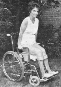
Footrests (or leg rests) are an important part of a wheelchair. They function to stabilize your legs for optimal hip and back posture, help promote redistribution of sitting pressures, and may promote circulation. Footrests come in a variety of options. Footrests may be fixed (as one plate or two), swing-away (i.e., can be moved away from the front or removed), or flip up (particularly in powered wheelchairs). Elevating footrests allow for the leg to be in a raised position, which may help alleviate some leg pain.

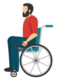
Having a foot rest that is too high may result in more pressure on your buttock.15

The length of the footrest hanger (i.e., the distance from the back of the knee to the heel) can impact how you are seated in the wheelchair. Having a footrest that is too short will push your knees upward, so that the bottom of your thighs are no longer in contact with the top of the seat cushion. As a result, more pressure is applied to your sit bones and there is a lessened ability to shift your weight backward if you slide forward. On the other hand, a footrest that is too long may result in sliding forward in the wheelchair. Consequently, someone with a footrest that is too long may tend to slide forward often in their chair, leading to a hunched back.

Some weak evidence suggests that footrests can affect how activities of daily living are performed, but not the types of activities performed. The use of footrest may help improve the sitting balance of individuals with a lumbar SCI, but not those with a thoracic SCI.

Arm rests on wheelchairs serve multiple purposes and offer more benefits than simply acting as a place to rest the arms. In addition to acting as an arm rest, this part is also used to help maintain posture, redistribute pressure, and to enhance functioning (e.g., transfers, stability). The benefit of using armrests is dependent on an individual’s level of injury and abilities. An individual with more control of their torso and arms will be less likely to need armrests. Some active individuals tend to find that arm rests get in the way.

Maintaining posture

Using armrests can help maintain a good seated posture for an individual using a wheelchair. By supporting the arms and forearms, weight is alleviated from the shoulders. Without armrests, the weight of your arms may pull your shoulder down, resulting in a hunched position. It is also important that your armrests are set to an appropriate height. Armrests that are too low may require the individual to lean forward to use the armrest, which may lead to hunching. If the armrests are too high, the shoulders may be pushed up too high, which can lead to discomfort.

Redistributing pressure

Armrests can act as a source of support for repositioning to alleviate pressure on the sit bones. Weak evidence suggests that individuals with paraplegia rely on armrests more than tetraplegics during weight shifting (9% of their body weight vs 5%). The researchers think that this may be the case because individuals with tetraplegia have weak arm extensor muscles, making weight relieving difficult. In addition to acting as a support to push off from, armrests can also help alleviate pressure on the sit bones by supporting the weight of your arms. By removing the weight of hanging arms, the hips are unloaded and pressure forces are redistributed.

Enhancing function

Armrests can be helpful for everyday activities such as transferring, picking up objects, and stability. When transferring, the armrests act as a source of support and are used to push up and off from, or they can be a firm object to hang onto when transferring back in. In addition, armrests may act as a source of stability when completing tasks that may challenge someone’s balance, such as picking up objects off the ground, reaching for high objects, and leaning.

There are a variety of seating accessories that can be used to optimize comfort and posture. Although accessories can be used to enhance your seating, the majority of your posture should be supported through your seating set up (i.e., the backrest, cushion, foot rest), and not through accessories.

Upper extremity accessories

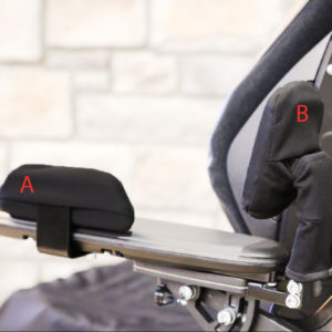
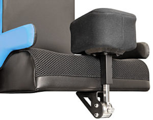
Arm supports to prevent the arm from falling off the side (A) and the elbow from sliding back (B).18

Seating accessories for the arms are more often found on power wheelchairs than manual wheelchairs. This group of accessories include armrest side supports and elbow blocks. Armrest side supports help prevent the forearm from falling off the side of the wheelchair, while elbow blocks prevent your elbows from sliding backwards. The use of these accessories can help keep your arm in place, especially when driving over rough terrain, and can help with repositioning.

Lower extremity support accessories

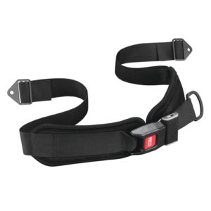
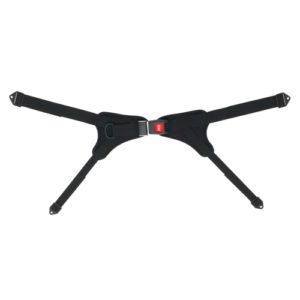
A two-point seatbelt (left) and a four-point seatbelt (right).19-20

Various accessories are available to help support and position all parts of the lower limb including: the hips and buttock, knees, lower legs, and feet. To support the hips and the buttock, positioning belts (sometimes referred to as seat belts) can be used. When used on wheelchairs, positioning belts can help prevent the hips from sliding forward and help to keep the hips properly aligned (i.e., not tilted or rotated). Different types of positioning belts are available depending on your needs: two-point belts or four-point belts. Four-point belts offer more support to the hips if required. It is also important to note that safety belts are not the same as positioning belts. Although both may contribute towards safety, positioning belts are more specialized to help maintain your hip posture.

A pommel connected to the wheelchair.21

There are also an assortment of accessories to address knees that press inward, splay outward, or are windswept. To address knees that press inward, pommels are cushions mounted either to the wheelchair or cushion, which go between the knees to keep them separated; however, pommels that are too big may interfere with transferring. To address knees that splay outward, adductor pads can be used. These pads are placed on the outer edges of the wheelchair cushion, which support the thigh and prevent the knees from falling outward.

Some accessories for the feet and lower leg include strapping and pads. Heel and toe straps can be connected to the footplate to help prevent the foot from moving forward or backward. After SCI, this can be particularly useful to help manage tone in the lower legs, and ensure the person remains stable in their wheelchair. To support the calves, pads can be attached to the footrest. Calf pads are particularly used with power wheelchairs, as they can provide support to the calves during tilting or reclining.

Trunk Support

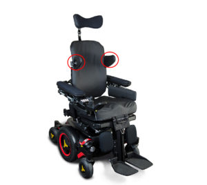
Lateral supports on a power wheelchair, circled in red.22

Lateral supports, straps, or upper body positioning belts are used by individuals who have difficulty maintaining an upright posture that may be caused by muscle weakness or other conditions. Lateral supports are square or rectangular pads that connect to the back of the wheelchair and rest against the trunk to promote balance and stability. Moreover, the lateral pads can also act as a clear indicator that you are out of alignment (e.g., when you notice you are heavily leaning on a lateral support, try to consciously correct your posture if possible). Chest supports, including chest straps, can help prevent tipping forward due to weakness in the abdominal and back muscles. When used properly, accessories can help with trunk support, stability, balance, and posture. Correct positioning and support of the trunk may prevent further progression of some spinal conditions, such as scoliosis. If placed incorrectly, these supports may be ineffective. For example, if they are placed too low they may not provide the support needed, while if they are placed too high they may irritate your shoulder, the nerves in your arm, or limit arm movement. When changing sitting positions, ensure to also readjust your trunk supports as they may shift as well.

Wheelchair seating is a complex procedure which includes multiple assessments by healthcare professionals to ensure that your wheelchair is best suited for your needs. Given that the majority of your day may be spent in a wheelchair, it is important to consider ways to relieve sitting pressures and to maintain your postures. To do so, some parts of your wheelchair that may be customized include the cushion, back, and leg rests. It is best to discuss all treatment options with your health providers to find out which treatments are suitable for you.

For a review of how we assess evidence at SCIRE Community and advice on making decisions, please see SCIRE Community Evidence.

SCIRE Community. “Powered Mobility Devices”. Available from: https://community.scireproject.com/topic/powered-mobility/

SCIRE Community. “Manual Wheelchairs”. Available from: https://community.scireproject.com/topic/manual-wheelchairs/

SCIRE Community. “Wheeled mobility video series”. Available from: https://community.scireproject.com/videos/wheeled-mobility/

Parts of this page has been adapted from the SCIRE Professional “Wheeled Mobility and Seating Equipment Following Spinal Cord Injury” Module:

Titus L, Moir S, Casalino A, McIntyre A, Connolly S, Mortenson B, Guilbalt L, Miles S, Trenholm K, Benton B, Regan M. (2016). Wheeled Mobility and Seating Equipment Following Spinal Cord Injury. In Eng JJ, Teasell RW, Miller WC, Wolfe DL, Townson AF, Hsieh JTC, Connolly SJ, Loh E, McIntyre A, editors. Spinal Cord Injury Rehabilitation Evidence. Version 6.0: p 1-178.
Available from: scireproject.com/evidence/wheeled-mobility-and-seating-equipment/ 

Evidence for “Why is proper seating important” is based on:

Bogie K, Wang X, Fei B, Sun J. New technique for real-time interface pressure analysis: Getting more out of large image data sets. Journal of rehabilitation and research development, 45, 5236.

Mao HF, Huang SL, Lu TW, Lin YS, Liu HM, Wang YH, et al. Effects of lateral trunk support on scoliotic spinal alignment in persons with spinal cord injury: a radiographic study. Archives of Physical Medicine and Rehabilitation, 87, 764-71.

Thomson, D., Tully, P., Blochlinger, S. (n.d.). Laying the foundation for proper positioning: introduction to positioning for functional ability and wheelchair seating. [Powerpoint]. Retrieved from: https://seatingsymposium.com/images/pdf/PS/PS2_Tully_Thomson_Blochlinger.pdf

Evidence for “Why are cushions important” is based on:

Sprigle, S. (2014). Measure it: proper wheelchair fit is key to ensuring function while protecting skin integrity. Advanced wound skin care, 27, 561-72.

Sprigle S, Delaune W. (2014) Factors That Influence Changes in Wheelchair Cushion Performance Over Time. Assistive Technology, 26, 61-68.

Evidence for “How can a cushion redistribute pressure” is based on:

Endsjo, A., Mullis, S, Sharpe, L. 2019. Wheelchair seating and positioning guide. Retrieved from: https://hub.permobil.com/wheelchair-seating-positioning-guide

Evidence for “What kinds of cushions are there” is based on:

Sprigle S, Chung KC, Brubaker CE. (1990) Reduction of sitting pressures with custom contoured cushions. Journal of rehabilitation research and development, 27, 135-40.

Stewart, D. (2019). Comfort wheelchair cushions. Retrieved from: https://mobilitybasics.ca/seating/basic

Stewart, D. (2019). Comfort wheelchair cushions. Retrieved from: https://mobilitybasics.ca/seating/basic

Wheelchair seat cushions. (2019). Retrieved from: https://www.wheelchair-works.com/wheelchair-seat-cushion.html

Waugh, K. (2014). Custom contoured seating: Ensuring Successful Outcomes. [Powerpoint slides]. Retrieved from: https://seatingsymposium.com/images/pdf/PS/PS3_Waugh.pdf

Evidence for “Backrest” is based on:

Fontein, J. (2017). What’s in a back? [Abstract]. Canadian seating and mobility conference.

Hong EK, Dicianno BE, Pearlman J, Cooper R, Cooper RA. (2016). Comfort and stability of wheelchair backrests according to the TAWC (tool for assessing wheelchair discomfort). Disability and Rehabilitation: Assistive Technology, 11, 223-227.

Samuelsson K., Bjork, M., Erdugan, A.M., Hansson, A.K., Rustner, B.  (2009). The effect of shaped wheelchair cushion and lumbar supports on under-seat pressure, comfort, and pelvic rotation. Disability and rehabilitation: assistive technology, 4, 329-336.

Schmeler, M.R., Buning, M.E. (1999). Wheelchair back supports. [Powerpoint slides]. Retrieved from: https://www.wheelchairnet.org/WCN_WCU/SlideLectures/MS/4SeatBacks.pdf

Yang YS, Koontz AM, Yeh SJ, Chang JJ. (2012). Effect of backrest height on wheelchair propulsion biomechanics for level and uphill conditions. Archives of physical medicine and rehabilitation, 93, 654-659.

Evidence for “Footrest” is based on:

Janssen-Potten YJ, Seelen HA, Drukker J, Spaans F, Drost MR. The effect of footrests on sitting balance in paraplegic subjects. Archives of Physical Medicine and Rehabilitation, 83, 642-8.

Sprigle, S. (2014). Measure it: proper wheelchair fit is key to ensuring function while protecting skin integrity. Advanced wound skin care, 27, 561-72.

Evidence for ” What accessories are used for seating?” is based on:

Stewart, D. (2019). Wheelchair seating products – accessories. Retrieved from: https://mobilitybasics.ca/seating/accessories

Image credits
  1. Wheelchair Posture. ©Shannon Sproule
  2. Sitting pressures. ©The SCIRE Community Team
  3. Offloading pressures ©The SCIRE Community Team
  4. Immersion ©The SCIRE Community Team
  5. Immersion and Envelopment ©The SCIRE Community Team
  6. Foam cushion ©The SCIRE Community Team
  7. Gel cushions ©The SCIRE Community Team
  8. Air cushions ©The SCIRE Community Team
  9. Honeycomb Cushion ©The SCIRE Community Team
  10. Jay basic back ©Sunrise Medical 2017
  11. Jay J2 Series Back ©Sunrise Medical 2017
  12. Ride designs custom 2 cushion ©Action Seating and Mobility
  13. ZR Back Right Product Detail ©Permobil 2020
  14. Wheelchair. ©George Hodan. CC0 1.0
  15. Modified from Disabled people set ©macrovector Freepik License
  16. Polio Wheelchair Lady ©jackcast2015, CC BY 2.0
  17. This was one of five images (PHIL #9170-9174), depicting the action of two different mobility-challenged women getting into a bathtub ©Richard Duncan, Public Domain
  18. Permobil original elbow & armrest side supports ©Permobil, 2020
  19. Hip belt, push button, 1” webbing, center pull, 6” pads. ©Adaptive Engineering Lab (AEL) 2015
  20. Hip stabilizing belt, push button, rear pull, large. ©Adaptive Engineering Lab (AEL) 2015
  21. Heavy duty flipdown abductor hardware. ©Therafin corporation
  22. BodiLink® Accessories. ©Permobil 2020


Disclaimer: This document does not provide medical advice. This information is provided for educational purposes only. Consult a qualified health professional for further information or specific medical advice. The SCIRE Project, its partners and collaborators disclaim any liability to any party for any loss or damage by errors or omissions in this publication.

Wheelchair Propulsion Assist Devices

By | | No Comments

Author: Sharon Jang | Reviewer: Jaimie Borisoff | Published: 15 May 2020 | Updated: ~

Wheelchair propulsion assist devices are pieces of equipment that can facilitate the use of a manual wheelchair. This page provides an overview of the different options available. SCIRE Community is not affiliated with and does not endorse any of the specific products mentioned on this page.

Key Points

  • Propulsion assist devices are technologies that attach to your manual wheelchair to facilitate propulsion
  • There is limited research on the effectiveness of these devices, but they are clinically well-accepted with perceived benefits associated with their use
  • There are options for both powered and non-powered propulsion assist devices, depending on your needs
  • Propulsion assist devices can be either front frame-mounted, replace your rear wheels, or rear frame-mounted
  • Propulsion assist devices are rapidly developing and changing; specific products in this article may no longer be available or unavailable in your area

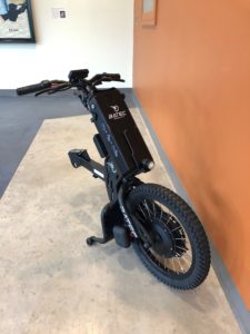
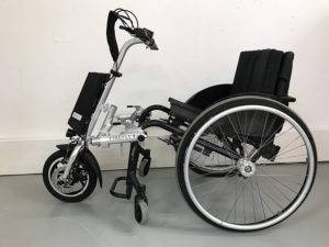
An example of a front-mounted propulsion assist device.1

Propulsion assist devices are pieces of equipment that can be attached to a manual wheelchair to facilitate the ease of using a wheelchair, or expand the capabilities of a manual wheelchair. These devices generally work by reducing the amount of effort required by the user to move their wheelchair. Use of these devices may help individuals go up hills, reduce the amount of force required to start moving the wheelchair, or travel over difficult terrain such as grass. Propulsion assist devices are normally added onto the front or back of the chair frame, but also sometimes replace the rear wheels. These devices can be further split up into two categories: powered and non-powered.

As technology is rapidly progressing, novel devices are being frequently released. The development of multiple devices to fill a niche market has resulted in some products becoming unavailable in certain areas, or leaving the market altogether. The quick changes and developments in this area make conducting research on propulsion assist devices difficult. Currently, there is limited research on the use of propulsion assist devices for manual wheelchair users and most of the research is focussed on the devices that replace the rear wheels.

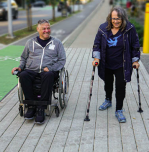
Using a propulsion assist device can be beneficial to manual wheelchair users. The man on the left is using a powered add-on.2

Power assist devices may be purchased by individuals with or without a prescription. Some indications for propulsion assist device use are limited strength in the upper body, or for those that are at a higher risk of an injury or pain to the arms and/or shoulders. These devices may improve participation in the community by enabling increased travel distance and access to more difficult environments.

In two studies (weak evidence), users of propulsion assist devices have reported benefits including:

  • Less strain on the cardiovascular and respiratory systems; this results in reduced overall feelings of fatigue.
  • Being able to go further with less effort (improved propulsion efficiency). This may help prevent overuse injuries.
  • Increased distance you are able to travel before feeling tired.
  • Travelling up and down hills.
  • Improved ability to travel on non-paved roads (e.g., grass, gravel, dirt), which increases the variety of spaces a wheelchair user may access.

Although there is limited research on propulsion assist devices, Pushrim Activated Power-Assist Wheelchairs (PAPAW) are the most researched device. Evidence has suggested that using PAPAWs have multiple benefits. One study provided moderate evidence for the use of PAPAWs to help individuals with SCI who have shoulder pain to propel their wheelchair further distances. The participants in this study also required less energy to wheel, and perceived that wheeling was easier with the use of PAPAWs. Other (weak) evidence has also shown that using a PAPAW may reduce the frequency of arm/shoulder injuries and the amount of energy required. Additional (weak) evidence suggests that individuals with tetraplegia using PAPAWs have improved ability to manoeuvre through a variety of textured surfaces and to complete activities of daily living.

Although propulsion assist devices can facilitate the use of a manual wheelchair, there are also some risks associated with their use. While this is not an exhaustive list, risks include:

Increased risk of wheelchair damage

Most manual wheelchair frames may not have been designed to withstand the different forces applied by propulsion assist devices. This may lead to premature wear and tear, or possible breakage, on some parts of the wheelchair, such as the footplate. Sudden breakage during use may lead to injury of the wheelchair user.

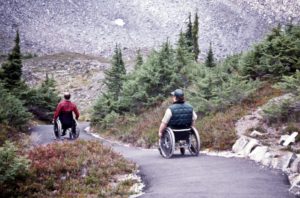
Using a propulsion assist device may increase the risk of tipping when going up or down hills.3

Decreased stability

The addition of a propulsion assist device either in front or behind the wheelchair may alter the center of gravity on the wheelchair. This may result in greater instability, increasing the risks of tipping backwards when going up and down steep hills. Forward stability may be compromised as well and induce tipping (e.g. when moving down at the bottom of a curb cut).

Greater impact forces when encountering an obstacle

Propulsion assist devices, especially motorized ones, allow wheelchair users to travel at a greater speed. However, hitting obstacles such as a rock or a curb in your wheelchair at a higher speed can result in greater forces, which may lead to a decrease in stability and increase the risk of falls.

Potential lateral stability issues

Propulsion assist devices that attach to the front pose a risk of tipping sideways. Particularly when travelling at higher speeds, sudden turns can result in tipping over.

Prior to obtaining a propulsion assist device, you should always consult your health professional. That said, some factors to consider when deciding whether to invest in an add-on include the following factors:

Upper limb function

Upper limb function is required to use these devices. For example, the individual may be responsible for attaching/detaching the device to the wheelchair, and some degree of hand function is needed for operation. Some products do have adapted controls for use by individuals with limited hand function.

Terrain

The device should facilitate the transportation of the user over surfaces that they frequently travel on, or wish to travel on. Some terrain considerations include indoor travel, outdoor travel, uneven surfaces, soft terrains e.g. grass, and hills.

Transfer ability

It is important to take into consideration how a device may impact transferring in and out of a wheelchair. Will it make transfers more difficult?

Transport

If a car is used for transportation, consideration of the weight of the propulsion device should be considered. Will the weight of the propulsion assist device and the wheelchair be manageable to lift into a car? Will the propulsion assist device fit into the car?

Weight

Is the device light enough for the user to remove by themselves? Will the weight make manual propulsion more difficult when not in use (e.g. if battery is drained before end of travel)?

Interaction with wheelchair

Propulsion assist devices require a certain part of a wheelchair to connect to, which may vary between types of wheelchair (e.g., rigid vs folding wheelchairs) and types of add-on. How the device mounts to a wheelchair should be considered, including the user’s capability for performing the attachment/detachment. Additional consideration may be needed if the device is going to be used between different wheelchairs.

Addressing casters

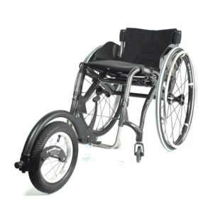
Casters help provide stability to a wheelchair, but become a limiting factor when a wheelchair user wishes to travel off a smooth even surface. There are some devices that connect to the front of the wheelchair via the foot plate and lift up the front caster wheels. This allows the wheelchair user to travel over uneven surfaces easier, such as dirt paths, sand, and gravel. An example of this kind of device is a FreeWheel.

Mechanical advantage

There are some devices that facilitate pushing by amplifying your efforts (a mechanical advantage) using gears or levers. Using a device with a mechanical advantage will allow you to go further with a given push. One example of a device that provides mechanical advantage is a lever-propelled wheelchair. There are some lever devices that can be connected to your wheel (others require replacement of the rear wheels) that allow the user to propel by pushing the lever forward. Using a lever has two purported benefits: 1) using a longer lever to propel a wheelchair requires less force, and 2) using a lever enables a more favorable movement pattern of the hands, wrists, and shoulder. Together these may reduce the risk of injury. An example of this device is the NuDrive.

Pushrim-Activated Power-Assist Wheelchairs (PAPAWs)

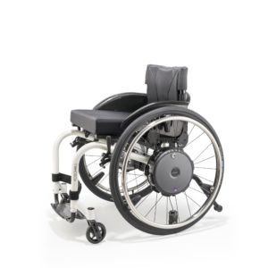
A PAPAW is a manual wheelchair with motorized rear wheels. The wheels are powered by a battery, which is attached to the back of the wheelchair. This type of wheelchair is controlled by normal pushing movements using the pushrims. PAPAWs assist individuals with limited strength or arm function by amplifying the force applied to the wheels. Each push on the pushrim by the user is sensed and proportionally amplified to increase the force to continue the forward movement. This also occurs for braking and turning (e.g., the wheels would detect a backwards force and apply a stronger braking force). This allows for users to go further with a given push, or to brake more efficiently with less force. When desired, the power assist function can be turned off. One limitation of using PAPAWs is the significantly increased weight of the wheelchair, which is especially noticeable when turned off. Another limitation is that the pushrims are damaged more easily, as this is where the sensors are located. Some examples of PAPAWs include the Alber E-motion and the Quickie Xtender.

Front mounted systems

These systems attach to the front of the wheelchair frame and typically lift up the front casters. Non–powered versions exist which are propelled via cranks and chains similar to handcycles. Most often powered, these devices are able to turn manual wheelchairs into a three-wheeled “scooter”. Front mounted systems are usually controlled with a throttle controller, which removes the need to manually propel the wheelchair. They are also able to reach significantly higher speeds in comparison to other power add-ons. A disadvantage to front mounted systems is that they add length to the wheelchair, which may impact the ability to move around indoors. In addition, the devices can be fairly heavy (e.g., the typical Batec system is 25 kg), and little to no customizations can be made. Examples of front mounted systems include the Rio Mobility Firefly, Triride, Klaxon, and Batec.

Rear mounted systems

Rear mounted propulsion device generally connect to the bar underneath the wheelchair seat (i.e., the camber tube). These devices can power the wheelchair so that no manual propulsion is required, although turning and braking still require constant user control. Currently, there are two models commercially available that are light, compact, and easily attached: the SmartDrive and the Smoov. The SmartDrive is controlled with a wristband worn by the user. A double tap of the wristband (or similar accelerometer-sensed motion of the hand/arm when tapping the wheel or other surface) will activate the motor and begin accelerating the wheelchair. Another tap sets the speed, while the next double tap will stop the motor. Gripping on the handrim is usually still needed to slow down and stop the chair. Users are not required to propel the wheelchair once it is in motion; they are only required to steer. Good reaction time is required to operate the device; a learning curve is involved when first using the device. The Smoov operates via a frame mounted control knob to set the desired speed. Starting and stopping the motor is accomplished through tapping the knob. Turning, slowing, and stopping are similar to the SmartDrive operation.

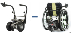
Other rear-mounted systems, such as the Spinergy ZX-1, convert manual wheelchairs into a power wheelchair. The user backs up into the device, which connects to the wheelchair with a push of the button. This add-on lifts up the rear wheels of the wheelchair, converting it into a power wheelchair that is controlled with a joystick.

The use of propulsion assist devices has the potential to protect your arms and shoulders from pain and overuse injuries, and allow you to travel further with less energy and more various terrain. Many different kinds of technologies are being rapidly developed and introduced to the market to facilitate manual wheelchair use. Thus, the world of powered propulsion assist devices is ever changing. Due to this factor, the devices discussed in this article may change, or may no longer be available for purchase.

Any reference to a specific product does not constitute or imply an endorsement by SCIRE Community. Professional advice should be sought before making any health care and treatment decisions.

For a review of how we assess evidence at SCIRE Community and advice on making decisions, please see SCIRE Community Evidence.

SCIRE Community. “Powered Mobility Devices”. Available from:  https://community.scireproject.com/topic/powered-mobility/

SCIRE Community. “Manual Wheelchairs”. Available from: https://community.scireproject.com/topic/manual-wheelchairs/

SCIRE Community. “Wheeled mobility video series”. Available from: https://community.scireproject.com/videos/wheeled-mobility/

SCIRE Community. “Wheelchair Add-ons”. Available from: https://community.scireproject.com/resources/products-and-devices/

Parts of this page has been adapted from SCIRE Project (Professional) “Wheeled Mobility and Seating Equipment Following Spinal Cord Injury” Chapter:

Titus L, Moir S, Casalino A, McIntyre A, Connolly S, Mortenson B, Guilbalt L, Miles S, Trenholm K, Benton B, Regan M. (2016). Wheeled Mobility and Seating Equipment Following Spinal Cord Injury. In Eng JJ, Teasell RW, Miller WC, Wolfe DL, Townson AF, Hsieh JTC, Connolly SJ, Loh E, McIntyre A, editors. Spinal Cord Injury Rehabilitation Evidence. Version 6.0: p 1-178.

Available from: https://scireproject.com/evidence/rehabilitation-evidence/wheeled-mobility-and-seating-equipment 

Evidence for “What are wheelchair propulsion devices” is based on:

Choukou, M.-A., Best, K. L., Potvin-Gilbert, M., Routhier, F., Lettre, J., Gamache, S., … Gagnon, D. (2019). Scoping review of propelling aids for manual wheelchairs. Assistive Technology, 0(00), 1–15. Retrieved from https://doi.org/10.1080/10400435.2019.1595789

Evidence for “What are the benefits of using a propulsion assist device” is based on:

Morgado Ramirez, D. Z., & Holloway, C. (2017). “But, I Don’t Want/Need a Power Wheelchair.” Proceedings of the 19th International ACM SIGACCESS Conference on Computers and Accessibility  – ASSETS ’17, 120–129. Retrieved from http://dl.acm.org/citation.cfm?doid=3132525.3132529

Evidence for “What are the risks of using a propulsion assist device” is based on:

Medicines and healthcare products regulator agency (2004). Guidance on the Stability of Wheelchairs, (March). Retrieved from http://www.unece.org/fileadmin/DAM/trans/doc/2004/wp29grsg/GRSG-ig-access-03-10.pdf

Ogilvie, C. (2019). Finite Element Analysis of a Wheelchair when Used with a Front-Attached Mobility Add-On by (November).

Evidence for “What should I consider when contemplating propulsion assist device” is based on:

Agency for clinical innovation (2019). Prescribing manual wheelchair with propulsion assist devices. Retrieved from: https://www.aci.health.nsw.gov.au/networks/spinal-cord-injury/spinal-seating/module-9/prescribing-manual-wheelchair-with-propulsion-assist-devices

Evidence for “What are the options for powered propulsion assist device” is based on:

Choukou, M.-A., Best, K. L., Potvin-Gilbert, M., Routhier, F., Lettre, J., Gamache, S., … Gagnon, D. (2019). Scoping review of propelling aids for manual wheelchairs. Assistive Technology, 0(00), 1–15. Retrieved from https://doi.org/10.1080/10400435.2019.1595789

Corfman TA, Cooper RA, Boninger ML, Koontz AM, Fitzgerald SG. Range of motion and stroke frequency differences between manual wheelchair propulsion and pushrim-activated power-assisted wheelchair propulsion. J Spinal Cord Med 2003;26(2):135-40

Nash MS, Koppens D, van Haaren M, Sherman AL, Lippiatt JP, Lewis JE. Power-assisted wheels ease energy costs and perceptual responses to wheelchair propulsion in persons with shoulder pain and spinal cord injury. Arch Phys Med Rehabil 2008;89:2080-5.

Image credits
  1. Batec Handbike. © SCIRE Community Team
  2. Moving in the community. © SCIRE Community Team
  3. Men in wheelchairs at heather lake, Mt. Baker Snoqualmie National Forest. © U.S. Forest Service – Pacific Northwest Region.
  4. Hand © Sandra. CC BY 3.0
  5. Mountain © iconcheese. CC BY 3.0
  6. Transfer © romzicon. CC BY 3.0
  7. Car © Priyanka, IN. CC BY 3.0
  8. Scales © Made, AU. CC BY 3.0
  9. Wheelchair © Satawat Anukul, TH. CC BY 3.0
  10. FreeWheel Wheelchair Attachment © FreeWheel 2020
  11. Stanley handling NuDrive Air © NuDrive Air 2020
  12. Alber E-Motion M25 © Manston mobility 2018
  13. Firefly Electric Attachable Handcycle for Wheelchair © Rio Mobility 2020
  14. Smart Drive © MAX Mobility, LLC 2019
  15. Spinergy ZX-1 © Spinergy 2020


Disclaimer: This document does not provide medical advice. This information is provided for educational purposes only. Consult a qualified health professional for further information or specific medical advice. The SCIRE Project, its partners and collaborators disclaim any liability to any party for any loss or damage by errors or omissions in this publication.

Stem Cells and Spinal Cord Injury

By | | No Comments

Author: Sharon Jang, Vanessa Mok | Reviewer: Peggy Assinck | Published: 13 May 2020 | Updated: ~

Since the discovery of stem cell therapy, researchers have attempted to determine whether it can be a safe and effective treatment for spinal cord injury (SCI). This page addresses what stem cell therapy is and its current place in SCI.

Key Points

  • Stem cells are cells that have the ability to self-renew indefinitely and specialize into different types of functioning cells
  • Stem cells treatment following SCI has been proposed to prevent further damage to the spinal cord, bridge lesion sites, and promote the regrowth of nerves
  • There is limited evidence that stem cell therapy is safe and effective in humans (across different disorders) and therefore more work needs to be done to confirm safety and potential effectiveness
  • There are currently no stem cell therapies approved for SCI. However, many clinics worldwide continue to offer cell transplantation-based treatments including stem cell therapies that have very little scientific evidence to back them up
  • It is important to stay well-informed about ongoing research and importantly the potential risks involved in stem cell-based treatments

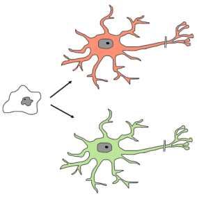
Stems cells have the ability to self-renew (blue arrow), and turn into different cell types such as muscle cells, skin cells, and nerve cells (pictured).1

All tissues and organs in the body originate from stem cells. Stem cells are unspecialized, blank cells that have the ability to turn into specialized cells (e.g., neural cells, muscle cells, etc) through chemical signals in the body. These cells are responsible for maintaining and repairing the tissue they are found in. Stem cells are defined by two characteristic features: 1) their ability to self-renew (i.e., make copies of themselves) and 2) their ability to differentiate into functioning cell types (i.e., they can turn into specialized cells). Some stem cells are only found in certain parts of the body or at certain stages of life. There are four main types of stem cells that are currently used in stem cell therapy or research.

Neurons extending from a group of embryonic stem cells.2

Embryonic stem cells

Embryonic stem cells are sourced from embryos that are about a week old. In Canada, embryos cannot be specifically created for research purposes. Instead, these stem cells originate from unused embryos from fertility clinics. Their use in research raises ethical controversies as the embryos are destroyed during the harvesting process. These cells are pluripotent which means that they can turn into any cell in the body except for the umbilical cord and placenta.

Adult stem cells

Adult stem cells (also known as somatic stem cells or tissue-specific stem cells) are sourced from organ tissues (e.g., the heart, bone marrow, fat) found in the bodies of infants, children, and adults. They can also be produced from discarded placentas and umbilical cords. They have less capability to self-divide and specialize into different cell types compared to embryonic stem cells. Rather, adult stem cells more commonly produce tissue types in which they are found, so are multipotent. For instance, blood stem cells only have the potential to generate red blood cells, white blood cells, and platelets.

Induced pluripotent stem cells

Advancements in technology have allowed researchers to reprogram specialized adult cells back into their pluripotent state to resemble embryonic stem cells. These cells are known as induced pluripotent stem cells. These cells are often obtained from taking a small sample from adult tissue, such as the skin. Because this process does not harm the donor nor does it involve embryos, the same ethical issues facing above-discussed cells are not encountered with induced pluripotent stem cells. Induced pluripotent stem cells can also reduce the risk of rejection by the body because the adult cells are derived from the person’s own body. Despite this exciting discovery, there are lots of obstacles to overcome before they are used in human studies.

Stem cell therapy is the use of stem cells to help replace or repair damaged tissue in the human body. In this type of therapy, stem cells are either differentiated into the cell of interest outside of the body prior to transplantation (exogenous), or directly transplanted into the site of injury (endogenous). In exogenous repair, scientists differentiate specific stem cell types in a laboratory and then implant the cells into the body. On the other hand, endogenous repair consists of directly transplanting stem cells into the body, and depends on the body’s ability to transform the cells into the specific type required for repair. These cells can be sourced from various places: autologous transplantation involves the use of stem cells from the person who is receiving the transplant, whereas allogenic transplantation involves receiving stem cells from a donor.

Cell transplantation versus stem cell transplantation

Cell transplantation is an umbrella term for stem cell transplantation. Different cells can be transplanted into the body including fat cells, bone marrow cells, and nose cells. Cell transplantation is the process of transferring cells into areas of the body that are damaged or injured. These transfers work in 2 main ways: 1) the cells can be transplanted into the injury site for direct repair, or 2) the cells can be transplanted into the blood where they can circulate around the body and cause system-wide changes. Most work in SCI focuses on putting a more specialized cell into the injury site (not just stem cells). For example, a stem cell can be pushed towards becoming a myelinating cell and then the myelinating cell could be transplanted into the injury site, or a Schwann cell could be harvested from the leg and then transplanted at the injury site.

Currently, only a small number of conditions are approved to be treated with stem cell therapy. Bone marrow transplants for leukemia have been performed for many years. Epithelial stem cell transplants are also done to regenerate cells in those who have suffered burns or disorders of the cornea. Stem cell therapy for other purposes, including SCI, is still relatively new and much of the research is still in its early stages. The figure below highlights important landmarks in the history of stem cells research.

Healthcare professionals preparing for a stem cell transplant.18

With SCI, stem cells could theoretically be used to replace injured or destroyed nerve cells or protect further attack by the immune system around the injury site. The main goal of stem cell therapy for SCI is to improve the conduction of information past the injury. As of yet, there are no stem cell treatments that can reverse or repair a SCI. While there is some thought that stem cells may be used to help recovery, researchers are still currently unsure of exactly how stem cells/cell transplantation can help recovery from a spinal cord injury. However, many hypotheses have been made:

  • Neuroprotection: Stem cells may help prevent further damage to the tissue in the area surrounding a spinal cord injury. By limiting the amount of damage, more function may be saved.
  • Supporting the host cell: Stem cells may help enhance the survival of nearby cells by secreting molecules that control inflammation and/or improve the regeneration of blood cells
  • Creating new blood vessels: Stem cells may help promote faster regrowth of new blood vessels and protection of the existing ones. Having more blood vessels may increase the amount of oxygen and nutrients available for surviving cells.
  • Regrowth of axons: Stem cells may help axons regrow through developing bridges across the site of the lesion and decreasing scars. However, it is important to note that the regeneration of axons may not always lead to functional benefits and could even lead to adverse effects such as increased pain.

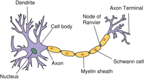
    The anatomy of a neuron.19

  • Regeneration of myelin: Myelin forms insulation around nerves, allowing electrical signals to transmit efficiently to other nerve cells. Stem ce­lls may also be used to replace the support cells that wrap myelin around nerves.

Refer to our article on Neuroprotection for more information

SCI research on stem cells has largely focused on laboratory and animal experiments. Human studies are in their initial phase.20

There are a number of ongoing and completed studies of weak to moderate evidence that evaluates the safety and efficacy of stem cells in people with SCI. Given that stem cells are still experimental, there is no specific stem cell type that is proven to be more effective over another. In research, studies have tested a large variation of stem cell sources, including blood, bone marrow, the nose, umbilical cords, and other nerves. While some of these stem cell sources come from fetuses, most studies have used stem cells from the person’s own body.

Care should be taken when interpreting these findings as they are derived from small trials with flaws in study design and documentation. Many more large-scale studies are required to determine whether stem cells have a role in treating people with SCI and if so, how transplantation should be done to minimize the health risks and maximize the potential of stem cell-based therapy.

Improved AIS score

In research, a common way to determine the success of any treatment is through comparing ASIA Impairment Scale (AIS) scores before and after transplantation. An individual’s AIS score consists of their muscle strength and sensation below their level of injury, in addition to anal sensation and contraction. Two weak evidence studies with a small number of participants (less than 10) found that stem cell treatments improved the AIS score of all participants. Conversely, two weak evidence studies found that only a portion (29-66.7%) of their participants saw an improvement in AIS scores.

Refer to our article on Spinal Cord Injury Basics for more information on AIS scores.

Natural recovery after SCI

Although some studies have shown improvements in movement and sensory AIS scores, it is also important to note that natural (or spontaneous) recovery can occur within the first year of a spinal cord injury. In individuals with SCI, natural recovery of movement and sensation may occur up to 12-18 months post injury. Some stem cell studies have been conducted with newly injured individuals (i.e., less than 1 year after sustaining a spinal cord injury). As a result, spontaneous recovery may have played a role in the findings of improved function and sensation, not stem cell therapy.

Improved sensation

There have been some mixed findings on the efficacy of stem cells on sensation. Some weak evidence research studies have seen improvements in sensation for some individuals, but no large-scale study has found improvements for their entire sample. A moderate evidence study found significant changes in sensation when compared to individuals who did not receive stem cell therapy. However, a second moderate evidence study found that sensation improved over a year, but the amount of improvement was not significantly different than individuals who did not receive stem cell therapy. Currently, it is hard to conclude whether the use of stem cells is effective for the restoration of sensation.

Improved motor function

There is some weak to moderate evidence showing that stem cells may help the recovery of motor function. The type of recovery in these studies has been quite diverse, ranging from a tetraplegic individual regaining the ability to hold their head up and move their upper limb, to the ability to walk with crutches and braces. However, the percentage of people who re-develop the ability to walk again in studies is low (i.e., 1-2 people out of 10-20 total participants). Moderate evidence suggests that stem cells may be more beneficial for movement in the arms and hands than the legs compared to people who did not receive stem cell therapy. Similar to sensation, no large-scale study has shown a high success rate in the recovery of motor functioning in response to stem cell therapy. In fact, there have been several studies that have found no improvements in motor function amongst their participants.

Improved neural connectivity

The signals relayed from the brain to the muscles are disrupted when a spinal cord injury occurs. To observe what connection remains, researchers apply stimulation to the brain and observe whether or not a signal is received at the arms/legs. If there is a signal present, researchers then note of how long it takes for the signal to get to the arms/legs, and the strength of the signal. As stem cells are supposed to theoretically help repair the spinal cord, it is thought that these signals would start to reappear/improve with stem cell therapy.

Motor evoked potentials

Motor evoked potentials are used to evaluate the connections between the brain and the muscles. Four low to moderate evidence studies have seen a reappearance of motor evoked potentials in individuals who were lacking them before, although the proportion of individuals who see a return is quite low (i.e., a range of 3-10 participants out of 20). Weak-moderate evidence research has also found improvements in the time it takes for signals to go through after stem cell therapy. It is important to note that these improvements are not always associated with improvements in AIS grades. Contrary to the previous findings, one research group found that the time taken for signals to travel from the brain to the muscle did not change, despite increases in AIS grade. This means that while functional improvements were seen, the connections between the brain and the muscle did not improve.

Somatosensory evoked potentials

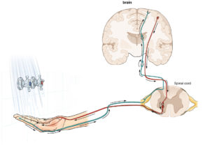
The brain normally interprets sensation through a pathway (in green) of nerves that travel from the limb, to the spinal cord, to the brain.24

are used to evaluate the sensory connections between the brain and the rest of the body. Several studies have found a reappearance of sensory signals in the wrist and/or ankles in a portion of their participants. For participants with sensory signals present prior to receiving stem cells, evidence shows that these signals may be strengthened, and that these signals may travel faster. Although these findings appear promising, the longevity of the strengthening effect is brought to light by one group of researchers who showed that the newly obtained sensory signals disappeared after one year. More research is required to determine how to consistently obtain these results, and to determine whether these effects are truly non-lasting.

Refer to our article on Understanding Research Evidence for more information on spontaneous recovery.

Bladder & Bowel

Individuals with spinal cord injury often experience a lack of sensation and control of their bowel and bladder. Some research suggests that stem cell therapy may restore some sensation of bladder fullness and anal sensation. There are also some positive reports on the restoration of voluntary bladder contractions and anal control. Moreover, one study (moderate evidence) indicates that stem cell therapy may further improve bladder function by allowing individuals to hold more urine in their bladder, to increase the rate of urine passage, and to empty urine with less effort from by the bladder. However, the proportion of people in these studies who receive these bladder benefits are low.

Functional Independence and Quality of Life

There is limited evidence that stem cell therapy has a positive impact on functional independence and quality of life. Participants from three studies (weak evidence) reported that their self-care ability has improved in areas including bowel/bladder management, grooming, feeding, transferring, dressing, and mobility.

Sexual functioning

Most of the research on the effects of stem cell therapy and sexual functioning has been conducted with men. One weak study found that 31% of the men had improved erections while another found that sexual functioning improved as a result of increased sensitivity in the genital area. More research is required to determine the consistency of the effects in addition to the impact on sexual functioning of women.

Evidence for no improvement

While there have been some successful findings from stem cell therapy use in SCI, there are trials that have found no effects. Three studies found that none of their participants experienced any changes in their sensory or motor function. Meanwhile, other studies with a small number of participants (e.g., 5-20) have found that stem cell therapy may have an effect for some individuals, but not others. The number of people who do not experience any positive benefits from stem cell therapy is unpredictable, and has ranged from 40-87%.

Even though Health Canada and USA Food and Drug Administration (FDA) have not yet approved of any stem cell therapies for SCI in Canada, some private clinics around the world are already selling these treatments before establishing safety and effectiveness. In Canada, offering stem cell therapies is illegal unless it is for research purposes. Before you decide, consider the following points below with a health care provider. Use these points to think critically about what is being advertised.

There is no consensus on whether stem cell therapy is effective.

As described in this article, there is a lack of evidence supporting the use of stem cell therapy for spinal cord injury. Stem cell therapy is being explored as a treatment for many types of medical conditions; however, there is only strong evidence that supports its use with blood or immune system disorders that are often experienced after cancer. Currently, scientists are unsure how to repeatedly produce positive effects with the use of stem cell therapy for individuals with SCI. In addition, there remain many uncertain aspects of stem cell therapy such as knowing which type of stem cell is most effective for replacing different tissues, how these stem cells should be manipulated to produce the required cells, and what is the best way to deliver stem cells to the target area.

There are a variety of risks associated with stem cell therapy

Safety

There are many safety risks associated with stem cell treatments, and these risks may be higher for individuals with spinal cord injuries. Some stem cell marketers may emphasize that a treatment is safer if the cells are coming from your own body; however, this does not guarantee safety. If the stem cells are grown and multiplied before being injected into your spinal cord, the cells may lose some important properties and traits in the process, such as the ability to control growth. This may lead to the development of various tumors and/or cancers. In addition, when the cells are removed from your body, it is possible that they be exposed to bacteria or viruses prior to being re-transplanted into your spine. There is also a chance that complications may arise which may lead to short- or long-term health problems. This may lead to your injury or symptoms becoming more difficult to manage.

In clinical trials with people with SCI, there have been multiple adverse events resulting from stem cell therapy. The majority of stem cell studies conclude that transplantation is relatively safe due to the lack of serious safety problems or complications observed. Mild adverse events are infrequently reported in studies, but some have been documented. Some examples that have been reported include urinary tract infections, pain, hypothermia, headaches, increased spasticity, pressure sores, and depression. Serious adverse events that have been noted in research studies include:

    • the worsening of motor functioning
    • development of a cyst on the spine (which causes a concern for cancer)
    • collection of fluid along the surgical site
    • meningitis, resulting in a decrease in AIS score
    • worsening of sensation (e.g., worsened symptoms of tingling, decrease in sensation)

However, it is important to note that these adverse effects were only monitored over the period of the study. Currently, long term effects and side effects of stem cell therapy is unknown. Further longitudinal research is required.

Financial

As the use of stem cell therapy is not currently approved by the government, most often government health programs and insurance companies will not cover the costs. The costs of receiving stem cell therapy can be very high. On top of the cost for the treatment (being in the tens of thousands of dollars itself), there is the cost of travel, accommodations, and other fees. In addition, travelling for stem cell therapy may require you to go through a medical procedure without the support of all your family and friends.

Research

Receiving an experimental treatment is not the same as participating in a clinical trial. If you choose to receive stem cell therapy from a private clinic, you may be ineligible to participate in future clinical trials. Participating in a clinical trial is advised over receiving stem cell therapy from an unregulated private clinic, as:

    • Clinical trials are overseen by an independent medical ethics committee. This group of people is there to protect your rights and safety as a participant.
    • Clinical trials require some safety and efficacy testing before trying it in humans. This ensures that the treatment is relatively safe and effective.
    • By participating in a clinical trial (research), you are contributing back to the pool of knowledge. When you partake in a research study, you help scientists learn more about stem cell therapy and the safety or effectiveness of it.
    • There is no cost for the treatment in a clinical trial, as participants are not required to pay to partake in research studies.

Stem cell marketing sites are likely misleading

Private stem cell clinics often use overly positive/ optimistic advertisements that may be misleading.31

Unapproved stem cell marking websites often feature many patient testimonials in addition to references to research studies. The use of testimonials may be very persuasive, but may be misleading. Some of the positive benefits that these individuals may be feeling may be the result of the placebo effect. Given that many people who go in for stem cell therapy treatment have a strong belief that it will work, they may perceive less pain or greater sensation after their treatment. Patient anecdotes should not be held equal to research evidence supporting the use of stem cell therapy.

Additionally, many private stem cell therapy clinics will try to sell the treatment by citing multiple research studies to build credibility. In reality, these research articles often have no direct association with stem cell therapy specific to spinal cord injuries. Currently, there is a lack of high-quality research studies on stem cell therapy. As a result, many of the human-based studies cited are low quality and are not specific to spinal cord injury, and some reports are based on animal trials.

Before taking part in a research trial, it is important to seek medical advice from your healthcare team. Your healthcare providers can direct you to SCI studies that are available, assess whether you are a suitable candidate, and provide you with educational resources. There are also online databases containing registered clinical trials such as SciTrialsFinder.net, ClinicalTrials.gov, ISRCTN Registry, and the International Clinical Trials Registry Platform (ICTRP).

Refer to our article on Understanding Research Evidence for more information about different types of research studies.

Ensure that the trial you wish to partake in is approved by:

  • An independent review committee such as an Institutional Review Board (IRB) or Ethics Review Board (ERB) to protect your rights as a participant
  • A regulatory agency such as Health Canada, USA Food and Drug Administration (FDA), or European Medicines Agency (EMEA) to make sure the treatment will be conducted in a safe manner

Experimental Treatments for Spinal Cord Injury: What you should know is a guide that outlines other important considerations to think about before agreeing to participate in a clinical trial for an experimental treatment.

Stem cells continue to be a hot topic in SCI research. Their therapeutic potential in preventing further damage to the spinal cord and promoting regrowth of nerve cells has inspired a vast number of studies. However, it is important to note that there is a lack of consistency in the research in regards to the effectiveness and reported benefits of stem cell therapy for individuals with SCI. While there has been some positive evidence for stem cell therapy in these studies, it remains unclear whether this treatment is safe and effective for people with SCI.

There are currently no approved stem cell treatments for SCI. Stem cell treatments have not been proven to be safe and effective in clinical trials. Even so, many clinics around the world offer stem cell therapies that do not have proper evidence to back up their claims. Although stem cells therapy is taking a long time to get approved as a regulated treatment, remember that the process by which research is translated into medical practice is in place to minimize harms and to maximize benefits and effectiveness. If you wish to undergo a stem cell treatment, the risks should be weighed against the benefits. It is best to discuss all treatment options with your health providers to find out which treatments are suitable for you.

For a review of how we assess evidence at SCIRE Community and advice on making decisions, please see SCIRE Community Evidence.

Evidence for “What are stem cells” is based on:

Canadian Stem Cell Foundation (2016). About Spinal Cord Injury. 1–4.

Evidence for “What are stem cells used for” is based on:

Insoo, H. (2010). The bioethics of stem cell research and therapy. Journal of Clinical Investigation. https://doi.org/10.1172/jci40435

International Society for Stem Cell Research. (2008). Patient Handbook on Stem Cell Therapies. 8.

Science learning hub. (2016). Stem cell research – timeline. Retreived from: https://sciencelearn.org.nz/resources/1967-stem-cell-research-timeline

What is biotechnology? (n.d.). Stem cells. Retrieved from: https://whatisbiotechnology.org/index.php/science/summary/stem/stem-cells-repair-tissues-and-regenerate-cells

New Scientist. (2014). Stem cell timeline: the history of a medical sensation. Retreived from: https://newscientist.com/article/dn24970-stem-cell-timeline-the-history-of-a-medical-sensation/

Evidence for “How can stem cells be used for SCI” is based on:

Assinck, P., Duncan, G. J., Hilton, B. J., Plemel, J. R., & Tetzlaff, W. (2017). Cell transplantation therapy for spinal cord injury. Nature Neuroscience, 20(5), 637–647. https://doi.org/10.1038/nn.4541

Evidence for “What stem cell research has been done in SCI” is based on:

Al-Zoubi, A., Jafar, E., Jamous, M., Al-Twal, F., Al-Bakheet, S., Zalloum, M., … Al-Zoubi, Z. (2014).  Transplantation of Purified Autologous Leukapheresis-Derived CD34 + and CD133 + Stem Cells for Patients with Chronic Spinal Cord Injuries: Long-Term Evaluation of Safety and Efficacy . Cell Transplantation, 23(1_suppl), 25–34. https://doi.org/10.3727/096368914×684899

Cheng, H., Liu, X., Hua, R., Dai, G., Wang, X., Gao, J., & An, Y. (2014). Clinical observation of umbilical cord mesenchymal stem cell transplantation in treatment for sequelae of thoracolumbar spinal cord injury.https://doi.org/10.1186/s12967-014-0253-7

Chotivichit, A., Ruangchainikom, M., Chiewvit, P., Wongkajornsilp, A., & Sujirattanawimol, K. (2015). Chronic spinal cord injury treated with transplanted autologous bone marrow-derived mesenchymal stem cells tracked by magnetic resonance imaging: a case report. https://doi.org/10.1186/s13256-015-0535-6

Cristante, A. F., Barros-Filho, T., Tatsui, N., Mendrone, A., Caldas, J. G., Camargo, A., … Marcon, R. M. (2009). Stem cells in the treatment of chronic spinal cord injury: evaluation of somatosensitive evoked potentials in 39 patients. Spinal Cord, 47, 733–738. https://doi.org/10.1038/sc.2009.24

Dai, G., Liu, X., Zhang, Z., Wang, X., Li, M., Cheng, H., … An, Y. (2013). Comparative analysis of curative effect of CT-guided stem cell transplantation and open surgical transplantation for sequelae of spinal cord injury. Retrieved from http://www.translational-medicine.com/content/11/1/315

Deda, H., Kürekçi, A. E., Kayıhan, K., Zgün, E. O. ¨, Stünsoy, U. ¨, & Kocabay, S. (2008). Treatment of chronic spinal cord injured patients with autologous bone marrow-derived hematopoietic stem cell transplantation: 1-year follow-up. Cytotherapy, 10(6), 565–574. https://doi.org/10.1080/14653240802241797

Fawcett, J.W., Curt, A., Steeves, J.D., Coleman, W.P.,Tuszynski, M.H., Lammertse, D…& Short, D. (2007). Guidelines for the conduct of clinical trials for spinal cord injury as developed by the ICCP panel: spontaneous recovery after spinal cord injury and statistical power needed for therapeutic clinical trials. Spinal cord, 45(3), 190-205. doi:10.1038/sj.sc.3102007

Frolov, A. A., & Bryukhovetskiy, A. S. (2012). Effects of Hematopoietic Autologous Stem Cell Transplantation to the Chronically Injured Human Spinal Cord Evaluated by Motor and Somatosensory Evoked Potentials Methods. Cell Transplantation, 21, 49–55. https://doi.org/10.3727/096368912X633761

Jarocha, D., Milczarek, O., Kawecki, Z., Wendrychowicz, A., Kwiatkowski, S., & Majka, M. (2014). Preliminary study of autologous bone marrow nucleated cells transplantation in children with spinal cord injury. Stem Cells Translational Medicine, 3(3), 395–404. https://doi.org/10.5966/sctm.2013-0141

Jarocha, D., Milczarek, O., Wedrychowicz, A., Kwiatkowski, S., & Majka, M. (2015). Continuous improvement after multiple mesenchymal stem cell transplantations in a patient with complete spinal cord injury. Cell Transplantation, 24(4), 661–672. https://doi.org/10.3727/096368915X687796

Kang, K.-S., Kim, S. W., Oh, Y. H., Yu, J. W., Kim, K.-Y., Park, H. K., … Han, H. (2005). A 37-year-old spinal cord-injured female patient, transplanted of multipotent stem cells from human UC blood, with improved sensory perception and mobility, both functionally and morphologically: a case study. International Society for Cellular Therapy, 7(4), 368–373. https://doi.org/10.1080/14653240500238160

Liu, J., Han, D., Wang, Z., Xue, M., Zhu, L., Yan, H., … Wang, & H. (2013). Clinical analysis of the treatment of spinal cord injury with umbilical cord mesenchymal stem cells. Cytotherapy, 15, 185–191. https://doi.org/10.1016/j.jcyt.2012.09.005

Moviglia, G. A., Fernandez Viña, R., Brizuela, J. A., Saslavsky, J., Vrsalovic, F., Varela, G., … Shuster, G. S. (2006). Combined protocol of cell therapy for chronic spinal cord injury. Report on the electrical and functional recovery of two patients. Cytotherapy. https://doi.org/10.1080/14653240600736048

Oh, S. K., Choi, K. H., Yoo, J. Y., Kim, D. Y., Kim, S. J., & Jeon, S. R. (2016). A Phase III Clinical Trial Showing Limited Efficacy of Autologous Mesenchymal Stem Cell Therapy for Spinal Cord Injury. Neurosurgery, 78(3), 436–447. https://doi.org/10.1227/NEU.0000000000001056

Oraee-Yazdani, S., Hafizi, M., Atashi, A., Ashrafi, F., Seddighi, A.-S., Hashemi, S. M., … Zali, A. (2016). Co-transplantation of autologous bone marrow mesenchymal stem cells and Schwann cells through cerebral spinal fluid for the treatment of patients with chronic spinal cord injury: safety and possible outcome. https://doi.org/10.1038/sc.2015.142

Park, H. C., Shim, Y. S., Ha, Y., Yoon, S. H., Park, S. R., Choi, B. H., & Park, H. S. (2005). Treatment of complete spinal cord injury patients by autologous bone marrow cell transplantation and administration of granulocyte-macrophage colony stimulating factor. Tissue Engineering, 11(5–6), 913–922. https://doi.org/10.1089/ten.2005.11.913

Park, J. H., Kim, D. Y., Sung, I. Y., Choi, G. H., Jeon, M. H., Kim, K. K., & Jeon, S. R. (2012). Long-term results of spinal cord injury therapy using mesenchymal stem cells derived from bone marrow in humans. Neurosurgery, 70(5), 1238–1247. https://doi.org/10.1227/NEU.0b013e31824387f9

Shin, J. C., Kim, K. N., Yoo, J., Kim, I. S., Yun, S., Lee, H., … Park, K. I. (2015). Clinical Trial of Human Fetal Brain-Derived Neural Stem/Progenitor Cell Transplantation in Patients with Traumatic Cervical Spinal Cord Injury. Neural Plasticity, 2015. https://doi.org/10.1155/2015/630932

Vaquero, J., Zurita, M., Rico, M. A., Bonilla, C., Aguayo, C., Montilla, J., … Reina, L. De. (2016). An approach to personalized cell therapy in chronic complete paraplegia: The Puerta de Hierro phase I/II clinical trial. Cytotherapy, 18(8), 1025–1036. https://doi.org/10.1016/j.jcyt.2016.05.003

Vaquero, J., Zurita, M., Rico, M. A., Bonilla, C., Aguayo, C., Fernández, C., … Fernández, M. V. (2017). Repeated subarachnoid administrations of autologous mesenchymal stromal cells supported in autologous plasma improve quality of life in patients suffering incomplete spinal cord injury. Cytotherapy, 19, 349–359. https://doi.org/10.1016/j.jcyt.2016.12.002

Yazdani, S. O., Hafizi, M., Zali, A. R., Atashi, A., Ashrafi, F., Seddighi, A. S., & Soleimani, M. (2013). Safety and possible outcome assessment of autologous Schwann cell and bone marrow mesenchymal stromal cell co-transplantation for treatment of patients with chronic spinal cord injury. Cytotherapy. https://doi.org/10.1016/j.jcyt.2013.03.012

Evidence for “What are the risks and considerations of using stem cells” is based on:

A Closer Look at Stem Cells. (2019). Nine things to know about stem cell treatments. Retreived from: https://closerlookatstemcells.org/stem-cells-medicine/nine-things-to-know-about-stem-cell-treatments/

Al-Zoubi, A., Jafar, E., Jamous, M., Al-Twal, F., Al-Bakheet, S., Zalloum, M., … Al-Zoubi, Z. (2014).  Transplantation of Purified Autologous Leukapheresis-Derived CD34 + and CD133 + Stem Cells for Patients with Chronic Spinal Cord Injuries: Long-Term Evaluation of Safety and Efficacy . Cell Transplantation, 23(1_suppl), 25–34. https://doi.org/10.3727/096368914×684899

Anderson, K. D., Guest, J. D., Dietrich, W. D., Bunge, M. B., Curiel, R., Dididze, M., … Levi, A. D. (2017). Safety of Autologous Human Schwann Cell Transplantation in Subacute Thoracic Spinal Cord Injury. Journal of Neurotrauma, 34, 2950–2963. https://doi.org/10.1089/neu.2016.4895

Assinck, P., Duncan, G. J., Hilton, B. J., Plemel, J. R., & Tetzlaff, W. (2017). Cell transplantation therapy for spinal cord injury. Nature Neuroscience, 20(5), 637–647. https://doi.org/10.1038/nn.4541

Blight, A., Curt, A., Ditunno, J. F., Dobkin, B., Ellaway, P., Fawcett, J., … Guest, J. D. (2009). Position statement on the sale of unproven cellular therapies for spinal cord injury: The international campaign for cures of spinal cord injury paralysis. Spinal Cord, 47(9), 713–714. https://doi.org/10.1038/sc.2008.179

Cheng, H., Liu, X., Hua, R., Dai, G., Wang, X., Gao, J., & An, Y. (2014). Clinical observation of umbilical cord mesenchymal stem cell transplantation in treatment for sequelae of thoracolumbar spinal cord injury. https://doi.org/10.1186/s12967-014-0253-7

Chotivichit, A., Ruangchainikom, M., Chiewvit, P., Wongkajornsilp, A., & Sujirattanawimol, K. (2015). Chronic spinal cord injury treated with transplanted autologous bone marrow-derived mesenchymal stem cells tracked by magnetic resonance imaging: a case report. ??? https://doi.org/10.1186/s13256-015-0535-6

Cristante, A. F., Barros-Filho, T., Tatsui, N., Mendrone, A., Caldas, J. G., Camargo, A., … Marcon, R. M. (2009). Stem cells in the treatment of chronic spinal cord injury: evaluation of somatosensitive evoked potentials in 39 patients. Spinal Cord, 47, 733–738. https://doi.org/10.1038/sc.2009.24

Curt, A., Hsieh, J., Schubert, M., Hupp, M., Friedl, S., Freund, P., … Guzman, R. (2019). Safety and preliminary efficacy of allogenic neural stem cell transplantation in chronic spinal cord injury: A translational phase I/IIa trial. Preprints with the Lancet, 73, 1–21.

Dai, G., Liu, X., Zhang, Z., Wang, X., Li, M., Cheng, H., … An, Y. (2013). Comparative analysis of curative effect of CT-guided stem cell transplantation and open surgical transplantation for sequelae of spinal cord injury. Retrieved from http://www.translational-medicine.com/content/11/1/315

Deda, H., Kürekçi, A. E., Kayıhan, K., Zgün, E. O. ¨, Stünsoy, U. ¨, & Kocabay, S. (2008). Treatment of chronic spinal cord injured patients with autologous bone marrow-derived hematopoietic stem cell transplantation: 1-year follow-up. Cytotherapy, 10(6), 565–574. https://doi.org/10.1080/14653240802241797

Dlouhy, B. J., Awe, O., Rao, R. C., Kirby, P. A., & Hitchon, P. W. (2014). Autograft-derived spinal cord mass following olfactory mucosal cell transplantation in a spinal cord injury patient. Journal of Neurosurgery: Spine, 21(4), 618–622. https://doi.org/10.3171/2014.5.SPINE13992

Fawcett, J. W., Curt, A., Steeves, J. D., Coleman, W. P., Tuszynski, M. H., Lammertse, D., … Short, D. (2007). Guidelines for the conduct of clinical trials for spinal cord injury as developed by the ICCP panel: Spontaneous recovery after spinal cord injury and statistical power needed for therapeutic clinical trials. Spinal Cord, 45(3), 190–205. https://doi.org/10.1038/sj.sc.3102007

Féron, F., Perry, C., Cochrane, J., Licina, P., Nowitzke, A., Urquhart, S., … Mackay-Sim, A. (2005). Autologous olfactory ensheathing cell transplantation in human spinal cord injury. Brain : A Journal of Neurology, 128(Pt 12), 2951–2960. https://doi.org/10.1093/brain/awh657

Foundation, C. S. C. (2016). About Spinal Cord Injury. 1–4.

Frolov, A. A., & Bryukhovetskiy, A. S. (2012). Effects of Hematopoietic Autologous Stem Cell Transplantation to the Chronically Injured Human Spinal Cord Evaluated by Motor and Somatosensory Evoked Potentials Methods. Cell Transplantation, 21, 49–55. https://doi.org/10.3727/096368912X633761

Insoo, H. (2010). The bioethics of stem cell research and therapy. Journal of Clinical Investigation. https://doi.org/10.1172/jci40435

International Society for Stem Cell Research. (2008). Patient Handbook on Stem Cell Therapies. 8.

International Society for Stem Cell Research. (2020). Nine Things To Know About Stem Cell Treatments – A Closer Look at Stem Cells. Retrieved April 9, 2020, from https://www.closerlookatstemcells.org/stem-cells-medicine/nine-things-to-know-about-stem-cell-treatments/

Jarocha, D., Milczarek, O., Kawecki, Z., Wendrychowicz, A., Kwiatkowski, S., & Majka, M. (2014). Preliminary study of autologous bone marrow nucleated cells transplantation in children with spinal cord injury. Stem Cells Translational Medicine, 3(3), 395–404. https://doi.org/10.5966/sctm.2013-0141

Jarocha, D., Milczarek, O., Wedrychowicz, A., Kwiatkowski, S., & Majka, M. (2015). Continuous improvement after multiple mesenchymal stem cell transplantations in a patient with complete spinal cord injury. Cell Transplantation, 24(4), 661–672. https://doi.org/10.3727/096368915X687796

Kang, K.-S., Kim, S. W., Oh, Y. H., Yu, J. W., Kim, K.-Y., Park, H. K., … Han, H. (2005). A 37-year-old spinal cord-injured female patient, transplanted of multipotent stem cells from human UC blood, with improved sensory perception and mobility, both functionally and morphologically: a case study. International Society for Cellular Therapy, 7(4), 368–373. https://doi.org/10.1080/14653240500238160

Lima, C., Escada, P., Pratas-Vital, J., Branco, C., Arcangeli, C. A., Lazzeri, G., … Peduzzi, J. D. (2010). Olfactory Mucosal Autografts and Rehabilitation for Chronic Traumatic Spinal Cord Injury. Neurorehabilitation and Neural Repair, 24(1), 10–22. https://doi.org/10.1177/1545968309347685

Lima, C., Pratas-Vital, J., Escada, P., Hasse-Ferreira, A., Capucho, C., & Peduzzi, J. D. (2006). Olfactory mucosa autografts in human spinal cord injury: A pilot clinical study. Journal of Spinal Cord Medicine, 29(3), 191–203. https://doi.org/10.1080/10790268.2006.11753874

Liu, J., Han, D., Wang, Z., Xue, M., Zhu, L., Yan, H., … Wang, & H. (2013). Clinical analysis of the treatment of spinal cord injury with umbilical cord mesenchymal stem cells. Cytotherapy, 15, 185–191. https://doi.org/10.1016/j.jcyt.2012.09.005

Mackay-Sim, A., Feron, F., Cochrane, J., Bassingthwaighte, L., Bayliss, C., Davies, W., … Geraghty, T. (2008). Autologous olfactory ensheathing cell transplantation in human paraplegia: a 3-year clinical trial. Brain, 131, 2376–2386.

Master, Z., & Caulfield, T. (2014). What you need to know about stem cell therapies. Retrieved from https://oirm.ca/sites/default/files/about-orim/sc_patient_booklet_feb_2014.pdf

Moviglia, G. A., Fernandez Viña, R., Brizuela, J. A., Saslavsky, J., Vrsalovic, F., Varela, G., … Shuster, G. S. (2006). Combined protocol of cell therapy for chronic spinal cord injury. Report on the electrical and functional recovery of two patients. Cytotherapy. https://doi.org/10.1080/14653240600736048

Oh, S. K., Choi, K. H., Yoo, J. Y., Kim, D. Y., Kim, S. J., & Jeon, S. R. (2016). A Phase III Clinical Trial Showing Limited Efficacy of Autologous Mesenchymal Stem Cell Therapy for Spinal Cord Injury. Neurosurgery, 78(3), 436–447. https://doi.org/10.1227/NEU.0000000000001056

Oraee-Yazdani, S., Hafizi, M., Atashi, A., Ashrafi, F., Seddighi, A.-S., Hashemi, S. M., … Zali, A. (2016). Co-transplantation of autologous bone marrow mesenchymal stem cells and Schwann cells through cerebral spinal fluid for the treatment of patients with chronic spinal cord injury: safety and possible outcome. https://doi.org/10.1038/sc.2015.142

Park, H. C., Shim, Y. S., Ha, Y., Yoon, S. H., Park, S. R., Choi, B. H., & Park, H. S. (2005). Treatment of complete spinal cord injury patients by autologous bone marrow cell transplantation and administration of granulocyte-macrophage colony stimulating factor. Tissue Engineering, 11(5–6), 913–922. https://doi.org/10.1089/ten.2005.11.913

Park, J. H., Kim, D. Y., Sung, I. Y., Choi, G. H., Jeon, M. H., Kim, K. K., & Jeon, S. R. (2012). Long-term results of spinal cord injury therapy using mesenchymal stem cells derived from bone marrow in humans. Neurosurgery, 70(5), 1238–1247. https://doi.org/10.1227/NEU.0b013e31824387f9

Rao, Y., Zhu, W., Liu, H., Jia, C., Zhao, Q., & Wang, Y. (2013). Clinical application of olfactory ensheathing cells in the treatment of spinal cord injury. Journal of International Medical Research. https://doi.org/10.1177/0300060513476426

Shin, J. C., Kim, K. N., Yoo, J., Kim, I. S., Yun, S., Lee, H., … Park, K. I. (2015). Clinical Trial of Human Fetal Brain-Derived Neural Stem/Progenitor Cell Transplantation in Patients with Traumatic Cervical Spinal Cord Injury. Neural Plasticity, 2015. https://doi.org/10.1155/2015/630932

Sipski, M. L., & Arenas, A. (2006). Female sexual function after spinal cord injury. Progress in Brain Research. https://doi.org/10.1016/S0079-6123(05)52030-2

Vaquero, J., Zurita, M., Rico, M. A., Bonilla, C., Aguayo, C., Fernández, C., … Fernández, M. V. (2017). Repeated subarachnoid administrations of autologous mesenchymal stromal cells supported in autologous plasma improve quality of life in patients suffering incomplete spinal cord injury. Cytotherapy, 19, 349–359. https://doi.org/10.1016/j.jcyt.2016.12.002

Vaquero, J., Zurita, M., Rico, M. A., Bonilla, C., Aguayo, C., Montilla, J., … Reina, L. De. (2016). An approach to personalized cell therapy in chronic complete paraplegia: The Puerta de Hierro phase I/II clinical trial. Cytotherapy, 18(8), 1025–1036. https://doi.org/10.1016/j.jcyt.2016.05.003

Yazdani, S. O., Hafizi, M., Zali, A. R., Atashi, A., Ashrafi, F., Seddighi, A. S., & Soleimani, M. (2013). Safety and possible outcome assessment of autologous Schwann cell and bone marrow mesenchymal stromal cell co-transplantation for treatment of patients with chronic spinal cord injury. Cytotherapy. https://doi.org/10.1016/j.jcyt.2013.03.012


Image credits

  1. Embryonic Stem Cell ©Gorkem Oner, CC BY 3.0 US
  2. Human Embryonic Stem Cells ©Russo E, CC BY 2.5
  3. Stem cell ©DailyPM, CC BY 3.0 US
  4. Rat ©Ayub Irawan, CC BY 3.0 US
  5. Waving ©Adrien Coquet, CC BY 3.0 US
  6. Spine ©Sahua D, CC BY 3.0 US
  7. Heart ©Alfa deisgn, CC BY 3.0 US
  8. Blind ©Adrien Coquet, CC BY 3.0 US
  9. Idea ©ibrandify, CC BY 3.0 US
  10. Bone ©Saeful Muslim, CC BY 3.0 US
  11. Liver ©Fauzan, CC BY 3.0 US
  12. Blood ©Evgeny Filatov, CC BY 3.0 US
  13. Eye ©Harpal Singh, CC BY 3.0 US
  14. White Cells ©Prosymbols, CC BY 3.0 US
  15. Brain ©Redouane Sayah, CC BY 3.0 US
  16. Person ©Andre Ru, CC BY 3.0 US
  17. Human ©Andrejs Kirma, CC BY 3.0 US
  18. Stem cells ©Bryan Jones, CC BY-NC-ND 2.0
  19. Neuron ©US National Cancer Institute’s Surveillance, Epidemiology, and End Results Program, CC BY-SA 3.0
  20. Research ©jarmoluk, CC0 1.0
  21. Neuropathy ©Servier Medical Art, CC BY 3.0
  22. Silhouette walking crutches man ©Mohamed Hassan, Pixabay
  23. Neuron brain nervous system neurons nerve cell CC0 1.0
  24. Sensory nervous system activation and response ©Openstax CC BY 4.0
  25. Bladder ©fauzan akbar, CC BY 3.0 US
  26. Large intestine ©BomSymbols, CC BY 3.0 US
  27. Transferring out of car. © The SCIRE Team
  28. Warning ©Oliver Silvérus, CC BY 3.0 US
  29. Money ©binpodo, CC BY 3.0 US
  30. Research ©Tezar Tantular, CC BY 3.0 US
  31. Youth active jump happy sunrise silhouettes two ©JillWellington, Pixabay


Disclaimer: This document does not provide medical advice. This information is provided for educational purposes only. Consult a qualified health professional for further information or specific medical advice. The SCIRE Project, its partners and collaborators disclaim any liability to any party for any loss or damage by errors or omissions in this publication.

Microbiome in Spinal Cord Injury, The

By | | No Comments

Authors: Sharon Jang, Vanessa Mok, Dominik Zbogar | Reviewer: Phillip Popovich | Published: 5 May 2020 | Updated: ~

The gut microbiome, also known as gut flora or gut microbiota, refers to the organisms that live in our digestive system. Research suggests that changes to the gut microbiome can affect the development of long-term complications and recovery following spinal cord injury (SCI).

Key Points

  • The microbiome is a community of organisms in the gut that contribute to the body’s day-to-day functions
  • Factors such as diet, medications, physical activity, sleep, smoking, and stress have been shown to affect the balance of the gut microbiome in the general population.
  • After SCI, the gut microbiome experiences unique challenges and changes. The implications of these changes are poorly understood.
  • Currently, very few studies exist regarding the gut microbiome in people with SCI.

Examples of bacteria that can be found in the digestive system.1-4

The microbiome refers to the trillions of viruses, fungi, and bacteria living all over the body. Although you may see the terms “microbiota” and “microbiome” sometimes used interchangeably, the microbiome technically refers to the genetic makeup (i.e. the DNA) of these organisms, while the microbiota refers to the organisms themselves. The core microbiota in humans is similar between people, however each of us has their own distinct variation of bacteria, viruses, and fungi that comprises their microbiota. These organisms exist in many areas of the body, including on the skin, in the nose, in the vagina, and in the bladder. However, microbiota have the highest density and variation in the large intestine.

Our past view of microbiota was focused on their potential to cause infection. However, more recent research has shown that microbiota play a substantial role in normal development and daily body functions. The greater the diversity or variation of the microbiota, the healthier and more resilient it is. The opposite has been associated with negative long-term effects on diseases later in life. Although research on the microbiome is still emerging, researchers have discovered that the microbiome is responsible for:

  • Preventing the growth of other harmful organisms
  • Stimulating the immune system to help fight off infections
  • Preventing the development of allergies
  • Food digestion and nutrient absorption
  • Sugar and fat metabolism
  • Brain development
  • Drug metabolism

In recent years, the microbiome has gained a lot of interest in research. This is partly due to new technology which allow scientists to observe the DNA of bacteria, resulting in a more specific analysis. Bacteria is primarily prevalent in the intestines, and amount to 10 times more than all the cells in the human body. This adds up to 1-3% of body mass, or 2-6 lbs of your weight. This article will focus on the gut microbiota, specifically the bacterial component of the microbiota, as most of the research on the microbiome thus far has focused on bacteria rather than fungi and viruses.

Development of the gut microbiome

While recent studies have shown that transmission of bacteria from the mother to the fetus is possible and occurs even before birth, it appears that much of the gut microbiome is established during delivery as the baby comes into contact with the microbes present in the mother’s birth canal and skin. Hence, a baby that is born through a C-section will have a different microbial composition. The gut microbiome continues to change due to organisms in the breastmilk; formula-fed infants will present with a different gut microbiome. The diversity of the microbiome increases until it resembles that of an adult around 3 years of age when a solid food diet is established.

Dysbiosis occurs when disease-causing organisms become more dominant than beneficial organisms.6

The microbiota is comprised of both good and bad bacteria, as well as bacteria that may be good or bad depending on the environment and circumstances. When your body is healthy, the good bacteria are able to keep pathogens in check, thus preventing illness. However, there are certain situations when disease-causing organisms become more prevalent than the beneficial bacteria.  Known as dysbiosis, this state in which the microbiota is not in balance may be caused by stress, antibiotic use, and dysfunction of the intestines. Traumatic SCI can cause neurological and psychological complications which require care that may predispose these individuals to dysbiosis. These include:

  • Psychological stress after injury and during adaptation
  • Having a neurogenic bowel/bladder
  • A weakened immune system, which creates a greater need for antibiotics

Dysbiosis can also lead to short-term negative effects such as food intolerances, stomach upset, and an increased risk of developing infections. It is also linked to chronic conditions such as:

  • Allergies
  • Psychiatric conditions (e.g. depression, anxiety)
  • Autoimmune diseases (e.g. rheumatoid arthritis, Crohn’s disease, inflammatory bowel disease)
  • Metabolic disorders (e.g. obesity, diabetes)
  • Neurologic conditions (e.g. pain, Alzheimer’s, neurogenic bowel dysfunction)
  • Non-alcoholic fatty liver disease

Many of these conditions are already common in people living with SCI. Hence, a healthy microbiome may be important in maintaining the regular functions of the microbiome and preventing the consequences of dysbiosis following SCI.

Movement of the gut slows as a consequence of neurogenic bowel, which is a common in SCI, especially with higher levels of injury.7

After SCI, one of the main group of nerves that innervates the gut, the sympathetic nervous system, becomes impaired. This can impact the gut microbiota in 3 ways: through slowing gut movement, through modifying the ability for microbiota to thrive, and through modifying the immune system in the gut. The slower movement of the gut contents, one of the results of impaired bowel function (known as neurogenic bowel dysfunction) after SCI, can impact the microbiota at the far end of the intestines by delaying the delivery of important nutrients. This is of concern because bacteria in the gut thrive by fermenting or breaking down foods such as starches and fibre, and produce metabolites such as butyrate. One study with weak evidence found that people with chronic (at least 12 months post-injury) complete SCI had significant decreases in butyrate-producing bacteria compared to the non-SCI population. As butyrate has anti-inflammatory effects on the nervous system, researchers believe that low butyrate levels can negatively affect long-term recovery following SCI due to increased inflammation.

Refer to our chapter on Bowel Changes After Spinal Cord Injury for more information about how the bowel changes after SCI.

Millions of bacteria grow on the mucus in the gut, forming a biofilm.8

Secondly, the sympathetic nervous system is responsible for stimulating mucus secretion within the gut. With a lack of input from the sympathetic nervous system, the production of mucus is decreased. This has important implications for the microbiota in the intestines, as mucus acts as a surface that allows for bacteria to bind to it, thus creating a biofilm. A biofilm is a group of bacteria that has formed a structured community on a surface. With a reduced amount of area for bacteria to thrive, the types of bacteria living in the gut may be altered.

Thirdly, the gut has a protective barrier to prevent bad bacteria from entering the body through the intestinal walls. This immune system within the gut is known as the gastrointestinal-associated lymphoid tissue (GALT) and is controlled by the sympathetic nervous system. With a lack of signaling from the sympathetic nervous system, functioning of the GALT may become compromised. In addition, chronic stress or trauma (which can be brought on by SCI) may change the permeability of the wall of the intestine, allowing harmful bacteria into the body. This may be a potential source of inflammation following SCI and may partly explain why people with SCI are more prone to long-term complications. While research with SCI rat models shows this compromise of the protective barrier and movement of bacteria into places it does not usually reside, (like the blood), it is not yet known if the same is true for humans.

Individuals with SCI are found to have different bacteria comprising their microbiota, compared to able-bodied individuals. For example, one study (weak evidence) found that the diversity of gut bacteria and number of overall bacteria of individuals with chronic tetraplegia were less than in able-bodied individuals. Conversely, another study found individuals with SCI to harbour greater bacterial diversity in their gut relative to people without SCI. However, the greater variation of bacteria in people with SCI consist of bacteria less commonly found in able-bodied individuals. The implications of these differences are unknown and have yet to be researched.

Comparing gut bacteria between different populations

Differences in bacterial composition have been noted in people with other health conditions (such as schizophrenia and diabetes), but whether these differences contributed to the progression of the disease or arose due to the condition is unclear. Also, differences between countries exist and may be attributed to differences in environmental conditions (e.g. diet, lifestyle). However, impacts of these alterations are largely unknown.

Previously, it was thought that urine was clean and sterile – that urine should be free from bacteria and white blood cells, which are both signs of infection. In recent years, researchers have discovered that healthy urine is not always sterile. In a conditioned called asymptomatic bacteriuria, non-harmful bacteria are found in the urine, but the individual does not experience any symptoms of a urinary tract infection (UTI) or other illness. Although certain strains of bacteria in urine may be healthy, after SCI, the proportion and composition of bacteria found in urine changes. One (weak evidence) study suggests that individuals with SCI and neurogenic bladders have microbiomes with more unhealthy bacteria, which may be a precursor for UTIs.

With the knowledge that pre-existing bacteria live in the bladder, some researchers have questioned whether it would be possible to modify the microbiota to prevent UTIs. Bacterial interference is a process whereby non-harmful bacteria are injected into the bladder. Ideally, the benign bacteria prevent the growth of harmful bacteria by creating competition for nutrients and space to colonize. Multiple studies (weak evidence) indicate bacterial interference may decrease the occurrence of UTIs in individuals with SCI, and may delay the recurrence of UTIs. However, there are barriers to using bacterial interference, including:

  • The process of injecting bacteria into the bladder, which is a cumbersome process that requires multiple administrations over consecutive days.
  • The maintenance of the injected bacteria, which may not successfully colonize the bladder.

Given these limitations to bacterial interference in the bladder, other researchers have attempted to change the microbiome through indwelling catheters. Individuals who use indwelling catheters are at risk for UTIs, as bacteria can grow in the catheter and make its way back up into the bladder. To counter this, some researchers have observed the impact of coating indwelling catheters with non-harmful bacteria in attempts to reduce rates of UTI. Using the concept of bacterial interference again, the purpose of coating an indwelling catheter is to prevent harmful bacteria from growing inside it, thus preserving the bladder microbiota. These studies (weak evidence) found this method decreased the average number of UTIs experienced per year, and that using bacterial interference in catheters is a successful strategy for preventing the growth of harmful bacteria.

Refer to our chapter on Urinary Tract Infections for more information on reducing UTI risk for individuals with SCI.

Changes to the vaginal microbiome after SCI

The vaginal microbiota is primarily dominated by Lactobacilli bacteria, which act as a first line of defence against harmful bacteria. This is done by Lactobacilli creating an acidic environment, competing for nutrients and growth sites, and stimulating the immune system. As a result, the vagina is protected from the growth of harmful bacteria, including those causing sexually transmitted infections (STIs). In one study (weak evidence), researchers found that women with SCI have less Lactobacilli bacteria and more bacteria associated with UTIs and yeast infections.

Overall, there is limited evidence demonstrating microbiome differences in the urinary and reproductive systems of people with SCI. More research is needed to determine the implications of these variations and whether intervention is beneficial or necessary.

Lifestyle factors can have a large impact on the bacterial composition in the gut. This in turn can cause biological changes that may predispose an individual to long-term diseases.11

The numerous factors known to affect the balance of the gut microbiome, and their relationship to SCI are discussed below.

Diet

Diet has a large impact on the composition of the bacteria in the gut. In particular, processed foods in the Western diet may contribute to decreases in microbiome function and diversity. Conversely, a diet that is comprised of a variety of whole foods and probiotics can promote a diverse community of bacteria. Many studies with weak to moderate evidence have demonstrated an association between the bacterial community in the gut and the development of diseases such as diabetes and obesity. However, there is still much to learn about the functions of the gut microbiome before we can find an optimal approach to diet that will control microbiome-induced chronic diseases.

Alcohol

Drinking alcohol can affect the integrity of the gut microbiome, but these effects may be dependent on the type of alcohol consumed. One study in able-bodied males found that those who consumed gin had decreased numbers of beneficial gut bacteria. On the other hand, those who consumed red wine had decreased numbers of harmful bacteria and increased numbers of beneficial bacteria. The researchers believed that moderate consumption of red wine provided a source of polyphenols which may explain differences seen between the two groups. Polyphenols are compounds found in plants that may have prebiotic-like effects. While the effects of alcohol on the microbiome in SCI are unknown, alcohol should be limited to avoid the known health risks on other body systems.

Medications

Antibiotics are a well-known class of drugs that can disrupt the growth of both harmful and beneficial organisms in the gut. This can effectively decrease the number and diversity of the bacteria. Susceptibility to urinary tract and lung infections and pressure ulcers is increased following SCI. Because antibiotics are frequently used to treat these conditions, people with SCI may experience further disruptions to an already disrupted microbiome. Some may also report fatigue, emotional, or neurological issues with antibiotic use, suggesting that what is happening in the gut can have an effect in other parts of the body.

Physical Activity

There is large variability in the amount of exercise performed by people with SCI, but most do not engage in any. Various studies on able-bodied individuals report a greater amount of certain healthy gut bacteria in physically active people compared to less active people. Thus, in SCI there may be a missed opportunity for physically inactive people to benefit from these healthy bacteria in their gut, some of which have anti-inflammatory effects and can protect from obesity.

Refer to our chapter on Exercise Guidelines to learn more about physical activity following SCI.

Sleep

Sleep problems occur more commonly in individuals with SCI compared with the general population. This is important because sleep deprivation can cause disruptions in cognition, immune function, and many other body functions. A recent study looked at how the lack of sleep impacted bacteria in the gut among the general population. It found that just two consecutive days of sleep deprivation lead to increased amounts of bacteria that are implicated in conditions such as weight gain and diabetes. These conditions are already prevalent in people living with SCI.

Smoking

Smoking can lead to unwanted effects on the gut microbiome.14

There is evidence that smoking can cause negative changes in the diversity of the gut microbiome. Notably, the bacterial composition changes caused by smoking appear to be similar to the changes brought about by conditions like inflammatory bowel disease and obesity. Not only does smoking upset the balance of the microbiome, but it can lead to many short-term and long-term consequences like pneumonia which is especially harmful in people with SCI who have pre-existing breathing problems.

Stress

Recent evidence suggests a link between stress and negative changes in the gut microbiome. Stress may play a role in gut bacteria disruptions following spinal cord injury as people with SCI experience both massive physical stress from the injury itself and significant psychological stress due to dramatic life changes.

There is limited data on interventions that affect the gut microbiome in the SCI population. Proposed interventions include the use of probiotics, prebiotics, and fecal transplantation.

Probiotics

Probiotics are live organisms that can be consumed to replenish the microbiome. Probiotics are available as supplements and are often present in cultured/fermented food products like yogurt, kefir, sauerkraut, kimchi, and miso. However, some of these food sources contain high levels of salt, saturated fat, or other ingredients associated with health risks, so ensuring moderation and variety of these types of food is important. Often times, many of these products to not contain large enough concentrations of probiotic bacteria to have a meaningful benefit. Also, production of supplements is not government regulated so there is no way to know if the amount and type of bacteria listed on a supplement label is actually found in that product.

Individuals with SCI frequently receive antibiotics given their increased risk of bacterial infections and this puts people with SCI at risk for antibiotic-associated diarrhea. One study with moderate evidence showed that probiotic drinks may prevent dysbiosis in people with SCI who are at risk of antibiotic-associated diarrhea. Another study with moderate evidence supported the use of probiotics after SCI as treatment for antibiotic-associated diarrhea by shortening the course of diarrhea by about 2 days. More research is needed to confirm these results and determine the safety and effectiveness of probiotics in people with SCI.

Antibiotic-associated diarrhea vs. C. difficile-associated diarrhea

The bacterium Clostridium difficile is responsible for C. difficile-associated diarrhea.16

Antibiotic-associated diarrhea is defined as the passing of three or more loose, watery stools per day due to taking oral antibiotics. This form of diarrhea generally requires little to no treatment and stops after discontinuation of the antibiotics.

Antibiotics can cause dysbiosis in the gut, leading to increased vulnerability to Clostridium difficile bacteria (also known as C. difficile or C. diff). This pathogenic bacterium releases a toxin that can cause diarrhea as well as other signs of infection such as fever and inflammation. This form of diarrhea, known as C. difficile-associated diarrhea requires treatment to get rid of the bacteria

Prebiotics

Prebiotics are dietary substances that are fermented or broken down by bacteria. In this way, prebiotics support the growth of beneficial gut bacteria by acting as a nutrient source. Foods high in fermentable fibres contain prebiotics. Examples of prebiotic-containing sources include whole grain products like barley as well as fruits and vegetables like bananas, onions, and asparagus.

Refer to our chapter on Dietary Fibre for more information about fibre intake and considerations for individuals with SCI.

The prebiotic lactulose is widely known as a treatment for constipation and hepatic encephalopathy (decreased brain function due to liver failure). Research suggests that prebiotics may also be a potential treatment for irritable bowel disease, but less is known about the use of prebiotics in other conditions. Although consuming 5-20 g/day of prebiotics was shown to significantly increase gut bacteria, there is insufficient data on the use of prebiotics in SCI to draw any conclusions at this time

Fecal transplantation

In one case study (weak evidence), a male with tetraplegia experiencing recurrent C. difficile infection was successfully treated with antibiotics following fecal transplantation (also known as fecal microbiota transplant or FMT). The researchers reported that transplants given through the oral route may be more feasible to avoid adverse effects related to transplants given through colonoscopies.

In another case study (weak evidence) of a male with tetraplegia, fecal transplantation resulted in resolution of C. difficile infection. Other noted benefits following his fecal transplant included a reduction in antibiotic-resistant organisms, episodes of sepsis (blood poisoning), infections, and antibiotic use.

What are fecal transplants

Stool can be freeze-dried and placed into capsules in preparation for fecal transplantation.17

Fecal transplantation involves the replacement of gut bacteria by transferring stool from a healthy donor into the digestive tract of the recipient. Delivery methods include enemas, oral capsules of frozen stool, colonoscopies, rectal tubes, or feeding tubes. Fecal transplants are currently used as a treatment option after multiple failed courses of antibiotics in people with recurrent infections due to C. difficile bacteria. It is believed that introducing stool containing organisms into a recipient’s gut enhances microbiome diversity and increases resistance to pathogenic organisms. However, fecal transplants are not without risk. Side effects that have been reported in studies include constipation, diarrhea, stomach upset, and fever. Other negative effects may result from the endoscopy procedure (e.g. bleeding) or the transmission of pathogenic organisms (ex. infection).

Fecal transplantation has been gaining attention as a potential management option for other conditions like inflammatory bowel disease, inflammatory bowel syndrome, hepatic encephalopathy, autism, metabolic syndrome, and obesity. Long-term safety data is not well-defined, but with the surge of ongoing studies, this gap may be filled in the near future.

Differences in bacterial composition have been identified in people with SCI and people without. As dysbiosis has been linked to various chronic diseases, maintaining a healthy gut microbiome may be a target to prevent or reduce the development of many long-term complications that develop after SCI.

There are currently limited studies on interventions affecting the gut microbiome in people with SCI. Diet has been demonstrated to have a significant role in shaping the microbiome and can be optimized by eating a healthy, plant-based diet. Probiotics may also be consumed to counteract some negative effects of antibiotics. Research in the able-bodied population also suggests that engaging in regular physical activity, achieving adequate sleep, avoiding smoking, and reducing stress may be key in supporting good bacteria and reducing bad gut bacteria.

We have only just begun to scratch the surface of how important the role of the microbiome is in various functions, and its relationship with genetics and the physical environment. Until more research evidence is available, the best course of action is to consult your health provider to find out how lifestyle factors can be modified to enhance the health of your microbiome.

For a review of how we assess evidence at SCIRE Community and advice on making decisions, please see SCIRE Community Evidence.

Evidence for “What is the gut microbiome” is based on:


Valdes, A. M., Walter, J., Segal, E., & Spector, T. D. (2018). Role of the gut microbiota in nutrition and health. BMJ (Online), 361, 36–44. https://doi.org/10.1136/bmj.k2179

Rescigno, M. (2014). Intestinal microbiota and its effects on the immune system. Cellular Microbiology, 16(7), 1004–1013. https://doi.org/10.1111/cmi.12301

Darouiche, R. O., & Hull, R. A. (2012). Bacterial interference for prevention of urinary tract infection. Clinical Infectious Diseases, 55(10), 1400–1407. https://doi.org/10.1093/cid/cis639

Rescigno, M. (2014). Intestinal microbiota and its effects on the immune system. Cellular Microbiology, 16(7), 1004–1013. https://doi.org/10.1111/cmi.12301

Ma, B.; Forney, L.; Ravel, J. (2013). The vaginal microbiome: rethinking health and diseases, 371–389.

Bull, M. J., & Plummer, N. T. (2014). Part 1: The Human Gut Microbiome in Health and Disease. Integrative medicine (Encinitas, Calif.), 13(6), 17–22

Bäckhed, F., Ding, H., Wang, T., Hooper, L. V, Koh, G. Y., Nagy, A., … Gordon, J. I. (2004). The gut microbiota as an environmental factor that regulates fat storage. Proceedings of the National Academy of Sciences of the United States of America, 101(44), 15718–15723. https://doi.org/10.1073/pnas.0407076101

Kigerl, K. A., Hall, J. C. E., Wang, L., Mo, X., Yu, Z., & Popovich, P. G. (2016). Gut dysbiosis impairs recovery after spinal cord injury. The Journal of Experimental Medicine, 213(12), 2603–2620. https://doi.org/10.1084/jem.20151345

Collins, S. M., Surette, M., & Bercik, P. (2012). The interplay between the intestinal microbiota and the brain. Nature Reviews Microbiology, 10(11), 735–742. https://doi.org/10.1038/nrmicro2876

Jandhyala, S., Talukdar, R., Subramanyam, C., Vuyyuru, H., Sasikala, M., & Reddy, D. (2015). Role of the normal gut microbiota. World Journal of Gastroenterology, 21(29), 8836–8847. https://doi: 10.3748/wjg.v21.i29.8787

Linsenmeyer, T. (2018). Catheter-associated urinary tract infections in persons with neurogenic bladders. Journal of Spinal Cord Medicine, 41(2), 132-141. https://doi.org/10.1080/10790268.2017.1415419

Tanaka, M., & Nakayama, J. (2017). Development of the gut microbiota in infancy and its impact on health in later life. Allergology International, 66(4), 515–522. https://doi.org/10.1016/j.alit.2017.07.010

Voreades, N., Kozil, A., & Weir, T. L. (2014). Diet and the development of the human intestinal microbiome. Frontiers in Microbiology, 5, 494. https://doi.org/10.3389/fmicb.2014.00494

Evidence for “What happens when the gut microbiome is out of balance?” is based on:

Kigerl, K.A., & Popovich, P.G. (2019). Gut Dysbiosis and Recovery of Function After Spinal Cord Injury. Oxford Research Encyclopedia of Neuroscience. https://doi.org/10.1093/acrefore/9780190264086.013.242

Cao, S., Feehley, T. J., & Nagler, C. R. (2014). The role of commensal bacteria in the regulation of sensitization to food allergens. FEBS Letters, 588(22), 4258–4266. https://doi.org/10.1016/j.febslet.2014.04.026

Foster, J. A., & McVey Neufeld, K.-A. (2013). Gut–brain axis: how the microbiome influences anxiety and depression. Trends in Neurosciences, 36(5), 305–312. https://doi.org/10.1016/j.tins.2013.01.005

Maeda, Y., & Takeda, K. (2017). Role of Gut Microbiota in Rheumatoid Arthritis. Journal of Clinical Medicine, 6(6). https://doi.org/10.3390/jcm6060060

Hold, G. L., Smith, M., Grange, C., Watt, E. R., El-Omar, E. M., & Mukhopadhya, I. (2014). Role of the gut microbiota in inflammatory bowel disease pathogenesis: what have we learnt in the past 10 years? World Journal of Gastroenterology, 20(5), 1192–1210. https://doi.org/10.3748/wjg.v20.i5.1192

Castaner, O., Goday, A., Park, Y.-M., Lee, S.-H., Magkos, F., Shiow, S.-A. T. E., & Schröder, H. (2018). The Gut Microbiome Profile in Obesity: A Systematic Review. International Journal of Endocrinology, 2018, 1–9. https://doi.org/10.1155/2018/4095789

Aw, W., & Fukuda, S. (2018). Understanding the role of the gut ecosystem in diabetes mellitus. Journal of Diabetes Investigation, 9(1), 5–12. https://doi.org/10.1111/jdi.12673

Tilg, H., Kaser, A. (2011). Gut microbiome, obesity, and metabolic dysfunction. Journal of Clinical Investigation, 121(6), 2126-32. https://doi.org/10.1172/JCI58109

Murri, M., Leiva, I., Gomez-Zumaquero, J. M., Tinahones, F. J., Cardona, F., Soriguer, F., & Queipo-Ortuño, M. I. (2013). Gut microbiota in children with type 1 diabetes differs from that in healthy children: a case-control study. BMC Medicine, 11(1), 46. https://doi.org/10.1186/1741-7015-11-46

Rousseaux, C., Thuru, X., Gelot, A., Barnich, N., Neut, C., Dubuquoy, L., … Desreumaux, P. (2007). Lactobacillus acidophilus modulates intestinal pain and induces opioid and cannabinoid receptors. Nature Medicine, 13(1), 35–37. https://doi.org/10.1038/nm1521

Jiang, C., Li, G., Huang, P., Liu, Z., & Zhao, B. (2017). The Gut Microbiota and Alzheimer’s Disease. Journal of Alzheimer’s Disease, 58(1), 1–15. https://doi.org/10.3233/JAD-161141

Zhang, C., Zhang, W., Zhang, J., Jing, Y., Yang, M., Du, L., … Li, J. J. J. (2018). Gut microbiota dysbiosis in male patients with chronic traumatic complete spinal cord injury. Journal of Translational Medicine, 16(1), 353. https://doi.org/10.1186/s12967-018-1735-9

Waldman, A. J., & Balskus, E. P. (2018). The Human Microbiota, Infectious Disease, and Global Health: Challenges and Opportunities. ACS Infectious Diseases, 4(1), 14–26. https://doi.org/10.1021/acsinfecdis.7b00232

Myers, J., Lee, M., & Kiratli, J. (2007). Cardiovascular Disease in Spinal Cord Injury. American Journal of Physical Medicine & Rehabilitation, 86(2), 142–152. Retrieved from http://www.ncbi.nlm.nih.gov/pubmed/17251696

Sauerbeck, A.D., Laws, J.L., Bandaru, V.V., Popovich, P.G., Haughey, N.J., & McTigue, D.M. (2015). Spinal cord injury causes chronic liver pathology in rats. Journal of Neurotrauma, 32(3), 159–169. https://doi.org/10.1089/neu.2014.3497

Boekamp, J. R., Overholser, J. C., & Schubert, D. S. P. (1996). Depression following a Spinal Cord Injury. The International Journal of Psychiatry in Medicine, 26(3), 329–349. https://doi.org/10.2190/CMU6-24AH-E4JG-8KBN

Elliott, T. R., & Frank, R. G. (1996). Depression following spinal cord injury. Archives of Physical Medicine and Rehabilitation, 77(8), 816–823. Retrieved from http://www.ncbi.nlm.nih.gov/pubmed/8702378

Mehta, S., Robert, M. A., Loh, E., Short, C., Frcpc, M. D., Wolfe, D. L., … Msc, H. (2013). Pain Following Spinal Cord Injury. Retrieved from www.scireproject.com

Evidence for “What changes occur in the gut microbiome after SCI?” is based on:

Kigerl, K.A., Zane, K., Adams, K., Sullivan, M.B., & Popovich, P.G. (2020). The spinal cord-gut-immune axis as a master regulator of health and neurological function after spinal cord injury.  Experimental Neurology, 323, 113085. https://doi.org/10.1016/j.expneurol.2019.113085

Gungor, B., Adiguzel, E., Gursel, I., Yilmaz, B., & Gursel, M. (2016). Intestinal Microbiota in Patients with Spinal Cord Injury. PLOS ONE, 11(1), e0145878. https://doi.org/10.1371/journal.pone.0145878

Choong, S., & Whitfield, H. (2000). Biofilms and their role in infections in urology. BJU International, 86, 935–941

Noller, C.M., Groah, S.L., Nash, M.S. (2017). Inflammatory Stress Effects on Health and Function After Spinal Cord Injury. Topics in Spinal Cord Injury Rehabilitation, 23(3), 207–217. https://doi.org/10.1310/sci2303-207

Liu, J., An, H., Jiang, D., Huang, W., Zou, H., Meng, C., Li, H. (2004). Study of bacterial translocation from gut after paraplegia caused by spinal cord injury in rats. Spine, 29(2):164–169. https://doi.org/10.1097/01.BRS.0000107234.74249.CD

Kigerl, K. A., Hall, J. C. E., Wang, L., Mo, X., Yu, Z., & Popovich, P. G. (2016). Gut dysbiosis impairs recovery after spinal cord injury. The Journal of Experimental Medicine, 213(12), 2603–2620. Retrieved from http://www.ncbi.nlm.nih.gov/pubmed/2781092

Zhang, C., Zhang, W., Zhang, J., Jing, Y., Yang, M., Du, L., … Li, J. (2018). Gut microbiota dysbiosis in male patients with chronic traumatic complete spinal cord injury. Journal of Translational Medicine, 16(1), 353. Retrieved from http://www.ncbi.nlm.nih.gov/pubmed/30545398

Jeffrey, E. (2018). Investigation of Spinal Cord Injury-Induced Gastrointestinal Dysfunction and Related Microbiota, Fungal, and Intestinal Alterations in a Rat Model and Humans with Spinal Cord Injury. University of Miami. Retrieved from https://scholarlyrepository.miami.edu/cgi/viewcontent.cgi?article=1738&context=oa_theses

Evidence for “Comparing gut bacteria between different populations” is based on:

Nguyen, T.T., Kosciolek, T., Maldonado, Y., Daly, R.E., Martin, A.S., McDonald, D., … Jeste, D.V. (2019) Differences in gut microbiome composition between persons with chronic schizophrenia and healthy comparison subjects. Schizophrenia Research, 204, 23–29. https://doi.org/10.1016/j.schres.2018.09.014

Sedighi, M., Razavi. S., Navab-Moghadam, F., Khamseh, M.E., Alaei-Shahmiri, F., Mehrtash, A., Amirmozafari, N. (2017). Comparison of gut microbiota in adult patients with type 2 diabetes and healthy individuals.  Microbial Pathogenesis, 111, 362–369. https://doi.org/10.1016/j.micpath.2017.08.038

Schnorr, S. L., Candela, M., Rampelli, S., Centanni, M., Consolandi, C., Basaglia, G., … Crittenden, A. N. (2014). Gut microbiome of the Hadza hunter-gatherers. Nature Communications, 5, 3654. https://doi.org/10.1038/ncomms4654

Martínez, I., Stegen, J. C., Maldonado-Gómez, M. X., Eren, A. M., Siba, P. M., Greenhill, A. R., & Walter, J. (2015). The gut microbiota of rural papua new guineans: composition, diversity patterns, and ecological processes. Cell Reports, 11(4), 527–538. https://doi.org/10.1016/j.celrep.2015.03.049

De Filippo, C., Cavalieri, D., Di Paola, M., Ramazzotti, M., Poullet, J.B., Massart, S., Collini, S., Pieraccini, G., Lionetti, P.. (2010). Impact of diet in shaping gut microbiota revealed by a comparative study in children from Europe and rural Africa. Proceedings of the National Academy of Sciences of the United States of America, 107, 33, 14691–14696. https://doi.org/10.1073/pnas.1005963107

Evidence for “What changes occur to the bladder microbiome after SCI?” is based on:

Groah, S. L., Pérez-Losada, M., Caldovic, L., Ljungberg, I. H., Sprague, B. M., Castro-Nallar, E., … Pohl, H. G. (2016). Redefining Healthy Urine: A Cross-Sectional Exploratory Metagenomic Study of People With and Without Bladder Dysfunction. Journal of Urology, 196(2), 579–587. https://doi.org/10.1016/j.juro.2016.01.088

Fouts, D. E., Pieper, R., Szpakowski, S., Pohl, H., Knoblach, S., Suh, M.-J., … Groah, S. L. (2012). Integrated next-generation sequencing of 16S rDNA and metaproteomics differentiate the healthy urine microbiome from asymptomatic bacteriuria in neuropathic bladder associated with spinal cord injury. Journal of Translational Medicine, 10, 174. https://doi.org/10.1186/1479-5876-10-174

Darouiche, R. O., & Hull, R. A. (2012). Bacterial interference for prevention of urinary tract infection. Clinical Infectious Diseases, 55(10), 1400–1407. https://doi.org/10.1093/cid/cis639

Bossa, L., Kline, K., McDougald, D., Lee, B. B., & Rice, S. A. (2017). Urinary catheter-associated microbiota change in accordance with treatment and infection status. PloS One, 12(6), e0177633. https://doi.org/10.1371/journal.pone.0177633

Darouiche, R. O., Green, B. G., Donovan, W. H., Chen, D., Schwartz, M., Merritt, J., … Hull, R. A. (2011). Infectious Diseases Multicenter Randomized Controlled Trial of Bacterial Interference for Prevention of Urinary Tract Infection in Patients With Neurogenic Bladder. Infectious Diseases, 78(2), 341–610. https://doi.org/1016/j.urology.2011.03.062

Hull, R., Rudy, D., Donovan, W., Svanborg, C., Wieser, I., Stewart, C., & Darouiche, R. (2000). Urinary tract infection prophylaxis using Escherichia coli 83972 in spinal cord injured patients. Journal of Urology, 163(3), 872–877.

Prasad, A., Riosa, S., Darouiche, R. O., & Trautner, B. W. (2009). A bacterial interference strategy for prevention of UTI in persons practicing intermittent catheterization. Spinal Cord, 47, 565–569. https://doi.org/10.1038/sc.2008.166

Sundén, F., Håkansson, L., Ljunggren, E., & Wullt, B. (2010). Escherichia coli 83972 Bacteriuria Protects Against Recurrent Lower Urinary Tract Infections in Patients With Incomplete Bladder Emptying. Journal of Urology, 184(1), 179–185. https://doi.org/10.1016/j.juro.2010.03.024

Trautner, B.W., Hull, R.A., & Darouiche, R.O. (2003). Escherichia coli 83972 inhibits catheter adherence by a broad spectrum of uropathogens. Urology, 61(5),1059–1062. https://doi.org/0.1016/s0090-4295(02)02555-4

Ceccarani, C., Foschi, C., Parolin, C., D’Antuono,A., Gaspari, V., Consolandi, C., …  Marangoni, A. (2019). Diversity of vaginal microbiome and metabolome during genital infections. Scientific Reports, 9, 14095. https://doi.org/10.1038/s41598-019-50410-x

Evidence for “Changes to the vaginal microbiome after SCI” is based on:

Pires, C.V., Linhares, I.M., Serzedello, F., Fukazawa, E.I., Baracat, E.C., & Witkin, S.S. (2016). Alterations in the Genital Microbiota in Women With Spinal Cord Injury. Obstetrics & Gynecology127(2), 273-278. https://doi.org/10.1097/AOG.0000000000001257

Evidence for “What affects the gut microbiome?” is based on:

Coyle, D. (2017). 8 Surprising Things That Harm Your Gut Bacteria. Retrieved February 12, 2019, from https://www.healthline.com/nutrition/8-things-that-harm-gut-bacteria

Makki, K., Deehan, E. C., Walter, J., & Bäckhed, F. (2018, June 13). The Impact of Dietary Fiber on Gut Microbiota in Host Health and Disease. Cell Host and Microbe. Cell Press.

Claesson, M. J., Jeffery, I. B., Conde, S., Power, S. E., O’Connor, E. M., Cusack, S., … O’Toole, P. W. (2012). Gut microbiota composition correlates with diet and health in the elderly. Nature, 488(7410), 178–184. Retrieved from http://www.ncbi.nlm.nih.gov/pubmed/22797518

Lozupone, C. A., Stombaugh, J. I., Gordon, J. I., Jansson, J. K., & Knight, R. (2012). Diversity, stability and resilience of the human gut microbiota. Nature, 489(7415), 220–230. Retrieved from http://www.ncbi.nlm.nih.gov/pubmed/22972295

Singer-Englar, T., Barlow, G., & Mathur, R. (2019, January 2). Obesity, diabetes, and the gut microbiome: an updated review. Expert Review of Gastroenterology and Hepatology. Taylor and Francis Ltd.

Górowska-Kowolik, K., & Chobot, A. (2019). The role of gut micorbiome in obesity and diabetes. World Journal of Pediatrics, 15(4), 332–340.

David, L. A., Maurice, C. F., Carmody, R. N., Gootenberg, D. B., Button, J. E., Wolfe, B. E., … Turnbaugh, P. J. (2014). Diet rapidly and reproducibly alters the human gut microbiome. Nature, 505(7484), 559–563. Retrieved from http://www.nature.com/articles/nature12820

Queipo-Ortuño, M. I., Boto-Ordóñez, M., Murri, M., Gomez-Zumaquero, J. M., Clemente-Postigo, M., Estruch, R., … Tinahones, F. J. (2012). Influence of red wine polyphenols and ethanol on the gut microbiota ecology and biochemical biomarkers. The American Journal of Clinical Nutrition, 95(6), 1323–1334. Retrieved from http://www.ncbi.nlm.nih.gov/pubmed/22552027

Evans, C. T., LaVela, S. L., Weaver, F. M., Priebe, M., Sandford, P., Niemiec, P., … Parada, J. P. (2008). Epidemiology of Hospital-Acquired Infections in Veterans With Spinal Cord Injury and Disorder. Infection Control & Hospital Epidemiology, 29(03), 234–242. Retrieved from http://www.ncbi.nlm.nih.gov/pubmed/18248306

Wolfe, D., McIntyre, A., Ravenek, K., Martin-Ginis, K., Cheung, A. L., Eng, J. J., … Hsieh, J. T. (2014). Physical Activity Participation Levels in SCI. Retrieved February 11, 2019, from https://scireproject.com/evidence/rehabilitation-evidence/physical-activity/increasing-physical-activity-participation-in-sci/physical-sci/

Zbogar, D., Eng, J. J., Miller, W. C., Krassioukov, A. V, & Verrier, M. C. (2016). Physical activity outside of structured therapy during inpatient spinal cord injury rehabilitation. Journal of Neuroengineering and Rehabilitation, 13(1), 99. Retrieved from http://www.ncbi.nlm.nih.gov/pubmed/27846851

Monda, V., Villano, I., Messina, A., Valenzano, A., Esposito, T., Moscatelli, F., … Messina, G. (2017). Exercise Modifies the Gut Microbiota with Positive Health Effects. Oxidative Medicine and Cellular Longevity, 2017, 1–8. Retrieved from https://www.hindawi.com/journals/omcl/2017/3831972/

Clarke, S. F., Murphy, E. F., O’Sullivan, O., Lucey, A. J., Humphreys, M., Hogan, A., … Cotter, P. D. (2014). Exercise and associated dietary extremes impact on gut microbial diversity. Gut, 63(12), 1913–1920. Retrieved from http://www.ncbi.nlm.nih.gov/pubmed/25021423

Bressa, C., Bailén-Andrino, M., Pérez-Santiago, J., González-Soltero, R., Pérez, M., Montalvo-Lominchar, M. G., … Larrosa, M. (2017). Differences in gut microbiota profile between women with active lifestyle and sedentary women. PLOS ONE, 12(2), e0171352. Retrieved from https://dx.plos.org/10.1371/journal.pone.0171352

Estaki, M., Pither, J., Baumeister, P., Little, J. P., Gill, S. K., Ghosh, S., … Gibson, D. L. (2016). Cardiorespiratory fitness as a predictor of intestinal microbial diversity and distinct metagenomic functions. Microbiome, 4(1), 42. Retrieved from http://microbiomejournal.biomedcentral.com/articles/10.1186/s40168-016-0189-7

Biering-Sørensen, F., Jennum, P., & Laub, M. (2009). Sleep disordered breathing following spinal cord injury. Respiratory Physiology and Neurobiology, 169(2), 165–170.

Benedict, C., Vogel, H., Jonas, W., Woting, A., Blaut, M., Schürmann, A., & Cedernaes, J. (2016). Gut microbiota and glucometabolic alterations in response to recurrent partial sleep deprivation in normal-weight young individuals. Molecular Metabolism, 5(12), 1175–1186. Retrieved from http://www.ncbi.nlm.nih.gov/pubmed/27900260

Savin, Z., Kivity, S., Yonath, H., & Yehuda, S. (2018). Smoking and the intestinal microbiome. Archives of Microbiology, 200(5), 677–684. Retrieved from http://link.springer.com/10.1007/s00203-018-1506-2

Stolzmann, K. L., Gagnon, D. R., Brown, R., Tun, C. G., & Garshick, E. (2010). Risk factors for chest illness in chronic spinal cord injury: a prospective study. American Journal of Physical Medicine & Rehabilitation, 89(7), 576–583. Retrieved from http://www.ncbi.nlm.nih.gov/pubmed/20463565

Clark, A., & Mach, N. (2016). Exercise-induced stress behavior, gut-microbiota-brain axis and diet: a systematic review for athletes. Journal of the International Society of Sports Nutrition, 13(1), 43. Retrieved from http://www.ncbi.nlm.nih.gov/pubmed/27924137

Tillisch, K., Mayer, E. A., Gupta, A., Gill, Z., Brazeilles, R., Le Nevé, B., … Labus, J. S. (2017). Brain Structure and Response to Emotional Stimuli as Related to Gut Microbial Profiles in Healthy Women. Psychosomatic Medicine, 79(8), 905–913. Retrieved from http://insights.ovid.com/crossref?an=00006842-201710000-00010

Knowles, S. R., Nelson, E. A., & Palombo, E. A. (2008). Investigating the role of perceived stress on bacterial flora activity and salivary cortisol secretion: A possible mechanism underlying susceptibility to illness. Biological Psychology, 77(2), 132–137. Retrieved from https://www.sciencedirect.com/science/article/pii/S0301051107001597

Kigerl, K. A., Hall, J. C. E., Wang, L., Mo, X., Yu, Z., & Popovich, P. G. (2016). Gut dysbiosis impairs recovery after spinal cord injury. Journal of Experimental Medicine, 213(12), 2603–2620.

Boekamp, J. R., Overholser, J. C., & Schubert, D. S. P. (1996). Depression following a Spinal Cord Injury. The International Journal of Psychiatry in Medicine, 26(3), 329–349. Retrieved from http://www.ncbi.nlm.nih.gov/pubmed/8976473

Elliott, T. R., & Frank, R. G. (1996). Depression following spinal cord injury. Archives of Physical Medicine and Rehabilitation, 77(8), 816–823. Retrieved from http://www.ncbi.nlm.nih.gov/pubmed/8702378

Evidence for “What treatments have a positive effect on the gut microbiome in people with SCI?” is based on:

Wong, S., Jamous, A., O’Driscoll, J., Sekhar, R., Weldon, M., Yau, C. Y., … Forbes, A. (2014). A Lactobacillus casei Shirota probiotic drink reduces antibiotic-associated diarrhoea in patients with spinal cord injuries: A randomised controlled trial. British Journal of Nutrition.

Curtin,P., Casella,G.D., & Turk, M.A. (2017). P7 Can probiotics shorten the duration of antibiotic associated diarrhea in spinal cord injury patients with neurogenic bowel? The Journal of Spinal Cord Medicine, 40(5), 605–625. Retrieved from http://www.ncbi.nlm.nih.gov/pubmed/28758546

Hod, K., & Ringel, Y. (2017). Treatment of Functional Bowel Disorders With Prebiotics and Probiotics. In The Microbiota in Gastrointestinal Pathophysiology: Implications for Human Health, Prebiotics, Probiotics, and Dysbiosis (pp. 355–364). Elsevier Inc.

Roberfroid, M. B. (2005). Introducing inulin-type fructans. British Journal of Nutrition, 93(S1), S13–S25.

Tuohy, K. M., Probert, H. M., Smejkal, C. W., & Gibson, G. R. (2003, August 1). Using probiotics and prebiotics to improve gut health. Drug Discovery Today.

Gibson, G. R., Probert, H. M., Loo, J. Van, Rastall, R. A., & Roberfroid, M. B. (2004). Dietary modulation of the human colonic microbiota: updating the concept of prebiotics. Nutrition Research Reviews, 17(2), 259–275.

Brechmann, T., Swol, J., Knop-Hammad, V., Willert, J., Aach, M., Cruciger, O., … Hamsen, U. (2015). Complicated fecal microbiota transplantation in a tetraplegic patient with severe Clostridium difficile infection. World Journal of Gastroenterology, 21(12), 3736–3740.

Crum-Cianflone, N. F., Sullivan, E., & Ballon-Landa, G. (2015). Fecal Microbiota Transplantation and Successful Resolution of Multidrug-Resistant-Organism Colonization. Journal of Clinical Microbiology, 53(6), 1986–1989. Retrieved from http://www.ncbi.nlm.nih.gov/pubmed/25878340

Khanna, S., Vazquez-Baeza, Y., González, A., Weiss, S., Schmidt, B., Muñiz-Pedrogo, D. A., … Kashyap, P. C. (2017). Changes in microbial ecology after fecal microbiota transplantation for recurrent C. difficile infection affected by underlying inflammatory bowel disease. Microbiome, 5(1), 55. Retrieved from http://microbiomejournal.biomedcentral.com/articles/10.1186/s40168-017-0269-3

Wang, J. W., Kuo, C. H., Kuo, F. C., Wang, Y. K., Hsu, W. H., Yu, F. J., … Wu, D. C. (2019, March 1). Fecal microbiota transplantation: Review and update. Journal of the Formosan Medical Association. Elsevier B.V.

Rossen, N. G., Fuentes, S., Van Der Spek, M. J., Tijssen, J. G., Hartman, J. H. A., Duflou, A., … Ponsioen, C. Y. (2015). Findings From a Randomized Controlled Trial of Fecal Transplantation for Patients With Ulcerative Colitis. Gastroenterology, 149(1), 110-118.e4.

Paramsothy, S., Paramsothy, R., Rubin, D. T., Kamm, M. A., Kaakoush, N. O., Mitchell, H. M., & Castaño-Rodríguez, N. (2017). Faecal microbiota transplantation for inflammatory bowel disease: A systematic review and meta-analysis. Journal of Crohn’s and Colitis, 11(10), 1180–1199.

Moayyedi, P., Surette, M. G., Kim, P. T., Libertucci, J., Wolfe, M., Onischi, C., … Lee, C. H. (2015). Fecal Microbiota Transplantation Induces Remission in Patients With Active Ulcerative Colitis in a Randomized Controlled Trial. Gastroenterology, 149(1), 102-109.e6.

Johnsen, P. H., Hilpüsch, F., Cavanagh, J. P., Leikanger, I. S., Kolstad, C., Valle, P. C., & Goll, R. (2018). Faecal microbiota transplantation versus placebo for moderate-to-severe irritable bowel syndrome: a double-blind, randomised, placebo-controlled, parallel-group, single-centre trial. The Lancet Gastroenterology and Hepatology, 3(1), 17–24.

Bajaj, J. S., Kassam, Z., Fagan, A., Gavis, E. A., Liu, E., Cox, I. J., … Gillevet, P. M. (2017). Fecal microbiota transplant from a rational stool donor improves hepatic encephalopathy: A randomized clinical trial. Hepatology, 66(6), 1727–1738.

Millan, B., Laffin, M., & Madsen, K. (2017, September 1). Fecal Microbiota Transplantation: Beyond Clostridium difficile. Current Infectious Disease Reports. Current Medicine Group LLC 1.

Kootte, R. S., Levin, E., Salojärvi, J., Smits, L. P., Hartstra, A. V., Udayappan, S. D., … Nieuwdorp, M. (2017). Improvement of Insulin Sensitivity after Lean Donor Feces in Metabolic Syndrome Is Driven by Baseline Intestinal Microbiota Composition. Cell Metabolism, 26(4), 611-619.e6.

Krajicek, E., Fischer, M., Allegretti, J. R., & Kelly, C. R. (2019, January 1). Nuts and Bolts of Fecal Microbiota Transplantation. Clinical Gastroenterology and Hepatology. W.B. Saunders.

Wang, Y., Wiesnoski, D. H., Helmink, B. A., Gopalakrishnan, V., Choi, K., DuPont, H. L., … Jenq, R. R. (2018). Fecal microbiota transplantation for refractory immune checkpoint inhibitor-associated colitis. Nature Medicine, 24(12), 1804–1808.


Image credits

  1. Modified from: Lactobacillus casei ©AJC1, CC BY-SA 2.0
  2. Modified from: Campylobacter bacteria ©Microbe World, CC BY-NC-SA 2.0
  3. Modified from: Koli Bacteria ©geralt geralt / 18959 images, CC0 1.0
  4. Modified from: jpg ©Lamiot, CC0 1.0
  5. Baby ©Nick Abrams, CC BY 3.0 US
  6. Modified from: Mazmanian SK, Lee YK. (2014). Interplay between intestinal microbiota and host immune system. Journal of Bacteriology and Virology, 44(1),1-9. CC BY-NC 3.0.
  7. Intestine segmentation ©Servier Medical Art, CC BY 3.0
  8. Modified from: Colon ©Servier Medical Art, CC BY 3.0
  9. The Earth seen from Apollo 17 ©NASA, Public Domain
  10. Modified from: Bacteria ©Maxim Kulikov, CC BY 3.0 US; urethra ©Prettycons, CC BY 3.0 US; and Zoom Out ©fahmionline, CC BY 3.0 US
  11. Modified from: Singh RK, Chang HW, Yan D, Lee KM, Ucmak D, Wong K. (2017). Influence of diet on the gut microbiome and implications for human health. Journal of Translational Medicine, 15, 73. CC BY 4.0.
  12. Red Wine bottle pouring ©Push Doctor, CC BY-NC 2.0
  13. Army Trials at Fort Bliss 160306-A-QR477-037 ©Adasia Ortiz, CC0 1.0
  14. Smoke ©Joffrey, CC BY-NC-ND 2.0
  15. yoghurt pack ©Oleksandr Panasovskyi, CC BY 3.0 US
  16. Clostridium difficile ©CDC, CC0 1.0
  17. Freeze-dried poop pills ©Patrik Nygren, CC BY-SA 2.0


Disclaimer: This document does not provide medical advice. This information is provided for educational purposes only. Consult a qualified health professional for further information or specific medical advice. The SCIRE Project, its partners and collaborators disclaim any liability to any party for any loss or damage by errors or omissions in this publication.

Wheelchair Provision

By | | No Comments

Author: Sharon Jang | Reviewer: Emma M. Smith | Published: 25 March 2020 | Updated: ~

Wheeled mobility devices like wheelchairs and scooters are an important part of independent living after spinal cord injury (SCI). This page provides an overview of the basics of the provision process and choosing a wheeled mobility device after SCI.

Key Points

  • Wheeled mobility devices such as wheelchairs and scooters are used to enhance mobility and independence for people with paralysis, weakness, or sensory loss resulting from an SCI.
  • There are many factors that play into device selection and set-up including the goal of the user, personal factors, and environmental factors.
  • The World Health Organization has outlined an 8-step wheelchair provision process to help ensure a smooth and successful transition into a using a new wheelchair.

Wheeled mobility devices are assistive technologies that are used to enhance mobility and independence following SCI. This may include a range of different wheeled devices, including manual wheelchairs, power wheelchairs and scooters.

Wheeled mobility devices are an important part of daily life for many people after SCI. They are used primarily to assist people with reduced mobility caused by weakness, paralysis, or loss of sensation, which can make it more difficult to move around independently. These devices assist individuals to move around and access their environment and community, which is an important part of being able to participate fully in life, including tasks of everyday living, work, social life, and recreation.

The main types of wheeled mobility devices typically used by people with SCI are manual wheelchairs and power wheelchairs; though there are also other options. The type of device is selected based on your physical abilities, needs, preferences, and available funding.

Manual wheelchairs

Manual wheelchairs are propelled by the user or pushed by another person. They do not typically have a battery or other power source. Manual wheelchairs are usually used by people who have enough muscle control and strength in the arms to propel the wheelchair forward on their own.

Refer to our article on Manual Wheelchairs for more information!

Power wheelchairs

Power wheelchairs are electrically powered devices that can be controlled by the user or another person. Depending on the level of movement control, they can be controlled by the hand, head, breath, or other specialized controls. Power wheelchairs are an alternative to manual wheelchairs and may be used when someone has limited arm control or strength, concerns with fatigue, pain, or injuries limiting their ability to propel a manual wheelchair, or a preference for powered mobility.

Refer to our article on Powered Mobility Devices for more information!

Other Devices

In addition to the many different types of wheelchairs that are available, there are several other power wheeled mobility devices such as mobility scooters and even Segways, which may be used in other circumstances, such as for people who are able to walk to some extent, but not for long periods of time.

Selecting a wheeled mobility device usually involves working together with your health team to find a device that fits best with your body, physical abilities, preferences, and lifestyle. Your source of funding is also an important factor, because mobility devices are often very expensive.

Your health team

Choosing a mobility device may involve working with several different health providers, which may include: occupational therapists, physiotherapists, equipment/assistive technology specialists, physicians, and sometimes equipment vendors like Durable Medical Equipment (DME) providers (people who sell medical equipment).

Assessment

A health history, physical examination, and interview will be completed to determine your functional abilities, wheelchair fit, and which device is most suitable for you. This will typically involve asking you questions about your health history, and lifestyle. In addition, your physical abilities and posture and alignment may be evaluated to determine which equipment will best suit your needs. Assessments may also involve special technologies for seating assessment, such as pressure mapping.

Refer to our article on Pressure Mapping for more information!

Factors to be considered

A wide range of factors must be considered when selecting a wheeled mobility device to ensure that it is safe and meets your needs. These factors may include:

Physical considerations

Physical considerations include your functional abilities (e.g., amount of arm/trunk control, strength, range of motion), body measurements (e.g., weight, height, and joint positioning).

Time since injury

Your potential for recovery, such as how long it has been since the injury and whether you are continuing to see improvements in function.

Medical considerations

The activities you wish to do in your wheelchair influence the selection of your wheeled mobility device. 6

Other medical conditions that may affect your movement, positioning, and wheelchair use, including pressure injuries, spasticity, shoulder injuries, and pain.

Lifestyle considerations

Lifestyle considerations include how much time you will be spending in the chair each day, what activities you will be doing in your chair, whether you will need to get the chair in or out of your car, or if you will be using the chair for sport.

Environmental considerations

Your home and work environment need to be considered in regards to what kind of mobility device is best for you. Some considerations include whether the location is carpeted, obstacles and barriers in the built environment, and the amount of space available for you to maneuver in.

Caregiver considerations

Will other people in your life who may provide assistance to you, such as paid caregivers, family members, or health providers be helping you push the mobility device? Will they help with folding it up?

Funding considerations

Your funding, such as whether you have medical coverage and insurance to reimburse the costs of equipment.

Your personal preference

It is also important to consider your preferences in device selection!

Your mobility needs may change over time

It is important to consider that your mobility needs may change over time. For example, changes that affect your functional status, the development of new medical conditions or the development of new technologies may result in a need to reassess your changing needs over time. Regular check-ins with your health team are an important part to take into consideration how your needs may change over time.

Wheelchair prescription and set-up is an important part of fitting a wheelchair appropriately. This requires consideration of several different factors. The configuration of the wheelchair will greatly affect the overall performance of the wheelchair in the community, as well as how a person functions in the wheelchair. Set-up is also important because complications may arise when inappropriate adjustments/selections are made. Choosing the right device is an important part of making sure that your day-to-day mobility is safe and meets your needs.

It impacts your safety and the prevention of health problems

The characteristics of your mobility device can impact your health in several ways. Ill-fitting or inappropriate equipment can contribute to health problems, such as:

  • Pressure injuries from rubbing, friction or areas of high pressure
  • Overuse injuries from poor positioning or too much resistance
  • Arm, shoulder, or back pain
  • Joint contractures or spasticity from poor positioning
  • Muscle imbalances or spinal deformities
  • Postural changes
  • Discomfort

Your device and how it is set-up can also affect your safety. For example, if a chair is set-up in a manner that makes it easy to tip over, it may lead to falls.

It impacts your mobility and everyday function

The characteristics of your mobility device can also affect how you function in day-to-day life. For example, small tweaks to your wheelchair set-up can make it easier or more difficult to maneuver and propel yourself. The characteristics of your device may also affect the environments and situations that you can use it in, such as whether it can be used outside, during sports, or can be put in and out of a car independently.

It affects your participation in a number of different activities and environments

There are a variety of styles of wheelchairs and scooters, each with their own benefits and drawbacks. Properties of various mobility devices, such as the turning radius, the length and width, and the stability of the device may impact the types of activities that a wheelchair user can participate in. For example, a wheelchair that has a wide turning radius may not be able to maneuver in smaller stores with narrow aisles. Additionally, some devices perform better in inclement weather than others. This may be a consideration for individuals living in areas where it is often rains or snows.
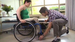
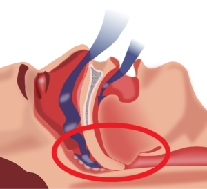
The process of selection is a complicated process that typically involves collaboration among people with spinal cord injury, their caregivers, device prescribers (Occupational Therapists or Physical Therapists), and vendors (sometimes referred to as DME Provider or Durable Medical Equipment Provider). The World Health Organization (WHO) identifies and breaks down this complicated process into 8 important steps. These include:

1. Referral and appointment

In the first step, you will be referred to a knowledgeable health provider. The referral process varies based on the services provided by each country. Some countries may have a self-referral process, while others may require a referral to a wheelchair/mobility device service.

2. Assessment

In this step, you will be accurately assessed to determine the most appropriate wheelchair and wheelchair components for you. Factors that are evaluated during an assessment include: physical functioning, posture, lifestyle/the environment it will be used in, and the tasks that are to be performed using the wheelchair.

3. Prescription

The prescription of a device that best matches the needs of the user then occurs. A vendor or therapist will work with you to choose the right wheelchair, cushion, and parts, and note down the many details and measurements of your wheelchair. In this step, you are encouraged to try various devices and seating set ups with a clinician or vendor to determine what is the most optimal set up for you.

4. Funding and Ordering

Once a detailed prescription has been created, it is then possible to determine an accurate quote for the cost of the wheelchair. A request for funding to the appropriate source with a letter of justification is provided by the prescribing therapist and physician. Determining a source of funding should be in place prior to ordering the wheelchair. Once that has been processed and approved the vendor/DME provider can order the equipment.

5. Product preparation or initial set-up

When the equipment arrives, it will be set-up and put together for the initial fitting as prescribed by the clinician. In addition, the wheelchair is inspected to ensure that it is safe and ready for use. This step is typically completed by the vendor/DME provider.

6. Fitting and adjusting

Getting your wheelchair fitted is important to make sure your chair is suited to your needs. 10

The therapist and/or the vendor will make an appointment to check on the wheelchair to ensure that it has been properly assembled. During this time, they will also make final adjustments so that it fits properly. Some things that are checked during this step include:

  • Making sure the wheelchair is the right size
  • Ensuring that the wheelchair is properly adjusted to your needs so that secondary complications are prevented
  • Confirming that any modifications made are fitting correctly.

7. User training

In this step, you will receive instructions on how to care for your wheelchair and training on wheelchair skills. This step allows you to use your device to its greatest potential, and to receive the most benefits from the device. Some key areas that should be covered in training include:

  • How to transfer in and out of your wheelchair
  • How to handle the wheelchair
  • Basic skills to use your wheelchair
  • How to prevent and watch for pressure sores
  • How to care for the wheelchair and seating components (e.g., cushion, backrest, etc.)
  • What to do if there is a problem.

8. Follow-up, maintenance and repairs

Follow-up is often required to fine tune the set up once the equipment has been used in a variety of environments. During follow-up sessions, the effectiveness of the wheelchair will be evaluated to maximize functioning, comfort, stability, and to make sure that the equipment has been properly maintained. There is no general guideline for how often a follow-up should occur; however, follow-ups and maintenance are beneficial and can contribute to the safe use of your device.

Read our article on Wheelchair Maintenance for more information.

There are many types of wheeled mobility devices that can help you get around after a spinal cord injury. Determining which device will best suit your needs is dependent on a variety of factors. These factors are often considered in partnership with a device prescriber (such as an occupational or physical therapist), who will also help ensure your device properly fits your body and needs.

For a review of how we assess evidence at SCIRE Community and advice on making decisions, please see SCIRE Community Evidence.

Parts of this page has been adapted from SCIRE Project (Professional) “Wheeled Mobility and Seating Equipment” Chapter:

Titus L, Moir S, Casalino A, McIntyre A, Connolly S, Mortenson B, Guilbalt L, Miles S, Trenholm K, Benton B, Regan M. (2016). Wheeled Mobility and Seating Equipment Following Spinal Cord Injury. In Eng JJ, Teasell RW, Miller WC, Wolfe DL, Townson AF, Hsieh JTC, Connolly SJ, Loh E, McIntyre A, Querée M, editors. Spinal Cord Injury Rehabilitation Evidence. Version 6.0: p 1-178.

Available from: https://scireproject.com/evidence/rehabilitation-evidence/wheeled-mobility-and-seating-equipment/

Armstrong, W., Borg, J., Krizack, M., Lindsley, A., Mines, K., Pearlman, J., … Sheldon, S. (2008). Guidelines on the provision of manual wheelchairs in less resourced settings. World Health Organization. https://doi.org/10.1007/978-1-349-13869-2_70

Frost, S., Mines, K., Noon, J., Scheffler, E., & Jackson-Stoeckle, R. (2012). Wheelchair service training package: Reference manual for participants. World Health Organization.

Mortenson, W. Ben, & Miller, W. C. (2008). The Wheelchair Procurement Process: Perspectives of Clients and Prescribers. Canadian Journap of Occupational Therapy, 75(3), 167–175.

Rehabilitation Engineering and Assistive Technology Society of North America (RESNA). (2011). Wheelchair service provision guide. Retreived from: https://resna.org/sites/default/files/legacy/resources/position-papers/RESNAWheelchairServiceProvisionGuide.pdf

Image credits:

  1. Image by the SCIRE Community Team
  2. Wheelchair etac cross ©Etac Sverige AB, CC BY-SA 3.0
  3. Pride Jazzy Select power chair ©Stephen B Calvert Clariosophic, CC BY-SA 3.0
  4. Welland Transportation ©Zdlpwebb, CC BY-SA 4.0
  5. Rehabilitation equipment ©satynek, Pixabay License
  6. Modifed from: wheelchair ©Saeful Muslim, CC BY 3.0 US
  7. Toillet ©Hadi, CC BY 3.0 US
  8. praying ©Hadi, CC BY 3.0 US
  9. Image by the SCIRE Community Team
  10. Image by the SCIRE Community Team


Disclaimer: This document does not provide medical advice. This information is provided for educational purposes only. Consult a qualified health professional for further information or specific medical advice. The SCIRE Project, its partners and collaborators disclaim any liability to any party for any loss or damage by errors or omissions in this publication.

Sleep Disordered Breathing After SCI

By | | No Comments

Author: Sharon Jang | Reviewer: Viet Vu | Published: 10 March 2020 | Updated: ~

Sleep disordered breathing is common after a spinal cord injury (SCI). This page explains what sleep disordered breathing is, why it occurs, what factors influence it, and current management options.

Key Points

  • Sleep disordered breathing is a family of conditions (including sleep apnea) that involve the interruption of air flow during sleep.
  • Symptoms of sleep disordered breathing include feeling tired during the day, snoring, and choking or gasping for air in your sleep.
  • Sleep disordered breathing is prevalent after SCI, and can be attributed to the level of injury, weight, sleep position, and medications.
  • Lifestyle modifications and the use of continuous positive airway pressure (CPAP) machines are the most common management strategies.

Obstructive sleep apnea occurs when the throat muscles relax (highlighted by the red circle), resulting in a blockage of your airway.1

Sleep disordered breathing is an umbrella term for conditions that involve an interruption of breathing throughout the night. In research, sleep disordered breathing is evaluated by observing two key factors:

  • Apnea, a loss of air flow for 10 seconds or more, and
  • Hypopnea, a partial blockage of an airway resulting in decreased air flow to the lungs and decreased oxygen in the blood.

When you sleep, the body normally goes into a state of hypoventilation, or a slow and shallow breathing. This results in a decrease in oxygen circulating in the blood. However, weak evidence suggests that after an SCI, hypoventilation becomes more prevalent during sleep when compared to able-bodied people. Among those with SCI, hypoventilation occurs more often in individuals with tetraplegia versus paraplegia.

The two most common disorders under sleep disordered breathing include:

  • Obstructive sleep apnea, which occurs when the throat muscles relax and temporarily block your airway, and
  • Central sleep apnea, which occurs when the brain is unable to properly send signals to the breathing muscles. This occurs when your unconscious breathing stops.

Of the two types of sleep apneas, obstructive sleep apnea is more prevalent among individuals with SCI of all levels. However, central sleep apnea is more prevalent among individuals with cervical level injuries. Narcotic use can also increase the risk of central sleep apnea. It is important to note that some patients have mixed sleep apnea, a combination of obstructive and central sleep apnea.

The prevalence of sleep apnea in individuals with spinal cord injury is two to five times greater than that in the non-SCI population. In the SCI population, research has found that sleep apnea rates vary from 27-82%. The variation in prevalence can be attributed to different diagnostic measures used in research studies (e.g., evaluating sleep apnea in a lab versus home setting) and the way each study defines sleep apnea. Level of injury and type of injury also influence the prevalence of sleep apnea in the SCI population.

A few hypotheses have been made by scientists as to why sleep apnea is prevalent in SCI. In general, sleep apnea is attributed to a complex interaction of a variety of factors:

Level of injury

Sleep disordered breathing is more prevalent in individuals with tetraplegia compared to those with paraplegia. Having a higher level of injury is usually associated with decreased muscle functioning and neural control over your organs. These impairments can create troubles with breathing, specifically with inhaling, exhaling, and the amount of air your lung can hold.

Refer to our article on Respiratory Changes After SCI for more information.

Changes in sensitivity to oxygen and carbon dioxide

After an SCI, your body becomes more sensitive to the amount of carbon dioxide circulating throughout your body. So, when there is a slight increase in carbon dioxide in the body, the brain senses it as a large change, which cues the body to hyperventilate, or rapidly breathe. However, since the change in carbon dioxide was small to start with, hyperventilation results in excess removal of carbon dioxide, resulting in very low carbon dioxide levels. During sleep, breathing is dependent on the amount of carbon dioxide circulating in the body. If this level drops below the level required for breathing, then central sleep apnea occurs. While some researchers believe this may be a cause of central sleep apnea, others note that there is currently only weak evidence to support this hypothesis.

Weight

Measures of body composition, including body mass index (BMI), neck circumference, and waist circumference, may be linked to the prevalence of sleep disordered breathing. Weak research evidence has suggested that a greater neck circumference or BMI can increase the odds of having sleep disordered breath. This is concerning for individuals with SCI, as 44-66% of this population are overweight or obese. However, other studies have found no relationship between BMI or neck circumference and sleep disordered breathing in SCI.

Sleeping on your back

One weak evidence study has suggested that there is more than a 50% increase in apneic events that occur when you sleep on your stomach or on your back, rather than on your side. More specifically, tetraplegics who sleep on their backs experienced more apneas and hypopneas per hour compared to those who slept in other positions. Despite this evidence, other researchers found that sleeping on your back enhances overall breathing functions after SCI. More research is required to determine the optimal sleeping position for health benefits, keeping in mind that bed mobility and turns overnight also help to maintain skin health.

Medications

Common medications used by individuals with SCI can also impact breathing during sleep. These include: narcotics, baclofen, benzodiazepines (lorazepam, diazepam, clonazepam), testosterone, and heart medications to treat high blood pressure or arrhythmias. Although medications can cause sleep disordered breathing, they are not likely to be the main contributor to the issue.

Increased Nasal Resistance

Individuals with tetraplegia may find it harder to breathe in with their nose (i.e., nasal resistance) because swelling of the blood vessels and a thickening of the mucus in the nose is a common effect of cervical spinal cord injury. One moderate evidence study found that individuals with tetraplegia experienced greater nasal resistance in comparison with able-bodied individuals. Since increases in breathing resistance can cause the airways to collapse, some researchers believe that this may contribute to the higher prevalence of obstructive sleep apnea in individuals with tetraplegia.

Some of the most common symptoms of sleep apnea in individuals with spinal cord injury include:
  • Feeling unrefreshed after a night’s sleep
  • Difficulty concentrating when you are awake
  • Feeling sleepy during the day, as assessed by the Epworth Sleepiness Questionnaire in clinics
  • Difficulties falling asleep
  • Awakening multiple times throughout the night
  • Snoring during sleep
  • Choking or gasping for air during sleep

Although the above are symptoms of sleep apnea, it is important to note that these same symptoms can result from secondary complications from a spinal cord injury (e.g., pain, spasticity, posture).

Continuous positive airway pressure machines

CPAP and BiPAP are powered by a machine which regulates the flow of air. These machines are connected via a tube to a mask worn over the nose and mouth..9

The first line of treatment for sleep disordered breathing generally includes lifestyle changes consisting of weight loss and the avoidance of alcohol and smoking. These lifestyle changes are normally done in conjunction with the use of a continuous positive airway pressure (CPAP) machine. CPAP machines act as a “pneumatic splint” that holds the airway open using a continuous pressure of air. To use a CPAP machine, a mask or nasal pillows are worn over the face/nose overnight.

CPAP machines are commonly used to address sleep apnea, and their effectiveness for the SCI population is supported by some weak evidence research studies. Although CPAP machines can help with breathing, multiple weak evidence research articles report poor adherence in using CPAP machines. Some of these reasons include:

  • Difficulties putting on the mask, especially among individuals with limited hand function
  • Mask discomfort
  • Feelings of claustrophobia
  • Decreased sleep quality/hard time falling asleep with it on

Although CPAP machines have the potential to help with sleep disordered breathing, more research is required to determine how helpful CPAP machines are to individuals with SCI, and how we can improve adherence to this treatment.

Bilevel positive airway pressure machines

Bilevel positive airway pressure (BiPAP) machines operate similarly to CPAP machines in that air pressure is acts as a “pneumatic splint”. however, BiPAP machines do not deliver a constant pressure of air. Exhaling when using a CPAP machine may be difficult, as breathing against an inflow of air requires effort. The BiPAP machine is unique as the pressures it exerts varies with inhalation/exhalation. Normally, the BiPAP machine will be set to a higher pressure for inhalation, and to a reduced pressure during exhalation to facilitate this process. While there have been some thoughts that BiPAP machines may benefit the able-bodied population, more research is required to determine the effectiveness of BiPAP machines in an SCI population. It is worth a discussion with your doctor if you are not tolerating a CPAP machine to trial a BiPAP machine to treat sleep apnea.

Dental appliances

An example of a dental appliance. A gap exists between the teeth to help promote airflow when sleeping.10

Dental appliances are sometimes an alternative to CPAP if an individual exhibits mild sleep disordered breathing. Dental appliances fit in the mouth like a mouth guard, and help pull the jaw and the tongue forward to prevent obstructive sleep apnea by opening up the airway. Although there has been a lot of literature supporting the use of dental appliances in an able-bodied population, more research needs to be conducted in an SCI population.

Invasive interventions

In the UVPPP surgical procedure, extra tissue is removed from the roof of your mouth and/or from your throat.11

Surgical interventions for sleep disordered breathing are often the last resort, after CPAP or BiPAP fail to work. There are a variety of surgical procedures that are used to aid obstructive sleep apnea, many of which involve reducing or repositioning the soft tissue at the back of the throat. One of the most common surgical procedures is an uvulopalatopharyngoplasty (UVPPP), whereby the soft tissues at the back of your mouth and throat are reduced and removed. However, the success rate for this procedure is variable, and there is only weak evidence to support this technique in able-bodied populations. Moreover, sleep disordered breathing in SCI may result from complex interactions between a variety of factors including level of injury, weight, sleep position, and medications. Although obstructive sleep apnea is common in SCI, researchers are unsure whether it stems from the structure of the throat or changes accompanied by an SCI. The evidence for surgical procedures to aid obstructive sleep apnea after SCI is sparse and requires more research.

Sleep disordered breathing, or a lack of breathing during sleep, is two to five times more prevalent in the SCI population compared to the able-bodied population. This increase in prevalence is believed to be related to a variety of factors including weight, level of injury, sleep position, and medications. While there are a variety of non-invasive and invasive procedures to manage sleep disordered breathing, more research is required to determine which treatment is most effective in an SCI population.

For a review of how we assess evidence at SCIRE Community and advice on making decisions, please see SCIRE Community Evidence.

Parts of this page have been adapted from the SCIRE Project “Respiratory management” Chapter:

Sheel AW, Welch J, Townson AF (2018). Respiratory Management Following Spinal Cord Injury. In: Eng JJ, Teasell RW, Miller WC, Wolfe DL, Townson AF, Hsieh JTC, Connolly SJ, Noonan VK, Loh E, Sproule S, Querée M, McIntyre A, editors. Spinal Cord Injury Rehabilitation Evidence. Version 6.0. Vancouver: p. 1-72.

Available from: https://scireproject.com/evidence/rehabilitation-evidence/respiratory-management/ 


Evidence for “What is sleep disordered breathing” is based on

Bascom, A. T., Sankari, A., Goshgarian, H. G., & Badr, M. S. (2015). Sleep onset hypoventilation in chronic spinal cord injury. Physiological Reports, 3(8), 1–10.

Castriotta, R. J., & Murthy, J. N. (2009). Hypoventilation after spinal cord injury. Seminars in Respiratory and Critical Care Medicine, 30(3), 330–338.

Chiodo, A. E., Sitrin, R. G., & Bauman, K. A. (2016). Sleep disordered breathing in spinal cord injury: A systematic review. Journal of Spinal Cord Medicine, 39(4), 374–382.

Fuller, D. ., Lee, K., & Tester, N. J. (2014). The impact of spinal cord injury on breathing during sleep, 27(4), 1–19.

Sankari, A., Vaughan, S., Bascom, A., Martin, J. L., & Badr, M. S. (2019). Sleep-Disordered Breathing and Spinal Cord Injury: A State-of-the-Art Review. Chest, 155(2), 438–445. Retrieved from https://doi.org/10.1016/j.chest.2018.10.002

Evidence for “Why is sleep disordered breathing common after SCI” is based on

Baydur A, Adkins RH, Milic-Emili J. (2001). Lung mechanics in individuals with spinal cord injury: effects of injury level and posture. Journal of applied physiology, 90, 405–411.

Berlowitz, D. J., Brown, D. J., Campbell, D. A., & Pierce, R. J. (2005). A longitudinal evaluation of sleep and breathing in the first year after cervical spinal cord injury. Archives of Physical Medicine and Rehabilitation, 86(6), 1193–1199.

Chiodo, A. E., Sitrin, R. G., & Bauman, K. A. (2016). Sleep disordered breathing in spinal cord injury: A systematic review. Journal of Spinal Cord Medicine, 39(4), 374–382.

Mason, M., Cj, C., & Smith, I. (2015). Effects of opioid, hypnotic and sedatingmedications on sleep- disordered breathing in adults with obstructive sleep apnoea (Review). Cochrane Database of Systematic Reviews, (7).

Fuller, D. ., Lee, K., & Tester, N. J. (2014). The impact of spinal cord injury on breathing during sleep, 27(4), 1–19.

McEvoy, R. D., Mykytyn, I., Sajkov, D., Flavell, H., Marshall, R., Antic, R., & Thornton, A. T. (1995). Sleep apnoea in patients with quadriplegia. Thorax, 50(6), 613–619.

Oksenberg, A., Silverberg, D. S., Arons, E., & Radwan, H. (1997). Positional vs nonpositional obstructive sleep apnea patients: Anthropomorphic, nocturnal polysomnographic, and multiple sleep latency test data. Chest, 112(3), 629–639.

Sankari, A., Vaughan, S., Bascom, A., Martin, J. L., & Badr, M. S. (2019). Sleep-Disordered Breathing and Spinal Cord Injury: A State-of-the-Art Review. Chest, 155(2), 438–445. Retrieved from https://doi.org/10.1016/j.chest.2018.10.002

Wijesuriya, N. S., Lewis, C., Butler, J. E., Lee, B. B., Jordan, A. S., Berlowitz, D. J., & Eckert, D. J. (2017). High nasal resistance is stable over time but poorly perceived in people with tetraplegia and obstructive sleep apnoea. Respiratory Physiology and Neurobiology, 235, 27–33. Retrieved from http://dx.doi.org/10.1016/j.resp.2016.09.014

Evidence for “What are the symptoms of sleep apnea” is based on

Chiodo, A. E., Sitrin, R. G., & Bauman, K. A. (2016). Sleep disordered breathing in spinal cord injury: A systematic review. Journal of Spinal Cord Medicine, 39(4), 374–382.

Fuller, D. ., Lee, K., & Tester, N. J. (2014). The impact of spinal cord injury on breathing during sleep, 27(4), 1–19.

Evidence for “What treatment is available for sleep disordered breathing” is based on

Burns, S. P., Little, J. W., Hussey, J. D., Lyman, P., & Lakshminarayanan, S. (2000). Sleep apnea syndrome in chronic spinal cord injury: Associated factors and treatment. Archives of Physical Medicine and Rehabilitation, 81(10), 1334–1339.

Burns, S. P., Rad, M. Y., Bryant, S., & Kapur, V. (2005). Long-term treatment of sleep apnea in persons with spinal cord injury. American Journal of Physical Medicine and Rehabilitation, 84(8), 620–626.

Fuller, D. ., Lee, K., & Tester, N. J. (2014). The impact of spinal cord injury on breathing during sleep, 27(4), 1–19.

Rotenberg, B. W., Vicini, C., Pang, E. B., & Pang, K. P. (2016). Reconsidering first-line treatment for obstructive sleep apnea: A systematic review of the literature. Journal of Otolaryngology – Head and Neck Surgery, 45(1), 1–9. Retrieved from http://dx.doi.org/10.1186/s40463-016-0136-4

Stockhammer, E., Tobon, A., Michel, F., Eser, P., Scheuler, W., Bauer, W., … Zach, G. A. (2002). Characteristics of sleep apnea syndrome in tetraplegic patients. Spinal Cord, 40, 286–294.

Tromans, A. M., Mecci, M., Barrett, F. H., Ward, T. A., & Grundy, D. J. (1998). The use of the BiPAP® biphasic positive airway pressure system in acute spinal cord injury. Spinal Cord, 36(7), 481–484.

Image credits
  1. Obstruction ventilation apnée sommeil © Habib M’henni, CC0 1.0
  2. Modified from Outlines. ©Servier Medical Art. CC BY 3.0
  3. visceral fat © Olena Panasovar, CC BY 3.0 US
  4. Bad breath © Mello, CC BY 3.0 US
  5. Sleeping on back © Sergio Filipe Cardoso Pires, CC BY 3.0 US
  6. Medication © Nikita Kozin, CC BY 3.0 US
  7. Nose © Rachel Healey, CC BY 3.0 US
  8. Black man sleeping at his desk cartoon vector ©Videoplasty, CC BY-SA 4.0
  9. CPAP and BiPAP by SCIRE
  10. Orthoapnea, oral appliance © Orthoapnea, CC BY-SA 3.0
  11. Modified from 4 figures. © Drcamachoent, CC BY-SA 4.0


Disclaimer: This document does not provide medical advice. This information is provided for educational purposes only. Consult a qualified health professional for further information or specific medical advice. The SCIRE Project, its partners and collaborators disclaim any liability to any party for any loss or damage by errors or omissions in this publication.

Powered Mobility Devices

By | | No Comments

Author: Sharon Jang | Reviewer: Emma M. Smith | Published: 4 March 2020 | Updated: ~

Powered wheelchairs and scooters are an important part of independent living after spinal cord injury (SCI). This page provides an overview of the basics of choosing a powered wheeled mobility device after SCI. For more general information on wheelchair provision and wheeled mobility devices, please see our page on the wheelchair provision process. Although this page mentions specific products, SCIRE Community is not affiliated with and does not endorse any of these products.

Key Points

  • Power wheelchairs may be used by people who need greater support and assistance for mobility.
  • Power wheelchairs are normally classified by their drive (rear-wheel drive, mid-wheel drive, front-wheel drive).
  • There are many adjustable parts on a power wheelchair to ensure your safety and comfort.
  • Other powered mobility options other than wheelchairs are available (i.e., scooters, Segways); however, they are not often used by individuals with SCI.

A typical power wheelchair includes a metal frame containing a motor and battery, a seat with a backrest, footrests, small caster wheels in the front and/or back, and two large rear wheels at the back. It is propelled and maneuvered using a joystick or other control function.1

Power wheelchairs are mobility devices powered by a battery and motor, and operated by the user or by another person, such as a caregiver. Power wheelchairs may be controlled in a number of different ways: by hand using a joystick, by head movements through head array systems, by the breath through sip-and-puff controllers, and by other specialty controls. This allows power wheelchairs to be used by individuals with high cervical SCI.

Power wheelchairs are typically used by those with limited control of the muscles at the elbow (complete injuries above C5) or other reasons which make it difficult to propel a manual wheelchair. Additional factors that may encourage the use of a power wheelchair over a manual wheelchair include:

  • Improved independence (if the individual is unable to effectively propel a manual wheelchair)
  • Avoidance of overuse injuries
  • Increased speed (if the individual has weak upper body strength)
  • Lengthening the amount of time that a person can spend in the wheelchair
  • Improves the ability to participate in important activities more easily

Types of power wheelchairs are described by the base. The base is the bottom portion of a power wheelchair that houses the motors, batteries, drive wheels, casters and electronics. The seating system sits on top of this base. The base is classified according to the drive wheel location relative to the system’s center of gravity. The three classifications of power wheelchairs are rear-wheel drive, mid-wheel drive, or front-wheel drive, each with their own benefits and drawbacks.

Rear-Wheel Drive.2

Rear-wheel drive

Rear-wheel drive wheelchairs have drive wheels located behind the user’s center of gravity, with casters in front. Benefits of rear-wheel drive wheelchairs include increased stability when travelling at higher speeds. However, rear-wheel drive wheelchairs are the most likely to tip backwards when going uphill, and have a large turning radius which may make it difficult to maneuver in indoor or tight spaces.

Mid-wheel drive

Mid-wheel drive wheelchairs have drive wheels located right below the user’s center of gravity, with caster wheels both in front and behind the drive wheels (Image 3). Benefits of a mid-wheel drive wheelchairs include:

Mid-wheel drive power wheelchair with tilt-in space capability.3

  • Having the smallest turning radius, thus making them most effective for indoor mobility
  • Being highly sensitive to change in direction
  • Being the most stable when going up hills and uneven ground
  • Given that mid-wheel drive wheelchairs have a total of 6 wheels as opposed to 4, the user may experience a bumpier ride; however, this can be offset with good quality suspensions. Additionally, mid-wheel drive wheelchairs are not the most efficient at going over uneven ground or soft terrain – users may get stuck when going over these surfaces.

Front-wheel drive

Front-wheel drive power wheelchair with recline capability.4

Front-wheel drive wheelchairs have the drive wheels located in the front of the chair, with caster wheels behind (Figure 4). Some benefits of this type of wheelchair include:

  • Being the most stable type of wheelchair when going on uneven terrain and up hills
  • Being the best at going over obstacles
  • Having the ability to turn around tight corners well
  • Allowing the user to pull up close to surfaces as the footplate is closer to the wheelchair since there are no casters in the way

Some drawbacks to front-wheel drive wheelchair include increased difficulty turning in small spaces due to a long back end, less stability when going at high speeds (may tend to fishtail, making it hard to drive in a straight line).

Drive controls

Drive controls for power wheelchairs come in a variety of forms and they are chosen depending on the physical capabilities of the user. Joysticks are used if you have enough arm movement to drive the wheelchair. There are different styles of joysticks available for use, depending on your hand function. This may include ball shaped controls, knob shaped both large and small, stick shaped, as well as U-shaped to name a few. Selecting an appropriate joystick according to your abilities will provide control and precision when driving. For people without the strength or endurance to drive with their arms they may choose to drive with their head, chin or use their mouth with a sip and puff control that is controlled by breath.

Various types of joystick controllers include U-shaped (A), stick (B), and ball (C).5-7


Novel drive systems

With the advancement of technology, more unique drive systems are becoming available to accommodate for a range of abilities. Recently, there has been emerging evidence for:

  • Tongue drive systems: This new system allows individuals to drive a power wheelchair with their tongues. For this to work, an individual will need to have their tongue pierced and have a titanium magnetic barbell fitted. Once in place, the user will wear a headset with magnetic sensors. This allows for individuals to move their tongues to certain teeth/spots in their mouth to drive their chair.
  • Eye gaze systems: This system uses an eye tracking device, which allows the user to drive a powered wheelchair with their eyes. To maneuver, the user simply looks left to go left, looks right to go right, and blinks for 1 second to start or stop the wheelchair. Weak evidence suggests that users are satisfied with the system and that it may be feasible to accurately drive a wheelchair with the eyes.
  • Facial drive systems: New programs allow individuals to drive their wheelchair with various facial movements/emotions as they choose. This includes movements including raised eyebrows, head movement to the left/right, or head movement up/down.

Ultimately, we must note that the tongue drive system is very new, and more research will need to be conducted before its use in practice.


Positioning functions

Power wheelchairs can have the added function of repositioning the SCI user. Repositioning helps shift weight to other parts of the body. This is helpful in providing comfort, supporting posture, and lessening pressure on certain parts of the body.

Tilting

Tilting a power wheelchair maintains the hip and knee angles by shifting both the seat and backrest together. This reduces the chances of rubbing on the skin when moving between positions. Generally, tilting with a power positioning device to a minimum angle of 30° is needed before the beneficial effects of weight shifting can be obtained, while a 55° tilt has been recommended. Additional benefits of tilting include assisting with respiratory function, providing a more comfortable position to rest in without having to transfer to a bed, and increasing stability and balance when travelling on uneven surfaces (e.g., hills).

Reclining

Reclining a power wheelchair consists of the backrest moving backwards/downwards while the seat remains stable. This increases the angle between the seat and the backrest. Some benefits of reclining include stretching out the hips, facilitating toileting and changing catheters, and facilitating transfers for caregivers. There is some weak research evidence that suggests that reclining to 120º may help reduce the pressure applied by the buttock, but reclining back this far also increases the amount of rubbing, which can lead to sores.

Repositioning and pressure sores

One recommended strategy to prevent the development of pressure sores includes weight shifting. Relieving weight off the buttock may help prevent pressure sores by allowing the tissues under pressure to regain blood flow. This is normally done using strategies such as leaning forward or lifting off the seat. If you have a higher level of injury, you may not be able to relieve pressure off your buttock independently. This is where the tilt function of a power wheelchair can come in handy – tilting backwards may help reduce the pressure applied on your bottom and allow for proper blood flow to occur. For more information on pressure relief and sores, check out our page on pressure sores.


Standing

Some wheelchairs can support standing in people with SCI. Not only does standing decrease pressure at the sit bones and coccyx, it also provides many other physiological advantages such as maintaining bone mass density, improving circulation, and enhancing functional tasks such as reaching for items on a shelf. It can also be helpful in managing spasticity and for social interaction or certain job functions.

Elevation

The elevation function of a power wheelchair raises the height of the seat. This is an option that can be added to the chair to enable individuals to reach and access things independently or without shoulder strain. It can also improve social interaction enabling the wheelchair to move to a height where they do not have to look up or be blocked in a room full of people.

Arm rests

Arm rests are multifunctional pieces that are located on the sides of the wheelchair. First, arm rests act as a support for the arm and the shoulder. When in motion, the arm rest help with balance during sudden stops, going up/down hills, and with balance in general. In addition, swelling in the arms may be reduced when the arms are kept elevated. Resting arms on the arm rest may also help maintain upper body posture as the weight of the arms is supported (versus hanging and pulling the upper body downward).

Individuals with cervical level injuries may require a specialized shaped armrest to provide support. 12

Secondly, arm rests can act as a source of stability when weight shifting to relief pressure. Individuals with a SCI can use their arm rest to push themselves up when shifting, or use the arm rest to act as a stabilizer when shifting their weight. For someone who adjusts their position often, gel padded arm rests may be used to increase comfort and support.

Arm rests can be highly adjustable based on various needs.

  • Having an arm rest that is too high up may push up the shoulders and may be uncomfortable
  • The use of adjustable arm rests may be helpful if the individual wants to be able to pull up to a table
  • Some arm rests come with a side panel built in to provide thigh support
  • Some arm rests have the function to be flipped up, while other types of arm rests may be completely removed from the wheelchair
  • A removable arm rest may be used to help facilitate transfers

Additionally, arm rests for individuals with injuries C4 or above may require special adjustments. Arm troughs (or a more formed arm rest) may be used to provide additional support to the arm to prevent it from moving. In addition, elbow blocks may be used to help support an upright position when tilting. In the case that an individual experiences spams in the arm, sheepskin or gel pads may be used to protect the skin when the arm rests in the trough.

Foot rests: footplates and leg rests

A footplate is generally a single plate (left), while foot rests consist of individual pieces (right). 13,14

Depending on the drive mode, power wheelchairs may have a footplate or leg rests. A foot plate is often connected to the base of the power wheelchair, and is one piece of metal that can be flipped up and down and supports both of the feet. In contrast, leg rests are two separate supports for each foot.

Where the foot rest sits is dependent on the drive base and the physical characteristics of the person. A rear wheel drive wheelchair has footplates the furthest away from the chair as there are caster wheels located on the front. This results in added length to the wheelchair, which may make it harder to turn. The use of a single centre-mounted footplate may help reduce the length of the wheelchair. Front wheel drive wheelchairs do not have front casters that interfere with the foot rests, as the caster wheels are located in the rear. As a result, the foot rests on front wheel drive wheelchairs do not interfere.

Some power wheelchairs may have an option for powered footrests, allowing the user to adjust the angle of the footplates through a motor. To prevent the legs from dangling when using the tilt or recline function, some power wheelchairs may have a foot rest elevation function. This function helps to increase the height and angle of the foot rest to elevate the legs when reclining, and lowers back into the default position when returning to an upright position.

The batteries on a power wheelchair are designed to run for generally 3-9 hours of continuous use, and are made to last for up to 5 years. Some factors that influence how far you can drive on one charge of your battery can be categorized into fixed and varying factors. Many of these factors impact each other, and changing one aspect may influence another.

Fixed factors

Fixed factors influence how long your battery lasts, but cannot be changed. Examples include:

  • Weight and size of the battery – a larger battery will allow for a greater driving distance, but adds to the overall weight of the wheelchair
  • The drive type of the wheelchair – rear wheel drive wheelchairs tend to use more power than a front or mid wheel drive
  • Weight of the wheelchair – a lighter chair will use less battery
  • Weight distribution – more weight applied on the caster wheels will use more battery power during manoeuvering

Varying factors

Varying factors are considerations that can influence your battery life in which you can have some control over to a certain extent. These factors include:

Using a powered wheelchair on smooth even ground in moderate temperatures can help prolong the battery life.15

  • The payload of the wheelchair – this is a combination of the user’s weight in addition to any items they may be carrying (e.g., respiratory products, groceries, battery charger, etc.). Most often the maximum payload the battery can handle is much greater than the average weight the power wheelchair carries.
  • Properties of the driving surface – driving on uneven, rough, slippery, or soft (e.g., gravel, grass) will use more battery power.
  • Temperature – using your power wheelchair in extreme weather (hot or cold) can impact how long the battery lasts for. Using your wheelchair in over 30 degrees or under 0 degrees Celsius will reduce the battery’s normal capacity.
  • Driving behaviour – performing a lot of stop and go actions, going up hills, and climbing curbs consume more battery
  • Low air pressures in the tires – if you are using pneumatic tires and they are underinflated, this will use up battery faster
  • Powered options – using the tilt/reclining functions or powered leg rest functions may reduce the driving range of your wheelchair

If you are using your power wheelchair every day, you should be charging your wheelchair daily. When charging your battery, it is better to charge it for longer amounts of time than in multiple short charges (i.e., less than 2 hours). A minimum charge time of 12 hours or longer has been recommended; power wheelchair batteries will not overcharge! If you are using a new power wheelchair, note that it may take 10-20 charges prior to the batteries reaching their fullest capacity.

While the majority of people who use wheeled mobility devices after SCI will use a manual or power wheelchair, other devices are also sometimes used.

Scooters

Scooters are powered devices that are typically used for getting around in the community. They come in a variety of sizes and in 3 wheeled and 4 wheeled designs. They are often used by individuals whose functional abilities do not require a full-time wheelchair, but may need support when moving around in the community. This support may help a person move greater distances, conserve energy, or get around safely if they have impaired balance, pain, or fatigue. Scooters are typically larger devices compared to wheelchairs, and are controlled by hand controls similar to a bike or motorcycle. They are usually cheaper than power wheelchairs but do not have as small a turning radius and also cannot facilitate complex seating systems. Due to their longer length, they are also more difficult to accommodate in wheelchair taxis or vans.

Additionally, there are tiller-like devices that can be added on to powered wheelchairs. This allows powered wheelchair users to operate their device similarly to a scooter. An example of this device is the JoyBar.

Segways

Segways are powered, self-balancing two-wheeled devices with arm bars that a person can stand on to move around in the community. While Segways are not intended specifically to be a rehabilitation device, some people with higher levels of functional abilities (people who are able to stand and balance effectively) may use these devices to assist with getting around in the community in a similar way to scooters. They are considered to be smaller, faster, and more manoeuvrable than power wheelchairs. Early research has suggested that Segways may be a potential mobility option for people with SCI who have limited walking abilities, although there may be some difficulties getting on or off the device.

In more recent times, several seated Segway-like devices have started entering the market. These devices (such as the Nino), allow individuals with upper trunk function to drive the wheelchair by leaning forward and to brake the wheelchair by leaning back. More research is required to determine how useful and practical it would be for individuals with spinal cord injury.

As described in this article, many factors play into the selection and set up of a powered mobility device. This article provides you with information on the various parts of a power wheelchair that can be adjusted to suit your needs. If you think that a feature needs to be adjusted or added, consult with your health care or wheelchair provider.

Any reference to a specific product does not constitute or imply an endorsement by SCIRE Community. Professional advice should be sought before making any health care and treatment decisions.

For a review of how we assess evidence at SCIRE Community and advice on making decisions, please see SCIRE Community Evidence.

Parts of this page has been adapted from SCIRE Professional “Wheeled Mobility and Seating Equipment” Module:

Titus L, Moir S, Casalino A, McIntyre A, Connolly S, Mortenson B, Guilbalt L, Miles S, Trenholm K, Benton B, Regan M. (2016). Wheeled Mobility and Seating Equipment Following Spinal Cord Injury. In Eng JJ, Teasell RW, Miller WC, Wolfe DL, Townson AF, Hsieh JTC, Connolly SJ, Loh E, McIntyre A, Querée M, editors. Spinal Cord Injury Rehabilitation Evidence. Version 6.0: p 1-178.
Available from: https://scireproject.com/evidence/wheeled-mobility-and-seating-equipment/ 


Evidence for “What types of power wheelchairs are there?” is based on:

Queensland Spinal Cord Injuries Services. (2019). Comparison of front, mid, and rear wheel drive power chairs. Retrieved from: https://www.health.qld.gov.au/__data/assets/pdf_file/0028/428482/pdwc-comparison.pdf

Evidence for “What parts of a power wheelchair should I know about?” is based on:

Agency for Clinical Innovation. (2020). Ke2p the big picture in mind. Retrieved from: https://www.aci.health.nsw.gov.au/networks/spinal-cord-injury/spinal-seating/module-10/keep-the-big-picture-in-mind

Boninger, M. and the Model Systems Knowledge Translation Center. (2019). The power wheelchair: what the spinal cord injury consumer needs to know. Retrieved from: https://msktc.org/sci/factsheets/wheelchairs/The-Power-Wheelchair

Giesbrecht EM, Ethans KD, & Staley D. Measuring the effect of incremental angles of wheelchair tilt on interface pressure among individuals with SCI. Spinal Cord 2011;49:827- 31.

Kim J, Park H, Bruce J, Sutton E, Rowles D, Pucci D, et al. The tongue enables computer and wheelchair control for people with spinal cord injury. Science translational medicine 2013;5(213):213ra166.

Kim J, Park H, Bruce J, Rowles D, Holbrook J, Nardone B, et al. Qualitative assessment of tongue drive system by people with high-level spinal cord injury. Journal of rehabilitation research and development 2014;51(3):451-466

Kim J, Park H, Bruce J, Rowles D, Holbrook J, Nardone B, et al. Assessment of the tongue- drive system using a computer, a smartphone, and a powered-wheelchair by people with tetraplegia. IEEE Transactions on Neural Systems and Rehabilitation Engineering 2016;24(1):68-78.

Laumann A, Holbrook J, Minocha J, Rowles D, Nardone B, West D, et al. Safety and efficacy of medically performed tongue piercing in people with tetraplegia for use with tongue- operated assistive technology. Topics in spinal cord injury rehabilitation 2015;21(1):61-76.

Queensland Spinal Cord Injuries Service. (2019). Powerdrive Wheelchair Features. Retrieved from: https://www.health.qld.gov.au/__data/assets/pdf_file/0024/425625/pdwc-features.pdf

Stewart, D. (2019). Wheelchair arm rests. Retrieved from: https://mobilitybasics.ca/wheelchairs/armrests

United Spinal Association (2019). Wheelchair armrests – what do they really do? Retrieved from: https://www.unitedspinal.org/wheelchair-armrests/

Evidence for “What impacts battery life” is based on

Invacare. (2020). How to charge and maintain a battery on a power wheelchair. Retrieved from: https://www.passionatepeople.invacare.eu.com/charge-maintain-battery-power-wheelchair/

Karman. (2020). Power wheelchair batteries: understanding how the battery works. Retrieved from: https://www.karmanhealthcare.com/power-wheelchair-batteries-understanding-how-the-battery-works/

Scootaround. (2019). How long do wheelchair batteries last? (and battery life tips!). Retrieved from: https://scootaround.com/en/how-long-do-wheelchair-batteries-last-and-battery-life-tips

Image credits:

  1. Modified from: Wheel isolated ©MBGX2, Pixabay License
  2. Mid wheel drive Image ©Br Yonten Phuntsok, Pixabay License
  3. Wheel isolated ©MBGX2, Pixabay License
  4. Front wheel drive ©Stephen B Calvert Clariosophic, CC BY-SA 3.0
  5. U-shaped joystick handle ©Bodypoint
  6. Large ball ©Permobil
  7. Joystick ©Wheelchair and Scooter Repair
  8. Midwheel drive power wheelchair with tilt-in-space capability ©Model systems knowledge translation center (MSKTC)
  9. Front wheel drive power wheelchair with recline capability ©Model systems knowledge translation center (MSKTC)
  10. Power elevating seat ©Model systems knowledge translation center (MSKTC)
  11. 2019 F5 Corpus VS ©Permobil
  12. Molded wheelchair arm rest ©Comfort Company
  13. Intrepid mid-wheel power wheelchair ©Intrepid
  14. Conventional footrest ©David Stewart
  15. Motorized wheelchair wheelchair elderly man ©Kevin Philips, Pixabay License
  16. Welland Transportation ©Zdlpwebb, CC BY-SA 4.0
  17. JoyBar © Joybar
  18. Segway ©Ivva,CC BY-SA 2.0 
  19. Nino Segway Wheelchair ©Gyronova


Disclaimer: This document does not provide medical advice. This information is provided for educational purposes only. Consult a qualified health professional for further information or specific medical advice. The SCIRE Project, its partners and collaborators disclaim any liability to any party for any loss or damage by errors or omissions in this publication.

Manual Wheelchairs

By | | No Comments

Author: Sharon Jang | Reviewer: Emma M. Smith | Published: 3 March 2020 | Updated: ~

Manual wheelchairs are a type of wheeled mobility device that are an important part of independent living after spinal cord injury (SCI). This page provides an overview of manual wheelchairs. SCIRE Community is not affiliated with and does not endorse any of the specific products mentioned on this page.

Key Points

  • Manual wheelchairs are typically used by people with enough movement control and strength in the arms to propel the wheelchair independently
  • A manual wheelchair consists of many different parts, most of which can be altered to suit your needs
  • Manual wheelchairs can be adjusted to prevent injury and to promote comfort
  • Multiple factors play into injuries related to manual wheelchair use, including the way you push your wheelchair, your wheelchair set up, and the surfaces you are wheeling on

Manual wheelchairs are wheelchairs that are propelled by the user or pushed by another person. They do not have a battery or other power source.

For the most part, manual wheelchairs are used by people who have enough muscle control and strength in their arms to propel the wheelchair forward on their own. For people with SCI, this typically means that a person needs to at least have function of the biceps (the muscle that bends the elbow), which is intact for people with complete injuries at C5 and below. However, it can be difficult to propel a wheelchair for individuals with C5 and C6 injuries, so only some individuals with these types of injuries will be able to do so. Use of a manual wheelchair is more common in people with control of the triceps (the muscle that straightens the elbow), which is intact in people with complete injuries at C7 and below. In some cases, a manual wheelchair can be pushed by another person or propelled using the legs.

As a bridge between the more common manual and power wheelchairs, power-assist add-ons combine features of manual and power wheelchairs to provide greater assistance for those who need it. Additionally, non-powered propulsion assist add-ons may also be used to facilitate wheeling. Propulsion assist options are available for manual wheelchairs and come in several different styles. The use of powered add-ons has been reported to improve efficiency and to reduce strain on the cardiovascular system. The following article will focus on manual wheelchairs that are propelled with the arms.

Refer to our chapter on Propulsion Assist Devices for more information!

A typical manual wheelchair contains a frame, seat with a backrest, footrests or footplate, two small caster wheels in the front, and two large rear wheels at the back. The rear wheels contain hand rims which are used to propel and maneuver the wheelchair. Wheel locks on the rear wheels help prevent the wheelchair from moving when it is not in use or when getting in or out of it.

There are many different styles of manual wheelchairs, most of which allow for the ability to adjust and change aspects of the set up. This can be important in the first few years that a person uses a wheelchair as a person’s skills and priorities may change. Nonetheless, it is important to try to get as lightweight and compact a wheelchair as possible while also ensuring it is safe, durable and flexible.

There are many different components of manual wheelchairs that can be customized and changed to find the best set-up for an individual. Below, we list some of the common options and changes that may be possible when setting up a manual wheelchair. In general, changes to the set-up of the manual wheelchair are a series of tradeoffs. Although changes are usually intended to achieve a desirable improvement (such as greater ease of wheeling), sometimes they need to be balanced with reduced stability and an increased risk of tipping over. Please speak to your health providers before making any major changes to your wheelchair set-up.

Frame design

Manual wheelchairs come in two basic frame designs, folding frames or rigid frames. Folding frames have a crossbar under the seat that allows the frame to fold in half. Rigid frames may also fold but in a different way, where the backrest folds down. In many manual wheelchairs, wheels can removed if they are being put in a vehicle.

Folding manual wheelchairs

Folding wheelchairs are designed to be folded vertically and take up minimal storage space. This allows for easy portability (such as fitting the wheelchair into a car). However, these wheelchairs also have many moving parts that may break down or loosen over time, and are heavier than rigid wheelchairs. Folding wheelchair often have flip up, swing away or swing in footrests so they may be used by individuals that do not use the wheelchair full time, can stand or take some steps, or by those who foot propel.
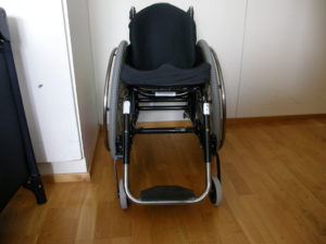
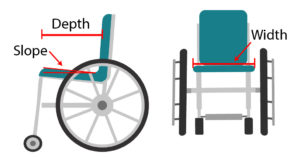
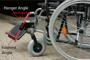
Rigid manual wheelchairs

Rigid wheelchairs tend to be lighter and more durable than folding wheelchairs, which makes them the common choice for people with complete SCI injuries. The rear tires often have to be removed to fit into a vehicle because they cannot fold inward. Some rigid manual wheelchairs can still be ordered with adjustability, which can be important if seating components change.


Frame materials

The majority of manual wheelchair frames are made of either aluminum or stainless steel. Ultralight and sports wheelchairs are often constructed from high performance aluminum, carbon fiber, chromium-molybdenum, nickel alloy steel, or titanium. Titanium and composites can be considerably more expensive but are more lightweight.

Seat width, depth and slope

It is important that the width, depth and slope (also known as dump) of the seat from front to back are well-fitted to the overall function and characteristics of the user. A wheelchair that is too narrow in width may cause skin problems due to rubbing, while a wheelchair that is too wide may hinder propulsion, and will interfere with the accessibility and maneuverability of the chair. A wheelchair that is too short in depth may provide insufficient support to the hips and stability to the bottom, while a wheelchair that is too long in depth may cause knee pain or a slouching posture.  In addition, an increased seat slope (i.e., one that is higher in the front than the back) makes transfers more difficult as one would need to lift up at an incline, but are better for people with a higher level of injury because this slope helps them find balance and stability to wheel.

Footrest angle and frame taper

The footplate is angled away from the frame of the chair and the post of the casters.6

When considering the footrest angle between the seat and the footplate hanger, the knee flexion range of movement and hamstring length needs to be considered as well as the visual field as the client may not be able to see their feet. For example, a greater frame angle improves wheelchair access by making the chair shorter but may make the chair more unstable in the forward direction. Some wheelchairs have a frame taper, which is the narrowing of the frame towards the front and continues down to the footplates. This improves access from the front of the wheelchair, making toilet transfers easier. However, the taper may not be compatible with the cushion and may impact the ability of the wheelchair to fold.

Rear Wheels

Spoked wheels (left) versus mag wheels (right).7,8

The rear wheels are set up with hand rims on them so that self-propelling is possible. Wheel material and the amount of air in the wheels affect how effective the wheels are in minimizing vibration or shock to prevent triggering of spasticity and to increase comfort. Wheel rims are made from a variety of materials including aircraft grade aluminum, plastic, fibreglass, reinforced nylon, titanium, carbon graphite, or steel. Types of rear wheels include: spoked wheels (made of metal, normally has more than 30 spokes), mag wheels (made out of synthetic material, less than 10 spokes), carbon-graphite mag wheels, and power assist wheels. The more spokes, the better the vibration absorption. The most common wheel size for adults is 60cm (24”) and for children it is 50cm (20”).

Tires

Airless (solid) tires tend to provide bumpier ride.9

Wheelchairs may have solid tires, foam-filled tires, or air (pneumatic) tires. Solid tires do not run the risk of popping, however, they often result in a bumpier ride. On the contrary, air tires provide a softer ride, but require regular maintenance of air pressure. A lack of air in the tires (less than 50% of the recommended PSI) makes propelling a wheelchair more difficult. It is recommended that tires be re-inflated every two months to keep the pressure above 50%. Some type of tires include: standard pneumatic tires, high pressure pneumatic tires, outdoor pneumatic tires, puncture proof tires, and solid plastic tires.

Hand rims/Push rims

Hand rims are attached to the rear wheel rim on wheelchairs that are intended for self-propulsion. This part is optional, and some users may opt not to use them. Hand rims are commonly made out of aluminum, and may be anodized or vinyl coated. Some downsides to using aluminum hand rims are that they may turn your hand black, and can become very sharp if they are damaged. Other models of hand rims have a rubber like material and assist individuals without full handgrip with their push stroke. In addition, flexible hand rims have been developed to adapt to the shape of the hand in order to reduce strain resulting from the bending action of the wrist and fingers.

Front caster wheels

The small front wheels are called caster wheels, and they help maneuver and steer the position of the wheelchair. Caster diameter, material, and position are important factors to consider that affect wheelchair balance and performance. Sizes available include 75mm (3”), 125mm (5”), 150mm (6”), 180mm (7”), 200mm (8”), and 250mm (10”). Bigger caster wheels are more stable, have a bigger turning radius, and easier to wheel over thresholds but are less maneuverable and responsive. A point of caution is that when caster wheels are trailing, a person’s weight will be more forward and may be in front of the caster wheel, which can cause tipping and is a frequent cause of falls in manual wheelchairs (see image below). Caster stem angle must always lie at right angles to the floor to ensure casters track correctly, or they may “flutter” when the wheelchair is pushed or can cause the front of the wheelchair to rise and fall during turning.

Forward casters (left) versus trailing casters (right).11

When choosing the type of caster, one should consider the most common type of surface they will be propelling on (e.g. outdoors or indoors), the desired front seat height, and the front frame angle of the wheelchair. The construction materials available for caster wheels include plastic, urethane and aluminum. Pneumatic caster tires, usually 150mm (6”)” and 200mm (8”) in diameter, have more shock absorption features making them ideal for outdoor wheeling, while 75mm (3”) and 125mm (5”) urethane caster tires are more common and are good for indoor propulsion and sport specific wheelchairs. Those who experience significant spasticity and/or discomfort when propelling wheelchairs often opt for pneumatic casters to achieve a smoother ride.

Wheel locks

Wheel locks are used on the large rear wheels for safety with transfers and when no movement is wanted. They vary in styles including push to lock, pull to lock, and scissor style locks. To decrease the force required to engage/disengage the wheel locks, extended brake levers can be applied, as they minimize the forward reach required to access the wheel locks. The type and position of the lock should be influenced by the user’s reaching ability, balance, strength, and hand function as well as the impact of wheel lock position on transfers. Some clients with good physical skills may not even require wheel locks at all.

Anti-tippers

Anti-tippers are an optional set of smaller wheels connected to a metal poles that are attached to the frame of your wheelchair 381 to 508mm (1.5 to 2″) above the ground. Anti-tippers are commonly found on rear of the chair to prevent a backwards fall, however they can also be found on the front of the wheelchair to prevent forward tipping. Anti-tippers may be especially useful for new wheelchair users, those who have recently switched to a new wheelchair, or if one’s health is declining. While they are considered a safety item, they also have drawbacks. Firstly, anti-tippers may interfere with wheelchair skills requiring you to lean backwards, such as climbing a curb or doing a wheelie. Secondly, if the anti-tippers are set too low, it may impede on going over obstacles or up hills.

There are many necessary considerations for setting up a manual wheelchair properly for mobility and function. It is important to work with a knowledgeable health care professional and vendor to ensure the correct decisions are made.

These considerations may include:

Various factors can influence your ideal wheelchair set up. In this photo, some considerations include having a dog and wheeling on grass.13

  • Activities of Daily Living: It is important to think about where you need to take your chair, how close you can get to or under surfaces, reach for things, and how easily you can remove components and put them back on.
  • Mobility: You need to consider what surfaces you will wheel on (carpet, flooring, snow, rain). How the chair will move over inclines and transitions as well as in confined spaces. Is it a primary chair or just an indoor chair or a transport chair?
  • Positioning: It is important to consider your posture, comfort and interacting with people and your environment.
  • Psychosocial: You want to consider how you look and feel and how you will interact with people and your environment.
  • Transfers: You need to look at the ability to get in and out of the wheelchair safely so the height, stability, ease of moving and weight are some of the things to consider.

The three most important adjustments to have in a manual wheelchair are back angle, axle position and seat to floor height. These adjustments are important as they can have an impact on pain, wounds, or postural issues. The wheelchair set-up is essential to allow for safety and balance of the chair.

Axle Position (vertical and horizontal)

The horizontal position of the rear wheel axle can affect how much energy is required to move the wheelchair. Studies have shown that placing the rear wheel axle closer to the front of the wheelchair can make it easier to propel. However, it can also affect the stability of the wheelchair, and make the wheelchair more likely to tip backwards. This is because there is more weight behind the axle. Decisions on where to place the axle will depend on the person’s wheelchair skills. The distance between the shoulder and the axle of the rear wheel can also affect efficiency. One study showed that greater distances may result in greater energy requirements. Adding adjustable axles to manual wheelchairs can help individuals customize their wheelchairs to improve propulsion. This can lead to reduced risks of upper body injuries specifically shoulder injuries.

Backrest height and angle

The appropriate backrest height is largely a trade-off between posture, comfort and freedom of movement. A backrest is usually 406mm (16″) tall, and often does not come up any higher than your shoulder blades – any higher and it will impede your ability to push your wheelchair. Most manual wheelchairs have the ability to modify the height of the backrest (so it can be modified as function increases or decreases). A higher backrest is seen to be more comfortable, and is often used by individuals with limited trunk functioning as it provides more support. On the contrary, a shorter back rest allows for more movement of the trunk, which can be functionally useful (e.g., it will allow you to reach for objects sideways and behind you).

Using a wheelchair with backrest height that is not optimal may have negative consequences. If the backrest is too low, there will be a lack of support which may lead to postural instability. However, most often individuals are using backrests that are too high. This may result in a limited reach (and thus reduction in functional activity), pain, and a slouching posture as a result of compensating for sliding forward.

Other Adjustments

Rear wheel lateral position (space from frame) and camber

The lateral position of the rear wheel is the distance the wheel is from the wheelchair frame. This distance can affect the overall width of the wheelchair and how accessible the wheel is.

Camber relates to the rear wheels being set on an angle from their axle position, where the distance between the top of the wheels is less than at the bottom of the wheels. The angle can range from 0º up to an extreme 12º, although the average camber for day wheelchairs varies from 0º to 4º. Some wheelchair models offer adjustability in camber angles within the same chair. Stability, wheeling efficiency, and turning maneuverability can be enhanced with wheel camber, especially when moving over side slopes. In addition, the hands are better protected against trauma because the wheels touch the floor spanning a wider area than the hands have in contact with the hand rims. Too much camber can make the overall width of the wheelchair wider so assessment of the spaces and doorways needed to access is essential when deciding on camber.

Camber and Sports Wheelchairs

A large camber is often seen on sport wheelchairs, measuring up to 15º. A greater camber is of beneficial use to wheelchair sports, as it provides more lateral support to prevent tipping over to the side when turning quickly and sharply. It also helps to create turns that are smoother and sharper when in motion.


Footrest height and length

Proper support of the feet and legs is important to the comfort and safety of the wheelchair user. If the foot and thigh is not properly supported it may result in the leg or foot moving excessively and may result in instability, pain, and spasticity. In addition, it can create areas of pressure on the leg or the foot, ultimately leading to pressure injuries. Support can be adjusted through altering the height and length of the footrest.

The foot rest should be 1-2 inches off the ground to allow for optimal clearance. If it is too low, it may catch on door threshold and other objects, and may cause difficulties when trying to go up or down hills or slopes. Should the footrest unexpectedly hit an obstacle, there is a high risk of the user falling out of their wheelchair.

Refer to our article on Wheelchair Seating for more information on footrest set-ups

Using a manual wheelchair can be tough on the user’s arms and shoulders. Research has shown that 25-80% of manual wheelchair users experience injuries in their wrists, elbows, and or/shoulders. There have been multiple (weak) studies that have identified multiple factors that can be addressed in order to reduce/prevent the chances of injury:

Muscles required for propulsion

Muscles in the chest, upper back, shoulders, and arms that are used in propelling a wheelchair.17

When pushing a manual wheelchair, many muscles in the upper back, chest, shoulder, and arms are used. Pushing a manual wheelchair is not the most efficient way of getting around, as only between 2-14% of force applied by the arms goes into propelling a wheelchair, depending on the level of injury and style of propelling. The shoulder muscles are particularly strained during wheelchair propulsion as they are relatively smaller muscles that are responsible for both stabilizing the shoulder and applying force to a wheelchair to push it forward . Shoulder pain occurs in 31-73% of manual wheelchair users, and weak evidence suggests that individuals with tetraplegia experience more pain due to greater forces being applied with their arms.


Kinematics: the technique you use when propelling a wheelchair can impact your risk of shoulder pain/injury. For example, pushing a wheelchair at increasing speeds/intensities may contribute to the development of shoulder pain. Additional factors to be considered include the angle of your joints (i.e., the elbows, wrist) when pushing, and the angle at which you push your tires at.

Propulsion pattern: the pattern you push your wheelchair with (e.g., where do your hands go when you push and after you’re done pushing) may also have an influence on your risk of injury. Some weak evidence suggests that using the semi-circular and double-loop-over may reduce the risk of nerve injury and are the most optimal ways to push your wheelchair. Further evidence (weak) has indicated that arcing may be more efficient for short bouts of high intensity pushing (like when going uphill).

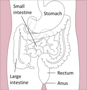
Body weight: Weak evidence suggests that having a higher body weight may be related to a higher risk of injury while propelling your wheelchair. This is due to the fact that moving a heavier body requires higher forces created by the shoulders. Body weight management is important in decreasing the amount of force created in your arms when pushing your wheelchair, and reduces the risk of injury.

Wheelchair set up: Having the rear wheels on a manual wheelchair placed in a forward axle position can help improve push rim biomechanics, reduce the amount of force put on your shoulders when propelling, and the frequency of propulsion (i.e., you do not have to push as much to go as far). Research has suggested the use of manual wheelchairs with adjustable axle positions so that the rear wheels can be optimally adjusted.

Wheeling on uneven surfaces: Some research suggests that wheeling across cross slopes (such as a driveway) can increase the forces on the arm and may lead to overuse injuries. In addition, more physiological effort  (i.e., heart rate, how much oxygen is being used, rating of perceived exertion) is required to go up slopes more than 2% incline, and more physiological and physical effort is required to go up slopes greater than 8%.

Manual wheelchairs are highly customizable pieces of equipment that can be tailored to your needs. In order to optimally use your wheelchair, certain adjustments to the backrest, frame, and tires can be made. Using manual wheelchairs requires upper arm function and strength. Improper set ups and techniques may lead to injuries.

It is best to discuss all treatment options with your health providers to find out which treatments are suitable for you.

For a review of how we assess evidence at SCIRE Community and advice on making decisions, please see SCIRE Community Evidence.

Parts of this page has been adapted from SCIRE Project (Professional) “Wheeled mobility and seating equipment following spinal cord injury” Chapter:

Titus L, Moir S, Casalino A, McIntyre A, Connolly S, Mortenson B, Guilbalt L, Miles S, Trenholm K, Benton B, Regan M. (2016). Wheeled Mobility and Seating Equipment Following Spinal Cord Injury. In Eng JJ, Teasell RW, Miller WC, Wolfe DL, Townson AF, Hsieh JTC, Connolly SJ, Loh E, McIntyre A, editors. Spinal Cord Injury Rehabilitation Evidence. Version 6.0: p 1-178.

Available from: https://scireproject.com/evidence/rehabilitation-evidence/wheeled-mobility-and-seating-equipment/ 


Evidence for “What are the components of a manual wheelchair?” is based on:

Boninger, M., and the Model Systems Knowledge Translation Center. (2019). The manual wheelchair: What the spinal cord injury consumer needs to know. Retrieved from: https://msktc.org/sci/factsheets/wheelchairs/The-Manual-Wheelchair

Spinal outreach team and The University of Queensland School of health and rehabilitation sciences. (n.d.). Manual wheelchairs: Information resource for service providers. Retrieved from: https://www.health.qld.gov.au/__data/assets/pdf_file/0026/429911/manual-wheelchairs.pdf

Evidence for “What adjustments can I make to my manual wheelchair?” is based on:

Sprigle, S. (2014). Measure it: proper wheelchair fit is key to ensuring function while protecting skin integrity. Advanced Skin Wound Care, 27(12), 561-72.

Evidence for “What are health concerns related to manual wheelchair use?” is based on:

Arva, J., Fitzgerald, S. G., Cooper, R. A., & Boninger, M. L. (2001). Mechanical efficiency and user power requirement with a pushrim activated power assisted wheelchair. Medical Engineering and Physics, 23(10), 699–705. https://doi.org/10.1016/S1350-4533(01)00054-6

Boninger, M. L., Souza, A. L., Cooper, R. A., Fitzgerald, S. G., Koontz, A. M., & Fay, B. T. (2002). Propulsion patterns and pushrim biomechanics in manual wheelchair propulsion. Archives of Physical Medicine and Rehabilitation, 83(5), 718–723. https://doi.org/10.1053/apmr.2002.32455

Curtis, K. A., Drysdale, G. A., Lanza, R. D., Kolber, M., Vitolo, R. S., & West, R. (1999). Shoulder pain in wheelchair users with tetraplegia and paraplegia. Archives of Physical Medicine and Rehabilitation, 80(4), 453–457. https://doi.org/10.1016/S0003-9993(99)90285-X

Gil-Agudo, A., Del Ama-Espinosa, A., Pérez-Rizo, E., Pérez-Nombela, S., & Pablo Rodríguez-Rodríguez, L. (2010). Upper limb joint kinetics during manual wheelchair propulsion in patients with different levels of spinal cord injury. Journal of Biomechanics, 43(13), 2508–2515. https://doi.org/10.1016/j.jbiomech.2010.05.021

Kulig, K., Newsam, C. J., Mulroy, S. J., Rao, S., Gronley, J. K., Bontrager, E. L., & Perry, J. (2001). The effect of level of spinal cord injury on shoulder joint kinetics during manual wheelchair propulsion. Clinical Biomechanics, 16, 744–751.

Rankin, J. W., Richter, W. M., & Neptune, R. R. (2011). Individual muscle contributions to push and recovery subtasks during wheelchair propulsion. Journal of Biomechanics, 44(7), 1246–1252. https://doi.org/10.1016/j.jbiomech.2011.02.073

Richter, W. M., Rodriguez, R., Woods, K. R., & Axelson, P. W. (2007). Consequences of a Cross Slope on Wheelchair Handrim Biomechanics. Archives of Physical Medicine and Rehabilitation, 88(1), 76–80. https://doi.org/10.1016/j.apmr.2006.09.015

Image credits

  1. Kuschall wheelchair model R33 ©Tim99~commonswiki, CC BY-SA 4.0
  2. Image modified from Different kinds of handicap equipments ©brgfx, Freepik License
  3. Wheelchair ©George Hodan, CC0 1.0
  4. Wheelchair parts (main pic) ©Memasa CC BY-SA 3.0
  5. The SCIRE Community Team
  6. Modified from disabled, stroller, the disease, wheelchair, disability, wheel, transportation, medical equipment, metal, mode of transportation, CC0 1.0
  7. Wheelchair disability paraplegic injured disabled ©stevepb, Pixabay License
  8. wheelchair, old, vintage, isolated, wheel, antique, transportation, white, retro, transport, CC0 1.0
  9. The SCIRE Community Team
  10. Wheelchair disabled person with reduced mobility man ©SGENET, Pixabay License
  11. Forward versus trailing casters ©Ian Denison
  12. Modified from Black and grey wheelchair, CC0 1.0
  13. Woman, dog, pet, friend, outdoors, grass, female, person, jacket, pal, CC0 1.0
  14. Axle Position by the SCIRE Community Team
  15. Modified from Disabled people set Free Vector ©Macrovector, Freepik License
  16. Euroleague – LE Roma vs Toulouse IC-27 ©Pierre-Selim, CC BY-SA 3.0
  17. Muscles that move the humerus ©Betts et al, CC BY-SA 4.0
  18. Stylized illustration of stroke pattern classification during wheelchair propulsion ©Emily Churton and Justin WL Keogh, CC BY 2.0


Disclaimer: This document does not provide medical advice. This information is provided for educational purposes only. Consult a qualified health professional for further information or specific medical advice. The SCIRE Project, its partners and collaborators disclaim any liability to any party for any loss or damage by errors or omissions in this publication.

Dietary Fibre

By | | No Comments

Authors: Vanessa MokDominik Zbogar | Reviewers: Janet ParkerGita Joshi | Published: 23 December 2019 | Updated: ~

Adequate fibre intake is important to ensure proper nutrition after spinal cord injury (SCI). This page outlines the current recommendations for dietary fibre and its role in managing conditions such as neurogenic bowel in SCI.

Key Points

  • Foods such as vegetables, fruits, legumes, and whole grains are great sources of dietary fibre.
  • Research in the general population shows that increasing dietary fibre reduces the risk for heart disease, obesity, diabetes, high blood pressure, and certain cancers.
  • Increasing dietary fibre in people with SCI may not have the same result as in the general population. It is important to continuously monitor how an increasing fibre intake affects you.
  • Increasing fibre in the diet should be done incrementally and may require increased fluid intake to prevent constipation.
  • An increased fibre intake may require a change in your bowel and bladder routine.

Dietary fibre is a kind of carbohydrate that is difficult for the body to break down, so it is not absorbed in the small intestines. Instead, it passes into the colon. Therefore, fibre supplies little to no calories.

Fibre can be classified into two types: soluble and insoluble. Both types of fibre play an important role in gut health and prevention of various health issues. Soluble fibre mixes with water in the intestine to form a gel-like substance that traps certain body wastes and moves them out of the body. Soluble fibre also decreases cholesterol, helps control blood sugar fluctuations, and feeds the good bacteria in the gut. Insoluble fibre absorbs and holds water, producing bulkier and uniform stool which helps with bowel movements, and it reduces the risk of colon cancer and diverticulitis among other benefits. Whole plant foods and fungi contain both types of fibre in varying amounts while animal food products contain no fibre.

Soluble fibreInsoluble fibre
  • Oatmeal/ oat bran
  • Seeds
  • Nuts
  • Legumes (e.g., beans, lentils, peas)
  • Fruits (e.g., oranges, blueberries)
  • Vegetables (e.g., broccoli)
  • Brown rice
  • Whole wheat bread
  • Whole grain cereal
  • Wheat bran
  • Fruits (e.g., bananas, avocados)
  • Vegetables (e.g., celery, carrots)Whole wheat bread, bananas, celery


A diagram of the digestive system showing the stomach, small intestine, large intestine, rectum, and anus.

Fibre aids in the proper movement of food through the intestines.7

When the spinal cord is injured, some or all of the nerve signals that would normally allow the brain and bowel (intestine or ‘gut’) to communicate are blocked. This can contribute to a number of bowel changes known as neurogenic bowel dysfunction. They include:

  • Reduced sensation
  • Slowed movement of stool through the bowel
  • Loss of bowel control

Depending on whether your injury is above or below T12, you may experience spastic bowel or flaccid bowel, respectively. Spastic bowel is characterized by increased muscle tone in the intestines and sphincters while flaccid bowel is characterized by decreased tone. This difference can play a role in how your body responds to changes in diet including increased fibre intake.

Dietary fibre is an important part of a bowel management program for neurogenic bowel. After a SCI, food moves through the bowels at a much slower pace. Slow movement means that food takes much longer to digest, which can lead to dry, hard stools and constipation. Too little fibre in the diet can worsen constipation, resulting in pain and difficulty when emptying the bowels. Fibre can increase bulk and soften stool. This stimulates bowel movements and makes stool easier to pass. Foods with high fibre content tend to be lower in calories too. As a result, diets that are low in fibre can contribute to uncontrolled weight gain and lead to less stable blood sugar levels after meals.

Line graph showing the relationship between risk of certain medical conditions (colorectal cancer, coronary heart disease, cardiovascular disease, stroke, and breast cancer) and dietary fibre intake.

Figure 1. In the general population, with increasing dietary fibre, the risk for various lifestyle diseases decreases in a dose-response manner.8

Research in the general population shows that adequate dietary fibre intake is associated with a decreased risk of developing numerous chronic diseases, including heart disease, high blood pressure, obesity, stroke, type 2 diabetes, and intestinal diseases (e.g. constipation, hemorrhoids).

As people with SCI are more prone to developing these health conditions due to factors including sedentary lifestyle and changes in metabolism, getting enough fibre in the diet may provide long-term health benefits.

Different species of gut bacteria

Examples of bacteria that can be found in the gut.9-12

There is emerging evidence suggesting that an imbalance of bacteria in the gut is linked to the progression of chronic conditions including diabetes, obesity, pain, and neurogenic bowel dysfunction. Additionally, recent studies have demonstrated that imbalances to the bacterial composition in the gut may result following SCI. Since fibre supports the growth of healthy bacteria in the gut, a fibre-rich diet may help to decrease these health risks in people with SCI.

Refer to our article on the Microbiome in SCI for more information!

Bar graph showing median fibre intake between females with SCI and males wih SCI in different age groups.

Figure 2. Graph showing median fibre intake in Canadians with SCI by gender and age group.13

A detailed look at fibre in the diet of individuals with SCI shows similarity between countries:

  • Canada (see the graph to the right): little variation between males and females or with age with values ranging from 15-23 g/day
  • Switzerland: people with acute SCI average 14.4 g/day and those with chronic SCI get 15.6 g/day
  • United States: 17.1 g/day
  • Iran:17.9 g/day
Individuals with SCI should not uniformly follow high fibre diets. For individuals with SCI, fibre should be increased slowly to avoid side effects and to assess tolerance. If symptoms of intolerance arise such as bloating or cramping, then one should try reducing or changing the type of fiber.

 
A box of All-Bran cerealIt is important to recognize that after a spinal cord injury, fibre can affect people differently and there is no agreed upon ideal amount of fibre for individuals with SCI. However, based on expert opinion, an initial diet containing no less than 15 g/day of fiber is recommended. One study (weak evidence) found that increasing dietary fibre from 25 g/day to 31 g/day with the addition of 40 g of “Kellogg’s All Bran” cereal a day to the diet worsened bowel function in 11 individuals with SCI. In fact, the higher fibre intake increased the time needed for food to move through the colon from 28 hours to 42 hours! While the Canadian recommendation for the general population is 25-38 grams per day, this study shows that individuals with SCI may respond differently to fibre. Further research is required on the effects of different types of fibre, as well as fluid intake on bowel function after an SCI.

Increase fibre and increase water

Glass of waterDrinking enough water during the day is important for your health and may help prevent complications such as urinary tract infections. There is a temptation to keep water intake low to reduce catheterization frequency or other disruptions to your bladder/bowel routine. But, when you increase fibre intake it is important to also increase fluid intake to compensate. Fibre sources such as fruits and vegetables naturally contain lots of water with all the fibre they provide. However, fibre supplements, cereals, or dried fruit, nuts, ground flax seed, and the like should be accompanied by increased water consumption. It is important to strike a balance between increased fluid intake, increased fibre, and the potential impacts on your bowel and bladder routine.

Interestingly, little to no attention is given in guidelines to how the source of fibre may affect the bowel. In one study in the general population, it was shown that doubling stool output could be accomplished with 10 g of grain or vegetable fibre while such a doubling required 25 g of fruit fibre. The interaction of different types of dietary fibre in SCI requires investigation.

The effect of increased fibre in individuals with SCI may differ depending on whether one has spastic or flaccid bowel. One study (weak evidence) interviewing individuals about their bowel routines noted that more individuals with flaccid bowel reported benefits by modifying their diet with high fibre foods than those with spastic bowel.

Given the benefits of dietary fibre in reducing a number of diseases, it makes sense to want to increase how much you get in your diet. Individuals with SCI should increase fibre in their diet slowly to avoid side effects. In addition, for some people, high amounts of fibre may not be tolerated and fibre should be reduced if it worsens their bowel function.

Assess how much dietary fibre you currently eatPen on top of a journal

  • Take a week-long diet history by writing down what and how much you eat and drink every day for a week. You can then calculate your fibre based on the foods you have eaten using an online fibre calculator.
  • At the same time, also record the effects of your current fibre intake on stool consistency (see the Bristol Stool Scale), frequency and duration of bowel movements.
  • With this information, you will know how much fibre you are currently getting and how it affects your bowel routine. Then, you may decide to stay where you are or to increase your fibre intake.

Increase dietary fibre slowly and continue to monitor your diet

  • If you decide to increase your fibre intake, proceed gradually and obtain fibre from a wide variety of sources as described in the beginning of this article.
  • Make one or two changes a week such as adding a daily serving of fruit or switching to whole grain pasta and continue to keep track of stool consistency, frequency of bowel movements, as well as any symptoms of intolerance. Keep track of how your body responds to more fibre, different sources of fibre-containing foods and more fluids to help you make informed decisions.
  • Recognize that some foods may not agree with you. These may be spicy foods that disrupt the digestive system or certain foods that cause more gas. Beans and cruciferous vegetables (like broccoli and Brussel sprouts) have a reputation for increasing flatulence, but this is usually temporary and your body adapts. Start off with small amounts of these foods and increase gradually. Recognize that you can develop a tolerance to certain foods.
  • Should symptoms of intolerance occur (e.g., bloating, cramping, and gas), reduce your dietary fibre intake or try a different source of fibre.
  • Balance the amount of water needed for optimal stool consistency with that needed for bladder management. Based on expert opinion, people with SCI should aim for a daily fluid intake of 500 mL more than the general population. This can be calculated by using the formula: 40 mL x body weight in kg + 500 mL.

Read our article on Bowel Changes After SCI for more information! 

Incorporating higher dietary fibre intake into your life

  • It is likely that increased dietary fibre and increased water in your diet will affect your bowel and bladder routine. Bowel movements may be required on a daily basis as opposed to every other day or less and more catheterizations may be required for the bladder.
  • These consequences of increased dietary fibre intake need to be weighed by you and your caregivers. If the changes are not acceptable, a new balance must be reached that works for your lifestyle.

Fruits and vegetables on half a plate, protein foods on a quarter of the plate, and whole grain foosd on the last quarter of the plate.

The main way to increase fibre is to make vegetables, fruit, whole grains, and plant-based protein foods the cornerstone of your diet, as per Canada’s Food Guide.

Eating a whole food plant-based diet to meet your fibre needs instead of supplements is preferred as whole foods provide nutritional benefits that fibre supplements do not. Indeed, studies which reduced the risk of disease with fibre used food and not supplements. It is not clear if fibre from supplements brings similar benefits. However, there may be situations where a fibre supplement is the best solution.

Below is an example from Dietitians of Canada of how a low fibre diet can be modified into a high fibre diet.

Chart comparing low and high fibre diet

Kombu seaweed

Kombu seaweed.22

Other simple ways to increase fibre in your diet include:

  • Choose whole fruit over fruit juices
  • Try a fruit or vegetable you’ve never heard of
  • Avoid peeling vegetables and fruits where appropriate
  • Add legumes such as lentils, beans, and peas to soups, salads, and other dishes
  • Add seaweed to soup
  • Switch white for 100% % whole grain bread, pasta and rice
    Ground flax seed

    Ground flax seed.23

  • Eat nuts and seeds as snacks or toppings to salads
  • Use whole grain flour when baking
  • Add ground flax seed to your morning oatmeal or smoothie
  • Read food nutrition labels and choose foods containing more fibre


Nutrition facts label on a box of crackersCalculating the percent daily value for fibre

A Daily Value meter showing 5% or less being a little and 15% or more being a lot.

The % Daily Value is found on the nutrition facts label on the packaging of many food products.25

The percent daily value (% DV) is a guideline to help you make informed food choices.

According to Canada’s Food Guide, food with 5% DV or less per serving size is considered little of a nutrient. On the other hand, food with 15% DV or more per serving size is considered a lot of a nutrient. To help reach the recommended amount of fibre in your diet, aim for food containing 15% DV or greater.

In Canada, the DV of fibre is 25 g/day. The box of crackers in the picture indicates that each serving size provides 2 g of fibre. The % DV would be calculated to be (2 g ÷ 25 g) x 100 = 8% DV.

Dietary fibre is an important part of a healthy bowel routine. Based on expert opinion, an initial diet containing at least 15 g/day of fiber is recommended in people with SCI. Increases in fibre should be individualized and done gradually. General population fibre guidelines may not be appropriate for people after SCI, and in some cases may worsen their bowel function.

Research in the general population shows that increasing fibre in the diet reduces the risk of many lifestyle diseases that individuals with SCI are at a higher risk of developing. If you are interested in increasing your dietary fibre, adding fibre requires an individualized approach.

The recommended individualized approach is to track your diet for a week before and then during the process of increasing fibre. With the information in the food journal, you can see out how much fibre and fluids you are getting on an average day and how increased fibre and fluids affect your bowel and bladder routine. Increases in dietary fibre should be progressed slowly and monitored closely by the individual and their health care provider.

For a review of how we assess evidence at SCIRE Community and advice on making decisions, please see SCIRE Community Evidence.

Parts of this page has been adapted from the SCIRE Professional “Bowel Dysfunction and Management” Module:

Coggrave M, Mills P, Williams R, Eng JJ, (2014). Bowel Dysfunction and Management Following Spinal Cord Injury. In Eng JJ, Teasell RW, Miller WC, Wolfe DL, Townson AF, Hsieh JTC, Connolly SJ, Noonan VK, Loh E, McIntyre A, editors. Spinal Cord Injury Rehabilitation Evidence. Version 5.0. Vancouver: p 1-48.

Available from: https://scireproject.com/evidence/bowel-dysfunction-and-management/

Evidence for “Why is fibre important in spinal cord injury?” is based on:

Lockyer, S., Spiro, A., & Stanner, S. (2016). Dietary fibre and the prevention of chronic disease – should health professionals be doing more to raise awareness? Nutrition Bulletin, 41(3), 214–231. https://doi.org/10.1111/nbu.12212

Slavin, J. L. (2008). Position of the American Dietetic Association: health implications of dietary fiber. Journal of the American Dietetic Association, 108(10), 1716–1731. Retrieved from http://www.ncbi.nlm.nih.gov/pubmed/18953766

Gungor, B., Adiguzel, E., Gursel, I., Yilmaz, B., & Gursel, M. (2016). Intestinal Microbiota in Patients with Spinal Cord Injury. PLOS ONE, 11(1), e0145878. https://doi.org/10.1371/journal.pone.0145878

Zhang, C., Zhang, W., Zhang, J., Jing, Y., Yang, M., Du, L., … Li, J. (2018). Gut microbiota dysbiosis in male patients with chronic traumatic complete spinal cord injury. Journal of Translational Medicine, 16(1), 353. https://doi.org/10.1186/s12967-018-1735-9

Data for Figure 1 “Risk of chronic diseases with dietary fibre intake” is based on:

Aune, D., Chan, D. S. M., Lau, R., Vieira, R., Greenwood, D. C., Kampman, E., & Norat, T. (2011). Dietary fibre, whole grains, and risk of colorectal cancer: Systematic review and dose-response meta-analysis of prospective studies. BMJ (Online). http://doi.org/10.1136/bmj.d6617

Threapleton, D. E., Greenwood, D. C., Evans, C. E. L., Cleghorn, C. L., Nykjaer, C., Woodhead, C., … Burley, V. J. (2013). Dietary fibre intake and risk of cardiovascular disease: systematic review and meta-analysis. BMJ (Clinical Research Ed.), 347, f6879. http://doi.org/10.1136/bmj.f6879

Threapleton, D. E., Greenwood, D. C., Evans, C. E. L., Cleghorn, C. L., Nykjaer, C., Woodhead, C., … Burley, V. J. (2013). Dietary Fiber Intake and Risk of First Stroke. Stroke, 44(5), 1360–1368. http://doi.org/10.1161/STROKEAHA.111.000151

Aune, D., Chan, D. S. M., Greenwood, D. C., Vieira, A. R., Navarro Rosenblatt, D. A., Vieira, R., & Norat, T. (2012). Dietary fiber and breast cancer risk: A systematic review and meta-analysis of prospective studies. Annals of Oncology. http://doi.org/10.1093/annonc/mdr589

Evidence for “How much fibre do we get?” is based on:

Walters, J. L., Buchholz, A. C., Martin Ginis, K. A., & SHAPE-SCI Research Group. (2009). Evidence of dietary inadequacy in adults with chronic spinal cord injury. Spinal Cord, 47(4), 318–322. https://doi.org/10.1038/sc.2008.134

Perret, C., & Stoffel-Kurt, N. (2011). Comparison of nutritional intake between individuals with acute and chronic spinal cord injury. The Journal of Spinal Cord Medicine, 34(6), 569–575. https://doi.org/10.1179/2045772311Y.0000000026

Tomey, K. M., Chen, D. M., Wang, X., & Braunschweig, C. L. (2005). Dietary intake and nutritional status of urban community-dwelling men with paraplegia. Archives of Physical Medicine and Rehabilitation, 86(4), 664–671. https://doi.org/10.1016/j.apmr.2004.10.023

Sabour, H., Javidan, A. N., Vafa, M. R., Shidfar, F., Nazari, M., Saberi, H., … Emami Razavi, H. (2012). Calorie and macronutrients intake in people with spinal cord injuries: An analysis by sex and injury-related variables. Nutrition, 28(2), 143–147. https://doi.org/10.1016/j.nut.2011.04.007

Data for Figure 2 “Median fibre intake in people with SCI” is based on:

Walters, J. L., Buchholz, A. C., Martin Ginis, K. A., & SHAPE-SCI Research Group. (2009). Evidence of dietary inadequacy in adults with chronic spinal cord injury. Spinal Cord, 47(4), 318–322. https://doi.org/10.1038/sc.2008.134

Evidence for “How much fibre should you get?” is based on:

Health Canada. (2019). Fibre. Retrieved January 2, 2019, from https://www.canada.ca/en/health-canada/services/nutrients/fibre.html

Consortium for Spinal Cord Medicine. (1998). Clinical practice guidelines: Neurogenic bowel management in adults with spinal cord injury. Retrieved from http://www.pva.org/media/pdf/cpg_neurogenic bowel.pdf

Cameron, K. J., Nyulasi, I. B., Collier, G. R., & Brown, D. J. (1996). Assessment of the effect of increased dietary fibre intake on bowel function in patients with spinal cord injury. Spinal Cord, 34(5), 277–283. Retrieved from http://www.ncbi.nlm.nih.gov/pubmed/8963975

de Vries, J., Birkett, A., Hulshof, T., Verbeke, K., & Gibes, K. (2016). Effects of Cereal, Fruit and Vegetable Fibers on Human Fecal Weight and Transit Time: A Comprehensive Review of Intervention Trials. Nutrients, 8(3), 130. https://doi.org/10.3390/nu8030130

Yim, S. Y., Yoon, S. H. S., Lee, I. Y., Rah, E. W., & Moon, H. W. (2001). A comparison of bowel care patterns in patients with spinal cord injury: Upper motor neuron bowel vs lower motor neuron bowel. Spinal Cord, 39(4), 204–207. https://doi.org/10.1038/sj.sc.3101131

Evidence for “How do you choose the right foods?” is based on:

Threapleton, D. E., Greenwood, D. C., Evans, C. E. L., Cleghorn, C. L., Nykjaer, C., Woodhead, C., … Burley, V. J. (2013). Dietary fibre intake and risk of cardiovascular disease: systematic review and meta-analysis. BMJ (Clinical Research Ed.), 347, f6879. https://doi.org/10.1136/bmj.f6879

Hartley, L., May, M. D., Loveman, E., Colquitt, J. L., & Rees, K. (2016). Dietary fibre for the primary prevention of cardiovascular disease. Cochrane Database of Systematic Reviews, (1), CD011472. https://doi.org/10.1002/14651858.CD011472.pub2

Dietitians of Canada. (2014). Healthy Eating Guidelines for Increasing your Fibre Intake. Retrieved from www.dietitians.ca

Health Canada. (2019). Percent daily value. Retrieved January 31, 2019, from https://www.canada.ca/en/health-canada/services/understanding-food-labels/percent-daily-value.html

Image credits

  1. Top view walnuts texture horizontal ©8photo, CC BY 2.0
  2. Almond almonds brazil nut ©David Stewart, CC BY 2.0
  3. Mr Beans ©Kenneth Leung, CC BY 2.0
  4. Vegan Nine Grain Whole Wheat Bread ©Veganbaking.net, CC BY-SA 2.0
  5. Banana © kimwang yip, CC0 1.0
  6. IMG_8230 1 ©Dennis Amith, CC BY-NC 2.0
  7. Modified from: Stomach Colon Rectum Diagram ©William Crochot, CC BY-SA 4.0
  8. Image ©SCIRE
  9. Modified from: Lactobacillus casei ©AJC1, CC BY-SA 2.0
  10. Modified from: Campylobacter bacteria ©Microbe World, CC BY-NC-SA 2.0
  11. Modified from: Koli Bacteria ©geralt geralt / 18959 images, CC0 1.0
  12. Modified from: jpg ©Lamiot, CC0 1.0
  13. Image ©SCIRE
  14. Kellogg’s Cereals #2 ©Like_the_Grand_Canyon, CC BY-NC 2.0
  15. Water ©rawpixel, CC0 1.0
  16. Image ©SCIRE
  17. Canada’s Food Guide ©Health Canada. All Rights Reserved. Adapted and reproduced with permission from the Minister of Health, 2019.
  18. sun ©Maxim Kulikov, CC BY 3.0 US
  19. sun ©johartcamp, CC BY 3.0 US
  20. sunset ©ruliani, CC BY 3.0 US
  21. Moon ©Three Six Five, CC BY 3.0 US
  22. Kombu ©Alice Wiegand, CC BY-SA 3.0
  23. Modified from: Ground flax seed ©Veganbaking.net, CC BY-SA 2.0
  24. Image ©SCIRE
  25. Daily Value meter ©Health Canada. All Rights Reserved. Adapted and reproduced with permission from the Minister of Health, 2019.


Disclaimer: This document does not provide medical advice. This information is provided for educational purposes only. Consult a qualified health professional for further information or specific medical advice. The SCIRE Project, its partners and collaborators disclaim any liability to any party for any loss or damage by errors or omissions in this publication.