Abdominal Binders

By | | No Comments

Author: Sharon Jang | Reviewer: Cathy Nevens | Published: 7 September 2019 | Updated: ~

Abdominal binders are simple pieces of equipment that are used to support the abdomen. This page outlines what abdominal binders are and how they are used after spinal cord injury (SCI).

Key Points

  • Abdominal binders are devices which apply pressure to the abdomen to help improve breathing and circulation in upright postures.
  • These may be elastic or non-elastic devices that wrap around the lower torso to apply pressure and mimic the function of the abdominal muscles.
  • Abdominal binders are considered safe with a minimal number of risks when used appropriately.
  • Research suggests that abdominal binders can help to reduce changes in blood pressure (orthostatic hypotension) when moving from a lying to standing position and may help to improve breathing after SCI.
A man in a wheelchair with an abdominal binder over his shirt

Abdominal binders wrap around to support the abdomen when the abdominal muscles are weak or paralyzed. They are normally worn under the shirt.1

An abdominal binder is an elastic piece of material that is placed normally placed around the lower torso to apply pressure to the abdomen. Abdominal binders should fit snugly around the torso and be tight enough to provide support, but should not be uncomfortable. Abdominal binders are typically worn under the shirt and are mainly used to improve circulation and breathing when in an upright position. They are also sometimes used to help maintain balance and stability of the trunk and to support sagging of the abdomen (sometimes called “quad belly”) that can happen when the abdominal muscles are weak.

There are two main types of abdominal binders: elastic and non-elastic binders. Each of these types will have a number of different models or designs available that may be used. Talk to your health providers about what type are most appropriate for you.

Elastic abdominal binders

The most common type of abdominal binder used by people with SCI is made from a stretchy elastic fabric that is placed around the abdomen and closed with Velcro. The material mimics the nature of the abdominal muscles by providing some pressure but also allowing the abdomen to expand and recoil while breathing. Some elastic abdominal binders have additional supports built into them that may be used to assist with balance and stability.

Non-elastic abdominal binders

Non-elastic abdominal binders include pieces of equipment such as a corset, girdle, straps or mechanical device to support the abdomen. These are made from a non-stretchy material that provides greater support. Non-elastic abdominal binders are not used as often after SCI because they have greater potential to injure the skin and may also restrict the abdomen while breathing, which may contribute to an abnormal breathing pattern.

Breathing and cough function

Cartoon lungsPeople with cervical and thoracic SCI may experience breathing problems because of a loss of nerve control to the diaphragm and other breathing muscles (including the abdominal muscles). This causes the diaphragm to sit too low in the abdomen so it cannot work optimally.

Abdominal binders are thought to mimic some of the function of the abdominal muscles to help support breathing. The binder compresses the abdomen, which increases pressure and may help to raise the diaphragm into a better position for breathing.

There is evidence that abdominal binding in people with tetraplegia can improve respiratory function. Studies have shown that the use of abdominal binders can improve an individual’s ability to inhale and exhale. Overtime, the use of an abdominal binder can strengthen the muscles that are used to inhale. The design of the abdominal binder may also influence its effectiveness. For example, one (weak evidence) study found that a custom girdle may cause individuals to perceive breathing as easier.

More research is required to find out how using an abdominal binder strengthens the diaphragm and whether this leads to easier breathing. Abdominal binding for people with SCI should be introduced gradually due to potential adverse effects on one’s ability to breathe.

Refer to our article on Respiratory Changes After SCI for more information!

Blood pressure and circulation

Cartoon electrocardiogram signal underneath a heart
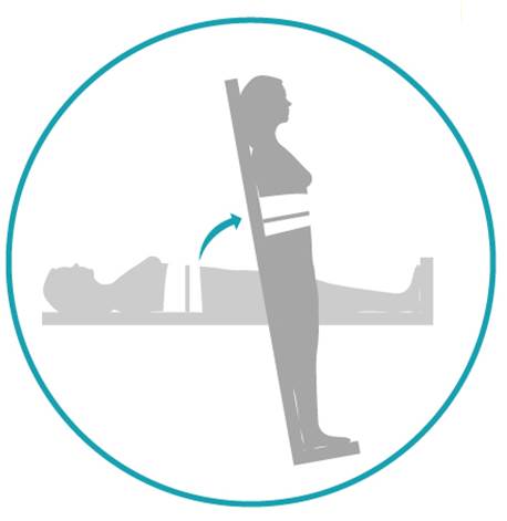
Many people with SCI experience a drop in blood pressure when moving from a lying or sitting position to an upright position. This is known as orthostatic hypotension. This condition happens because a loss of nerve function can impair the body’s ability to tighten (constrict) the blood vessels and change heart rate, which is an important part of maintaining blood pressure in different positions.

Because abdominal binders wrap around and compress the abdomen, they may help to increase pressure in the abdominal area. This may help to prevent blood pooling in the blood vessels in the abdomen when upright, which may help maintain blood pressure and allow better circulation.

There is conflicting evidence based on limited research that abdominal binders have any effect on cardiovascular responses in people with SCI. One study found that abdominal binders do not have any effect on average blood pressure or other cardiovascular responses. However, other studies suggest that abdominal binders in combination with leg stockings may have an effect on cardiovascular responses during lower intensity arm exercise.

Refer to our article on Orthostatic Hypotension for more information!

Speech

Cartoon sound waves coming out of a person's mouthThere is some weak evidence for the use of an abdominal binder to improve speech. One study found that participants with difficulty speaking due to cervical level injuries were able to produce more natural sounding speech, were able to speak louder, and improved overall voice quality with the use of an abdominal binder. Meanwhile, another study has shown that using an abdominal binder can extend the length that sound is produced for.

Balance, stability, and appearance

A profile of a man on a wheelchair with a protruded belly on the left. A profile of a man on a wheelchair with a flattened belly wearing an abdominal binder on the right.

Abdominal binders may be used to support the abdomen to reduce the appearance of “quad belly”.5

Some individuals use an abdominal binder as they perceive it helps to support their trunk or assist with sitting balance. In addition, some individuals find that it helps them balance when performing two-handed exercises. However, no research has been done to support this. Some individuals may use a binder to reduce the appearance of the abdomen sagging forward, sometimes called “quad belly”.

Abdominal binders are considered safe for most people. In all cases, the skin under the binder should be regularly checked for pressure sores. However, there are situations in which abdominal binders may not be appropriate and carry possible risks. Please consult a health provider for detailed safety information.

Abdominal binders should be used with caution in the following situations:

  • Do not use directly over areas prone to pressure injuries or over current injuries, wounds, or sores
  • Caution is advised if a person has a stoma (colostomy or urostomy bag) on the front of their abdomen. There is the possibility of the binder causing irritation or interference to the stoma site. A customized binder may be required to ensure no irritation to the area.

Risks of using abdominal binders may include:

  • Pressure injuries, if the equipment is too tight and creates too much pressure or cuts into the skin.
  • If the abdominal binder is tightened too much, it can get in the way of the abdomen and lower ribs expanding, which may make breathing difficult.
  • Also, an overly tight abdominal binder could cause an increase in spasticity or trigger autonomic dysreflexia in some people.

Refer to our articles on Pressure Injuries, Spasticity, and Autonomic Dysreflexia for more information!

Please consult a health provider for detailed instructions and safety information for your unique needs.

Abdominal binders are a physical treatment that supports the abdomen when the abdominal muscles are weak or paralyzed.

The support from the binder can improve cardiovascular and respiratory responses including blood pressure and breathing.

Some individuals may use a binder to help with balance and stabilizing the trunk but there is no research evidence to support this.

Some individuals may use a binder to reduce the appearance of abdominal muscles that bulge out following paralysis.

For a review of how we assess evidence at SCIRE Community and advice on making decisions, please see SCIRE Community Evidence.

This page has been adapted from SCIRE Professional “Respiratory Management” and “Orthostatic Hypotension” Modules:

Sheel AW, Reid WD, Townson AF (2018). Respiratory Management Following Spinal Cord Injury. In: Eng JJ, Teasell RW, Miller WC, Wolfe DL, Townson AF, Hsieh JTC, Connolly SJ, Noonan VK, Loh E, Sproule S, Querée M, McIntyre A, editors. Spinal Cord Injury Rehabilitation Evidence. Version 6.0. Vancouver: p. 1-72.
Available from: scireproject.com/evidence/respiratory-management/

Krassioukov A, Wecht JM, Teasell RW, Eng JJ (2014). Orthostatic Hypotension Following Spinal Cord Injury. In Eng JJ, Teasell RW, Miller WC, Wolfe DL, Townson AF, Hsieh JTC, Connolly SJ, Noonan VK, Loh E, McIntyre A, editors. Spinal Cord Injury Rehabilitation Evidence. Version 5.0. Vancouver: p 1- 26.
Available from: scireproject.com/evidence/orthostatic-hypotension/non-pharmacological-management-of-oh/ 

Evidence for “Are abdominal binders safe to use?”

Cornwell, P., Ward, E., Lim, Y., & Wadsworth, B. (2014). Impact of an Abdominal Binder on Speech Outcomes in People With Tetraplegic Spinal Cord Injury: Perceptual and Acoustic Measures. Topics in Spinal Cord Injury Rehabilitation, 20(1), 48–57. https://doi.org/10.1310/sci2001-48

Wadsworth, B. M., Haines, T. P., Cornwell, P. L., Rodwell, L. T., & Paratz, J. D. (2012). Abdominal Binder Improves Lung Volumes and Voice in People With Tetraplegic Spinal Cord Injury. YAPMR, 93(12), 2189–2197. https://doi.org/10.1016/j.apmr.2012.06.010

Image credits:

  1. Image by SCIRE
  2. Lungs ©karina, CC BY 3.0 US
  3. Blood Pressure ©Ricardo Moreira, CC BY 3.0 US
  4. speak ©Gregor Cresnar, CC BY 3.0 US
  5. Image by SCIRE


Disclaimer: This document does not provide medical advice. This information is provided for educational purposes only. Consult a qualified health professional for further information or specific medical advice. The SCIRE Project, its partners and collaborators disclaim any liability to any party for any loss or damage by errors or omissions in this publication.

Acupuncture

By | | No Comments

Author: SCIRE Community Team | Reviewer: Amrit Dhaliwal | Published: 5 July 2019 | Updated: ~

Acupuncture is a common complementary therapy used for various symptoms and conditions. This page outlines what acupuncture and dry needling are and their uses after spinal cord injury (SCI).

Key Points

  • Acupuncture is a treatment where small thin needles are inserted into specific points on the body to treat health conditions. Acupuncture is a complementary and alternative medicine treatment based on traditional Chinese medicine.
  • Acupuncture has been studied as a treatment for pain, bladder problems, and to aid functional recovery after SCI.
  • Scientists are not entirely sure how acupuncture might work. Its effects on pain, bladder function, and functional recovery after SCI are likely related to influences on the nervous system and/or circulation.
  • Overall, there is moderate evidence suggesting that acupuncture (including electroacupuncture) may be effective for treating neuropathic pain and bladder problems after SCI; and may aid functional recovery after SCI. The evidence for treating shoulder pain is unclear. Further studies are needed to confirm these findings.
Thin acupuncture needle inserted into cloth.

Acupuncture needles are thin needles that are inserted into acupuncture points on the body.1

Acupuncture is a complementary and alternative medicine (CAM) practice that has been used for thousands of years as a component of traditional Chinese medicine (TCM). Acupuncture involves the insertion of small thin needles into specific points on the body called acupuncture points or acupoints.

Acupuncture is used to treat many different symptoms and conditions. For people with SCI, acupuncture is used to treat pain, manage bladder problems, and possibly aid functional recovery.


Dry needling

Dry needling, also known as intramuscular stimulation (IMS), involves the use of similar thin needles that are inserted into trigger points. Trigger points are tight, irritable bands in the muscles and fascia that are a common cause of musculoskeletal pain. Dry needling typically elicits a small muscle twitch that may help to reduce muscle tension. Acupuncture and dry needling differ in both the theories that underlie their use and in how they are practiced.

Acupuncture is performed by health providers such as physiotherapists, physicians, chiropractors, and acupuncturists. In many regions, health providers need special training and a license to practice acupuncture.

Before the treatment

If you are considering trying an acupuncture treatment, it is important to discuss with your health providers to make sure that acupuncture is safe for you. Before starting a treatment, your health provider will perform an assessment and provide information about the treatment, its risks, and any other information you need to decide whether to proceed with an acupuncture treatment.

During the treatment

Person's lower leg with several acupuncture needles inserted into the skin

Acupuncture points are located at very specific points on the body.2

Acupuncture needles are thin, single-use, sterile needles that are solid and cannot be used to inject or withdraw fluids from the body. The needles are inserted into the surface of the skin at locations called acupuncture points. Acupuncture points are specific points on the body that are thought to influence the body systems. When the needles are inserted into the skin, they can cause minimal pain and/or bleeding.

Once the acupuncture needles are inserted, they may be left in for a specific amount of time determined by the therapist (usually 20 minutes or longer) before removal. Your response will be monitored during and after the treatment. While the needles are inserted, some practitioners choose to twist or shallowly plunge the needles into the skin or apply other stimulation in the form of heat or electricity to the needles. Acupuncture treatments are usually scheduled anywhere from a few days to a week apart.

Traditional Chinese medicine explanation

ancient illustration of man with acupuncture meridian labelled on his body

Ancient illustration of the acupuncture meridians based on Traditional Chinese Medicine.3

Traditional Chinese medicine (TCM) is based on the belief that illness happens because of imbalances in energy flow in the body. This energy flow is known as Qi (pronounced ‘chee’) and is thought to flow along lines of energy in the body called meridians. Traditional acupuncture points are located where these lines are believed to pass close to the surface of the skin. Thus, stimulating acupuncture points with needles is thought to promote balance of the body’s energy and treat health conditions.

Modern explanations

Traditional explanations for how acupuncture works do not align well with modern science. Scientists are not entirely sure how acupuncture might work, but its effects are likely related to influences on the nervous system and/or circulation.

Pain

Scientists have proposed several possible explanations for how acupuncture could work to reduce pain:

  • By blocking pain from traveling in the nerves
  • By causing the body to release substances that prevent pain (such as endorphins)
  • By altering blood circulation in important areas of the body
Bladder problems

Acupuncture may affect bladder function by influencing nerve signals or control centers for urination in the brain and spinal cord.

Functional recovery

Acupuncture has been proposed as a treatment to improve recovery of function after SCI. This is not well understood, but some scientists have proposed that it may be related to reducing damage caused by the after-effects of the injury.

There are certain situations in which acupuncture may not be safe to use. This is not a complete list; please consult a health provider for detailed safety information before using this treatment.

Acupuncture should be used with caution in the following situations:

Hand inserting a thin acupuncture needle into another person's arm.

It is important to consider whether acupuncture will be safe for you.4

  • By certain groups of people, such as children, pregnant women, and people with medical conditions (such as heart conditions, osteoporosis, or weakened immune systems)
  • Near major organs (such as certain places on the torso or neck)
  • By people who are prone to fainting or have a fear of needles
  • By people who are prone to autonomic dysreflexia
  • By people who are at risk of bleeding (including people taking anticoagulants)
  • By people who are unable to follow instructions or provide accurate feedback

Acupuncture should not be used in the following situations:

  • By people with metal allergies
  • In areas with open, infected, inflamed skin or recent surgery
  • Near tumors

Even for people who are not restricted from using acupuncture (see above), there may be risks and side effects with the use of this treatment. The common side effects of acupuncture are usually mild and serious complications are rare. However, it is important to discuss these possibilities in detail with your health provider before using this treatment.

Common risks and side effects of acupuncture may include:

  • Bruising, bleeding, and skin irritation
  • Nausea, vomiting, and diarrhea
  • Headaches
  • Sweating
  • Dizziness and fainting
  • Worsening of symptoms (like increased pain or muscles spasms)

For people with SCI (especially those with injuries above the level of T6), acupuncture needles may be a cause of irritation to the body if they are placed below the level of injury. This could increase the risk of autonomic dysreflexia in some people.

Rare complications of acupuncture may include:

  • Puncture of the lung (pneumothorax) or other internal organs
  • Nerve injury
  • Infection or spread of infectious diseases (such as Hepatitis B)
  • Needles breaking after they are inserted and becoming embedded in the skin
  • Convulsions

Many of the rare complications of acupuncture can result from improper acupuncture technique. Technique is a very important part of ensuring safety, and there can be major risks if acupuncture is performed incorrectly. For example, improper needle placement and not using properly sterilized needles or sterile technique can put a person at risk of complications. Because of these risks, it is important that acupuncture is only performed by a trained health provider.

Acupuncture for pain after SCI

White computer generated figure with a red spot representing pain over their back.

Acupuncture is a common alternative treatment for pain, although research is currently limited.5

Research has studied acupuncture for the treatment of several different types of pain after SCI, including neuropathic pain and shoulder pain.

Shoulder pain

The evidence is unclear about whether acupuncture helps to reduce shoulder pain after SCI. Two studies have compared acupuncture to other treatments, including a sham treatment and a movement therapy called Trager therapy. Although both of these studies found that acupuncture helped with shoulder pain after SCI, it was not more effective than the comparison treatments. Further research is needed to determine effectiveness.

Neuropathic pain

Moderate evidence from three studies suggests that acupuncture may reduce neuropathic pain after SCI. However, two of these studies were low quality so further research is needed to confirm this.

Acupuncture for bladder problems after SCI

Three studies have studied acupuncture as a treatment for bladder problems after SCI. These studies provide moderate evidence that electroacupuncture used together with conventional therapies may help people with SCI to develop effective bladder management earlier after injury.

Another small study provides weak evidence that regular needle acupuncture may help with bladder incontinence caused by hyperreflexic bladder.

Acupuncture for improving functional recovery after SCI

One study has investigated acupuncture for improving functional recovery after SCI. It provides moderate evidence that acupuncture helps to improve functional recovery early after SCI. However, other researchers have debated the quality of the study and whether its conclusions were accurate. More studies are needed to confirm whether acupuncture has any effects on the recovery of function after SCI.

Overall, there is moderate evidence suggesting that acupuncture (including electroacupuncture) may be effective for treating neuropathic pain, bladder problems, and possibly for improving functional recovery after SCI. The evidence for shoulder pain is unclear. Further studies are needed to confirm these findings.

There have not been any studies on whether dry needling is effective for treating people with SCI.

Acupuncture needs to be used with caution in certain situations, but overall is a safe treatment when performed by a trained practitioner. Until more research is done, it is best to discuss this treatment with your health provider to find out more about if it is a suitable treatment option for you.

For a review of how we assess evidence at SCIRE Community and advice on making decisions, please see SCIRE Community Evidence.

Parts of this page has been adapted from SCIRE Project (Professional) “Pain Management”, “Bladder Management”, and “Upper Limb” Chapters:

Mehta S, Teasell RW, Loh E, Short C, Wolfe DL, Hsieh JTC (2014). Pain Following Spinal Cord Injury. In Eng JJ, Teasell RW, Miller WC, Wolfe DL, Townson AF, Hsieh JTC, Connolly SJ, Noonan VK, Loh E, McIntyre A, editors. Spinal Cord Injury Rehabilitation Evidence. Version 5.0: p 1-79.

Available from: https://scireproject.com/evidence/rehabilitation-evidence/pain-management/

Hsieh J, McIntyre A, Iruthayarajah J, Loh E, Ethans K, Mehta S, Wolfe D, Teasell R. (2014). Bladder Management Following Spinal Cord Injury. In Eng JJ, Teasell RW, Miller WC, Wolfe DL, Townson AF, Hsieh JTC, Connolly SJ, Noonan VK, Loh E, McIntyre A, editors. Spinal Cord Injury Rehabilitation Evidence. Version 5.0: p 1-196.

Available from: https://scireproject.com/evidence/rehabilitation-evidence/bladder-management/

Connolly SJ, McIntyre A, Mehta, S, Foulon BL, Teasell RW. (2014). Upper Limb Rehabilitation Following Spinal Cord Injury. In Eng JJ, Teasell RW, Miller WC, Wolfe DL, Townson AF, Hsieh JTC, Connolly SJ, Noonan VK, Loh E, McIntyre A, editors. Spinal Cord Injury Rehabilitation Evidence. Version 5.0: p 1-77.

Available from: https://scireproject.com/evidence/rehabilitation-evidence/upper-limb/

Evidence for “Acupuncture for pain after SCI” is based on:

Shoulder pain

[1] Dyson-Hudson TA, Shiflett SC, Kirshblum SC, Bowen JE, Druin EL. Acupuncture and trager psychophysical integration in the treatment of wheelchair user’s shoulder pain in individuals with spinal cord injury. Arch Phys Med Rehab 2001;82:1038-46.

[2] Dyson-Hudson TA, Kadar P, LaFountaine M, Emmons R, Kirshblum SC, Tulsky D et al. Acupuncture for chronic shoulder pain in persons with spinal cord injury: a small-scale clinical trial. Arch Phys Med Rehab 2007;88:1276-83.

Neuropathic pain

[1] Norrbrink C, Lundeberg T. Acupuncture and massage therapy for neuropathic pain following spinal cord injury: An exploratory study. Acupunc Med 2011;29:108-15.

[2] Rapson LM, Wells N, Pepper J, Majid N, Boon H. Acupuncture as a promising treatment for below-level central neuropathic pain: A retrospective study. J Spinal Cord Med 2003;26:21-6.

[3] Nayak S, Shiflett SC, Schoenberger NE, Agostinelli S, Kirshblum S, Averill A et al. Is acupuncture effective in treating chronic pain after spinal cord injury? Arch Phys Med Rehab 2001;82:1578-86.

References for Acupuncture for bladder problems after SCI:

[1] Cheng P-T, Wong M-K, Chang P-L. A therapeutic trial of acupuncture in neurogenic bladder of spinal cord injured patients-A preliminary report. Spinal Cord 1998;36(7):476-480.

[2] Honjo H, Naya Y, Ukimura O, Kojima M, Miki T. Acupuncture on clinical symptoms and urodynamic measurements in spinal-cord-injured patients with detrusor hyperreflexia. Urol Int. 2000;65(4):190-5.

[3] Liu Z, Wang W, Wu J, Zhou K, Liu B. Electroacupuncture improves bladder and bowel function in patients with traumatic spinal cord injury: results from a prospective observational study. Evid Based Complement Alternat Med. 2013;2013:543174

[4] Gu XD, Wang J, Yu P, Li JH, Yao YH, Fu JM, Wang ZL, Zeng M, Li L, Shi M, Pan WP. Effects of electroacupuncture combined with clean intermittent catheterization on urinary retention after spinal cord injury: a single blind randomized controlled clinical trial. Int J Clin Exp Med. 2015 Oct 15;8(10):19757-63.

References for Acupuncture for functional recovery after SCI:

[1] Wong AM, Leong CP, Su TY, Yu SW, Tsai WC, Chen CP. Clinical trial of acupuncture for patients with spinal cord injuries. Am J Phys Med Rehabil. 2003 Jan;82(1):21-7.

Other references:

Ma R, Liu X, Clark J, Williams GM, Doi SA. The Impact of Acupuncture on Neurological Recovery in Spinal Cord Injury: A Systematic Review and Meta-Analysis. J Neurotrauma. 2015 Dec 15;32(24):1943-57.

Dorsher PT, McIntosh PM. Acupuncture’s Effects in Treating the Sequelae of Acute and Chronic Spinal Cord Injuries: A Review of Allopathic and Traditional Chinese Medicine Literature. Evid Based Complement Alternat Med. 2011;2011:428108.

Wang J, Zhai Y, Wu J, Zhao S, Zhou J, Liu Z. Acupuncture for Chronic Urinary Retention due to Spinal Cord Injury: A Systematic Review. Evid Based Complement Alternat Med. 2016;2016:9245186.

Shin BC, Lee MS, Kong JC, Jang I, Park JJ. Acupuncture for spinal cord injury survivors in Chinese literature: a systematic review. Complement Ther Med. 2009 Oct-Dec;17(5-6):316-27.

NIH consensus conference. Acupunc JAMA 1998;280:1518-24.

Pomeran ZB. Scientific basis of acupuncture. In: Stux G, Pomeran (Eds.). Basis of acupuncture (pp. 6-72). 4 Rev Ed. Springh-Verlag. 1998.

Wong JY, Rapson LM. Acupuncture in the management of pain of musculoskeletal and neurologic origin. Phys Med Rehab Clin North Am 1999;10:531-45.

Zhang T, Liu H, Liu Z, Wang L. Acupuncture for neurogenic bladder due to spinal cord injury: a systematic review protocol. BMJ Open. 2014 Sep 10;4(9):e006249.

Lee MHM, Liao SJ. Acupuncture in physiatry, in Kottke FJ, Lehmann JF (eds). Krusens Handbook of Physical Medicine and Rehabilitation, ed. 4. Philadelphia: Saunders 1990:402-32.

Chung A, Bui L, Mills, E. Adverse effects of acupuncture. Which are clinically significant? Canadian Family Physician. 2003;49:985–989.

White A. A cumulative review of the range and incidence of significant adverse events associated with acupuncture. Acupunct Med. 2004 Sep;22(3):122-33.

Ansari NN, Naghdi S, Fakhari Z, Radinmehr H, Hasson S. Dry needling for the treatment of poststroke muscle spasticity: a prospective case report. NeuroRehabilitation. 2015;36(1):61-5.

Salom-Moreno J, Sánchez-Mila Z, Ortega-Santiago R, Palacios-Ceña M, Truyol-Domínguez S, Fernández-de-las-Peñas C. Changes in spasticity, widespread pressure pain sensitivity, and baropodometry after the application of dry needling in patients who have had a stroke: a randomized controlled trial. J Manipulative Physiol Ther. 2014 Oct;37(8):569-79.

Dunning J, Butts R, Mourad F, Young I, Flannagan S, Perreault T. Dry needling: a literature review with implications for clinical practice guidelines. Phys Ther Rev. 2014 Aug;19(4):252-265.

Averill A, Cotter AC, Nayak S, Matheis RJ, Shiflett SC. Blood pressure response to acupuncture in a population at risk for autonomic dysreflexia. Arch Phys Med Rehabil. 2000 Nov;81(11):1494-7.

Gattie E, Cleland JA, Snodgrass S. The Effectiveness of Trigger Point Dry Needling for Musculoskeletal Conditions by Physical Therapists: A Systematic Review and Meta-analysis. J Orthop Sports Phys Ther. 2017 Mar;47(3):133-149.


Image credits:

  1. ‘Acupuncture’, ©Magali M , CC BY-NC-ND 2.0
  2. By thepismire, ‘her handiwork’, CC BY-NC-ND 2.0
  3. Acupuncture meridian illustration: This image is in the public domain in its country of origin and other countries, and is identified as being free of known restrictions under copyright law, including all related and neighboring rights.
  4. Acupuncture on an arm: Released into the public domain (by the author). There is no copyright associated with this file, and the website has released all ownership to the public domain.
  5. Stock image of back pain, ©3dman_eu, CC0.


Disclaimer: This document does not provide medical advice. This information is provided for educational purposes only. Consult a qualified health professional for further information or specific medical advice. The SCIRE Project, its partners and collaborators disclaim any liability to any party for any loss or damage by errors or omissions in this publication.

Cannabis (Marijuana) and Cannabinoids

By | | No Comments

Authors: SCIRE Community Team | Reviewer: Andrea Townson and Janice Eng | Published: 2 April 2019 | Updated: Apr 20, 2020

Cannabis (marijuana) is an alternative treatment option for pain and spasticity after spinal cord injury (SCI). This page outlines basic information about cannabis and its use after SCI.

Key Points

  • “Cannabis” refers to products derived from the cannabis plant, such as marijuana. The natural cannabinoids or compounds found in cannabis can also be made synthetically.
  • Cannabis may be inhaled as a smoke or vapour or taken by mouth as a capsule or spray.
  • Smoking cannabis is not recommended due to the risks associated with inhaling smoke.
  • The safety of cannabis products for use after SCI is not known. Please consult your health providers for detailed safety information.
  • Research on cannabis use after SCI is in its early stages. Studies done so far show that cannabis products may have beneficial effects on pain and are unclear about its effects on spasticity. More research is needed to establish if cannabis is a safe and effective treatment after SCI.
A photograph of leaves of a cannabis plant

Leaves of a cannabis plant.1

Cannabis is a term that refers to the products of cannabis (hemp) plants, a group of plants from central Asia that are now cultivated around the world. Cannabis sativa, Cannabis indica, and Cannabis ruderalis are three well-known types of cannabis, but many strains or varieties exist, both pure and hybrid types. Common preparations of cannabis include marijuana, which is the dried leaves and flowering tops of the plant, and hashish, which is its condensed resin. Cannabis has been used for thousands of years as a medicine and recreational drug.

Currently, cannabis is a controlled substance in most regions because of its psychoactive effects. However, exceptions are made in some places for approved medical or spiritual uses. In addition to medical use, in Canada recreational use of cannabis has also been made legal as of October 2018. Here, the sale of recreational cannabis was originally limited to dried cannabis and oils, but as of October 2019 edibles and concentrates are also legal for sale.

Cannabis has been studied as a treatment for conditions as diverse as nausea associated with cancer chemotherapy, loss of appetite in people with HIV, and spasticity associated with multiple sclerosis.

Cannabis has its unique properties because of naturally-occurring chemical compounds within the plant called cannabinoids. Cannabinoids act on receptors on the surface of cells called cannabinoid receptors, causing effects on body processes like pain, memory, appetite, and immune responses.

Diagram categorizing cannabinoids into endocannabinoids (produced in the body), phytocannabinoids (produced by the cannabis plant), and synthetic cannabinoids (synthesized in a lab)

There are various sources of cannabinoids, both natural and synthetic.2

Cannabinoids occur naturally within the body (endocannabinoids), in cannabis plants (phytocannabinoids), and can also be synthesized in a lab (synthetic cannabinoids). There are more than 60 cannabinoids present in cannabis, with the most well-known being Delta-9-tetrahydrocannabinol (commonly known as THC), which is responsible for many of the psychoactive effects for which cannabis is known such as creating a “high” or sense of euphoria. Other cannabinoids, like Cannabidiol (also known as CBD), are not psychoactive and may have different effects such as improving mental health concerns and preventing oxidative damage although evidence for this is currently not conclusive. Because of these benefits over THC as well as the reduced health risks, CBD is believed to be the component of cannabis that gives rise to its medicinal potential and opposes the negative psychiatric effects associated with THC.

The chemical structures of THC and CBD

The chemical structures of THC and CBD.3

Cannabis/Cannabinoids, whether plant-derived or human-made, may be used for medicinal or recreational purposes in a variety of ways.

Medical cannabinoid products

Medical cannabis

The laws and regulations required to get approval for medical marijuana differ by country and region. In Canada, use of medical cannabis requires authorization for use from a physician.

Prescription synthetic cannabinoids

In some countries, certain synthetic cannabinoids are available for therapeutic use and require a prescription from a physician. Like other medications, these products are registered with a Drug Identification Number (DIN) in Canada or with the Food and Drug Administration (FDA) in the United States. Prescription synthetic cannabinoids are carefully regulated and monitored for their composition and effects on the body and are developed to minimize accompanying intoxication.

Recreational cannabis products

An indoor grow op with rows of cannabis plants in pots.

There are various environmental and health risks associated with unlicensed grow-ops.4

Recreational use of cannabis is legal in Canada, but still subject to provincial or territorial restrictions. Recreational use outside these restrictions is illegal. Like medical cannabis, the production and distribution of recreational cannabis is regulated to ensure safety and quality. There are various concerns with the use of cannabis that is not regulated or produced legally. These cannabis products may include harmful contaminants (e.g., mold, bacteria, and pesticides) or have much greater variation in their chemical composition than cannabis products intended for medical use. It can be difficult to know exactly what dose you are receiving and the risks and side effects for using these products may be unknown. Another issue with cannabis sourced from illegal grow-ops include its negative impact on the environment as these sites may misuse toxic pesticides and may divert water supply away from lakes or rivers, threatening plant, wildlife, and human health. Unregulated cannabis products are not recommended for treating symptoms of SCI.

Illegal synthetic cannabinoids

A hand holding a jar of synthetic cannabinoids mixed with shredded cannabis plant material.

Illegal synthetic cannabinoids may be sold to look like cannabis.5

Even though synthetic cannabinoids act on the same receptors as the phytocannabinoids found in the cannabis plant, they may produce different effects on the body. Some non-prescription synthetic cannabinoids are made to imitate the psychoactive effects of THC, making them potentially dangerous especially since their actions on the body can be unpredictable. Known by names like “Spice” and “K2,” these compounds are often combined with plant-based products and sold as “alternatives” to marijuana. However, all activities associated with non-prescription synthetic cannabinoids (e.g., production, distribution, use) are illegal in Canada. Besides the fact that illegal synthetic cannabinoids have not been tested in humans, their product composition can vary greatly and may be laced with other unknown and potentially deadly substances. Synthetic cannabinoids also more potent than plant-derived THC. This means that they bind more strongly to the cannabinoid receptors, increasing the risk of overdose.

Cartoon image showing different dosage forms of cannabis (vape pen, capsules, cream, and oil).

Photograph of a female smoking a joint.

Smoking is not a recommended method of using cannabis.10

Cannabis products are usually inhaled or taken by mouth. Smoking is the most common method among the general population as well as within the SCI population. However, there are serious concerns about the negative health effects to the user and those nearby associated with inhaling and exhaling smoke, which contains many of the same harmful compounds as tobacco smoke. People with SCI, in particular, should avoid smoking cannabis as respiratory issues including compromised breathing and pneumonia are already prevalent in the SCI population. Vaporization is another method where the cannabis leaves are heated to form a vapour that is then inhaled. While vaping prevents the cannabinoids from burning which decreases the amount of toxic by-products produced compared to smoking, it is not without risks and has recently been associated with vaping-associated pulmonary injury (VAPI). After a sharp increase in VAPI cases in August and September of 2019, emergency department visits continue to decline. This is thought to be due to the removal of vitamin E acetate from most products, increased public awareness of the risks associated with THC containing e-cigarettes or vaping devices, and law enforcement actions related to illicit products in the US. Canadian extracts for vaping that contain THC are not allowed to have any added vitamins, minerals, nicotine, sugars, flavouring or colouring agents.

Cannabis can also be taken by mouth in the form of food items or other products like oils, capsules, and mouth sprays. Other less common methods cannabis may be delivered include through the skin (e.g. creams, lotions, balms, patches, etc.), through the rectum, or into the veins


A bottle of CBD oil with a dropper above.

Cannabis can be prepared by extracting the cannabinoids from the plant and dissolving it in oil.11

Cannabidiol oil

CBD oil is becoming more popular among people who wish to gain the health benefits of cannabis and avoid the psychoactive effects of THC. Although many people use CBD oil for a range of ailments, there is limited safety and efficacy data (and no research in SCI) to support its use for these conditions. Recently, positive results from three clinical trials with strong evidence have led the Food and Drug Administration (FDA) in the United States to approve the use of CBD oil for two rare forms of epilepsy in June 2018.

Prescription synthetic cannabinoids

Prescription synthetic cannabinoids often use isolated cannabinoid compounds or combinations of cannabinoids. This includes products such as:

  • Nabilone (Cesamet), a synthetic cannabinoid similar to THC that is taken by mouth as a capsule.
  • Dronabinol (Marinol), synthetic THC that is taken by mouth as a capsule. Please note that dronabinol is no longer available in Canada.
  • Nabiximols (Sativex), a mix of cannabis plant-derived THC and CBD that is taken as a mouth spray.
A jar of dried marijuana including the flowers and leaves.

Marijuana is the dried flowers and leaves of cannabis.12

There are currently no standard cannabis dosing regimens for SCI-related conditions. Dosing for medical cannabis varies based on factors such as method of delivery, past cannabis use, and the medical condition being treated. Additionally, the amount of THC and CBD in marijuana is not always the same. Thus, the effects of different marijuana products are not always the same. Levels of THC and CBD in a product can change based on the strain of the plant used as well as how the plant was grown and prepared.

Especially for those who have never used cannabis in the past, it is recommended that they start on low doses before slowly increasing the dose until their therapeutic goals are met. To minimize negative side effects related to THC and maximize symptom control, a strain with low THC and high CBD may be used initially. Immediately discontinue use if any intolerable side effects occur.

People who use cannabis for medicinal purposes consume an average of 1-3 g/day or 10-20 g/week. Even with equal grams of the same cannabis strain, the amount of cannabis the body actually absorbs differs depending on the method of delivery. For example, people who wish to switch from inhaling cannabis to taking cannabis by mouth may need to increase in their daily cannabis use by 2.5 times to get an equivalent dose. Each different form and method of cannabis use will change how quickly the drug produces an effect and how long it lasts in the body. For example, inhalation of cannabis will generally lead to a faster onset of action and longer-lasting effect than oral ingestion.

Inhalation Oral ingestion
Onset of action Few minutes 30 minutes (up to 3-4 hours)
Peak of effect 30 minutes 3-4 hours
Duration of effect 2-4 hours (up to 24 hours) 8 hours (up to 12-24 hours)

It is important that you closely follow the directions of your health providers and consult with them before making any changes to your cannabis use. Speak to your health provider for more detailed information.

The safety of medical cannabis use after SCI is not yet known. However, a number of risks and side effects of cannabis use in the general population are known. Many of the short-term side effects of cannabis have been reported to be mild to moderately severe and related to the dose of the drug taken. Uncommon but serious adverse effects may also exist. Furthermore, the risks to long-term users are not well known and some side effects may be related to regular use over time.

This is not a complete list. Speak to your health provider for detailed information about the risks and side effects of cannabis use.

Short-term side effects of cannabis may include:

Diagram of the human body showing the different side effects cannabis can have on the body.

Cannabis can cause many side effects to different body systems.13

  • Dizziness and lightheadedness
  • Dry mouth, throat irritation, and cough
  • Drowsiness
  • Altered judgment and attention
  • Anxiety and agitation
  • Hallucinations
  • Disorientation and confusion
  • Increased heart rate
  • Impaired coordination and balance
  • Impaired short-term memory
  • Headache
  • Paranoia and psychosis
  • Reddening of the eyes
  • Decreased intra-ocular pressure (pressure within the eyes)
  • Muscle relaxation
  • Interactions with other medications

Because cannabis lingers in the body long after use, task performance may be impaired for up to 24 hours. It is recommended to avoid operating heavy machinery or performing dangerous activities for 3-4 hours after inhaling cannabis, 6 hours after oral ingestion of cannabis, and 8 hours if a “high” is experienced. Examples of high-risk activities may include performing transfers and participating in physical therapy sessions.

Long-term cannabis use may be associated with:

  • Addiction and withdrawal
  • Airway problems like chronic bronchitis
  • Possible increased risk of mental disorders like anxiety, depression, schizophrenia, and psychosis in people at risk for these conditions
  • Possible increased cancer risk with long term smoking, although this is not yet clear

An emerging concern is the effects that cannabis use may have on adolescents and young adults. Studies have suggested that cannabis use early in adolescence may alter brain development and could be related to the development of psychotic disorders as adults.

Overdosage of cannabinoids

A cartoon cannabis leaf with an up arrow on the top left and a warning sign on the bottom right.Overdoses of cannabis, although not common, have been reported. The risk increases when both oral and inhaled forms of cannabinoids (prescription or recreational) are combined. The signs and symptoms of overdose are generally tolerable and overlap with the effects of THC such as dizziness, drowsiness, and sensory impairment. More severe complications including psychosis and convulsions occur rarely.

Unlike cannabis, synthetic cannabinoids carry a greater risk of overdose because they are more potent than THC. The clinical presentation of toxicity will depend on the specific synthetic cannabinoid used, but can be severe and even result in death. Since its introduction into the United States in 2008, there have been cases of adverse reactions in all 50 states. There is currently no antidote to synthetic cannabinoids, making the illegal use of these drugs an emerging public health threat. If you or someone you know experiences an overdose, seek medical attention immediately.

A cartoon cannabis leaf with a thumbs up and thumbs down above.

Studies show that cannabis is mostly used by patients with SCI for (chronic) pain and spasm relief, as well as for anxiety, stress and depression, bowel and bladder management, nausea, to increase appetite, to improve sleep, to decrease other medication use and for pleasure, recreation and relaxation. However, research has only studied the use of cannabinoid products in the treatment of pain and spasticity after SCI.

Pain

Early research provides moderate evidence that smoked and vapourized cannabis may help to reduce neuropathic pain. There is also weak evidence that oral plant-derived cannabinoid sprays may help to reduce neuropathic pain. Moderate evidence from two other studies indicates no benefit with synthetic cannabinoids. In one, dronabinol was no different than diphenhydramine (an anti-allergy medication with no pain-relieving properties) for reducing neuropathic pain. In the other, a synthetic cannabinoid called Normast showed no benefit. These last two studies were specific to people with SCI, while the other studies above also included people with other neurological conditions. Further research specific to people with SCI is needed to determine if cannabis and synthetic cannabinoids are safe and effective for pain after SCI.


Hear Matt describe his experience with synthetic and non-synthetic marijuana for pain management.

Spasticity

Research on cannabinoid products for spasticity after SCI has been conflicting. Four studies provide moderate evidence that synthetic cannabinoids and vapourized cannabis may help with spasticity after SCI. However, two other studies with moderate evidence have been inconclusive about whether cannabinoid products helped.

Overall, these studies show that cannabinoid-based treatments may have benefits in the treatment of spasticity, but further research through larger and more rigorous studies are needed before conclusions can be drawn about how effective they are.

There is early evidence that cannabinoid products may help to treat neuropathic pain after SCI and conflicting evidence about whether they help to treat spasticity after SCI. More studies are needed to confirm these findings.

It is not known whether cannabis is safe to use after SCI, especially over the long term, since cannabis use is associated with a number of potential risks and side effects. Until more research is done, it is important that you discuss this treatment option with your health providers in detail to find out if it is a suitable and safe treatment option for you.

For a review of how we assess evidence at SCIRE Community and advice on making decisions, please see SCIRE Community Evidence.

Parts of this page has been adapted from SCIRE Project (Professional) “Pain Management” and “Spasticity” Chapters:

Mehta S, Teasell RW, Loh E, Short C, Wolfe DL, Hsieh JTC (2014). Pain Following Spinal Cord Injury. In Eng JJ, Teasell RW, Miller WC, Wolfe DL, Townson AF, Hsieh JTC, Connolly SJ, Noonan VK, Loh E, McIntyre A, editors. Spinal Cord Injury Rehabilitation Evidence. Version 5.0: p 1-79.

Available from: https://scireproject.com/evidence/rehabilitation-evidence/pain-management/

Hsieh JTC, Wolfe DL, Townson AF, Short C, Connolly SJ, Mehta S, Curt A, Foulon BL (2012). Spasticity Following Spinal Cord Injury. In Eng JJ, Teasell RW, Miller WC, Wolfe DL, Townson AF, Hsieh JTC, Connolly SJ, Noonan V, Mehta S, Sakakibara BM, Boily K, editors. Spinal Cord Injury Rehabilitation Evidence. Version 4.0.

Available from: https://scireproject.com/evidence/rehabilitation-evidence/spasticity/


Evidence for “What is cannabis” is based on:

Atakan, Z. (2012). Cannabis, a complex plant: different compounds and different effects on individuals. Therapeutic Advances in Psychopharmacology, 2(6), 241–254. https://doi.org/10.1177/2045125312457586

Baker, D., Pryce, G., Croxford, J. L., Brown, P., Pertwee, R. G., Huffman, J. W., & Layward, L. (2000). Cannabinoids control spasticity and tremor in a multiple sclerosis model. Nature, 404(6773), 84–87. https://doi.org/10.1038/35003583

Ben Amar, M. (2006). Cannabinoids in medicine: A review of their therapeutic potential. Journal of Ethnopharmacology, 105(1–2), 1–25. https://doi.org/10.1016/j.jep.2006.02.001

Birdsall, S. M., Birdsall, T. C., & Tims, L. A. (2016). The Use of Medical Marijuana in Cancer. Current Oncology Reports, 18(7), 40. https://doi.org/10.1007/s11912-016-0530-0

Evidence for “What are cannabinoids?” is based on:

Aizpurua-Olaizola, O., Elezgarai, I., Rico-Barrio, I., Zarandona, I., Etxebarria, N., & Usobiaga, A. (2017). Targeting the endocannabinoid system: future therapeutic strategies. Drug Discovery Today, 22(1), 105–110. https://doi.org/10.1016/j.drudis.2016.08.005

Zerrin 2012

Crippa, J. A., Guimarães, F. S., Campos, A. C., & Zuardi, A. W. (2018). Translational Investigation of the Therapeutic Potential of Cannabidiol (CBD): Toward a New Age. Frontiers in Immunology, 9, 2009. https://doi.org/10.3389/fimmu.2018.02009

National Academies of Sciences, Engineering, and Medicine. 2017. The health effects of cannabis and cannabinoids: The current state of evidence and recommendations for research. Washington, DC: The National Academies Press. doi: 10.17226/24625.

Whiting et al. (2015) Cannabinoids for Medical Use. A Systematic Review and Meta-Analysis. JAMA 313(24): 2456-2473.

Mücke M, Phillips T, Radbruch L, Petzke F, Häuser W.(2018) Cannabis-based Medicine for chronic neurophathic pain in adults. Cochrane Database of Systematic Reviews, Issue 3. Art. No: CD012182 DOI: 10.1002/14651858.CD012182.pub2

Evidence for “How are cannabinoids used?” is based on:

Drossel, C., Forchheimer, M., & Meade, M. A. (2016). Characteristics of Individuals with Spinal Cord Injury Who Use Cannabis for Therapeutic Purposes. Topics in Spinal Cord Injury Rehabilitation, 22(1), 3–12. https://doi.org/10.1310/sci2201-3

Sheel, A. W., Welch, J. F., & Townson, A. (n.d.). Respiratory Management Following Spinal Cord Injury. Retrieved from www.scireproject.com

Health Canada (2018) Information for health care professionals. Cannabis (marihuana, marijuana) and the cannabinoids. Ottawa; Health Canada publications.

Center for Disease Control (2020) Outbreak of Lung Injury Associated with the Use of E-Cigarette, or Vaping, Products. Retrieved on 13-02-2020 from: https://web.archive.org/web/20200213002533/https://www.cdc.gov/tobacco/basic_information/e-cigarettes/severe-lung-disease.html

Evidence for “Cannabidiol oil” is based on:

Devinsky, O., Cross, J. H., Laux, L., Marsh, E., Miller, I., Nabbout, R., … Wright, S. (2017). Trial of Cannabidiol for Drug-Resistant Seizures in the Dravet Syndrome. New England Journal of Medicine, 376(21), 2011–2020. https://doi.org/10.1056/NEJMoa1611618

Devinsky, O., Patel, A. D., Cross, J. H., Villanueva, V., Wirrell, E. C., Privitera, M., … Zuberi, S. M. (2018). Effect of Cannabidiol on Drop Seizures in the Lennox–Gastaut Syndrome. New England Journal of Medicine, 378(20), 1888–1897. https://doi.org/10.1056/NEJMoa1714631

Thiele, E. A., Marsh, E. D., French, J. A., Mazurkiewicz-Beldzinska, M., Benbadis, S. R., Joshi, C., … Wilfong, A. (2018). Cannabidiol in patients with seizures associated with Lennox-Gastaut syndrome (GWPCARE4): a randomised, double-blind, placebo-controlled phase 3 trial. The Lancet, 391(10125), 1085–1096. https://doi.org/10.1016/S0140-6736(18)30136-3

Shannon, S., & Opila-Lehman, J. (2016). Effectiveness of Cannabidiol Oil for Pediatric Anxiety and Insomnia as Part of Posttraumatic Stress Disorder: A Case Report. The Permanente Journal, 20(4), 16-005. https://doi.org/10.7812/TPP/16-005

Evidence for “What is the suggested dosing of cannabis?” is based on:

Health Canada. (2013). Information for Health Care Professionals Cannabis (marihuana, marijuana) and the cannabinoids. Retrieved from https://www.canada.ca/content/dam/hc-sc/migration/hc-sc/dhp-mps/alt_formats/pdf/marihuana/med/infoprof-eng.pdf

Evidence for “What are the risks and side effets of cannabis? Is based on:

Grant, I., Atkinson, J. H., Gouaux, B., & Wilsey, B. (2012). Medical marijuana: clearing away the smoke. The Open Neurology Journal, 6, 18–25. https://doi.org/10.2174/1874205X01206010018

Volkow, N. D., Baler, R. D., Compton, W. M., & Weiss, S. R. B. (2014). Adverse health effects of marijuana use. The New England Journal of Medicine, 370(23), 2219–2227. https://doi.org/10.1056/NEJMra1402309

Zhang, M. W., & Ho, R. C. M. (2015). The Cannabis Dilemma: A Review of Its Associated Risks and Clinical Efficacy. Journal of Addiction, 2015, 1–6. https://doi.org/10.1155/2015/707596

Health Canada. (2013). Information for Health Care Professionals Cannabis (marihuana, marijuana) and the cannabinoids. Retrieved from https://www.canada.ca/content/dam/hc-sc/migration/hc-sc/dhp-mps/alt_formats/pdf/marihuana/med/infoprof-eng.pdf

Evidence for “What are cannabinoids used for after spinal cord injury?” is based on:

Cardenas DD, Jensen MP. (2006) Treatments for chronic pain in persons with spinal cord injury: A survey study. The journal of spinal cord medicine 29:109-117.

Shroff FM. (2015) Experiences with Holistic Health Practices among Adults with Spinal Cord Injury. Rehabilitation Process and Outcome 4:27-34.

Drossel C, Forchheimer M, Meade MA. (2016) Characteristics of Individuals with Spinal Cord Injury Who Use Cannabis for Therapeutic Purposes. Top Spinal Cord Inj Rehabil;22:3-12.

Government of Canada (2019) Final regulations: Edible cannabis, cannabis extracts, cannabis topicals. Retrieved on 13-02-2020 from: https://www.canada.ca/en/health-canada/services/drugs-medication/cannabis/resources/regulations-edible-cannabis-extracts-topicals.html

Andresen SR, Biering-Sorensen F, Hagen EM, Nielsen JF, Bach FW, Finnerup NB. (2017) Cannabis use in persons with traumatic spinal cord injury in Denmark. J Rehabil Med 49:152-160.

Bruce D, Brady JP, Foster E, Shattell M. (2018) Preferences for Medical Marijuana over Prescription Medications Among Persons Living with Chronic Conditions: Alternative, Complementary, and Tapering Uses. Journal of alternative and complementary medicine (New York, NY) 24:146-153.

Hawley LA, Ketchum JM, Morey C, Collins K, Charlifue S. (2018) Cannabis Use in Individuals With Spinal Cord Injury or Moderate to Severe Traumatic Brain Injury in Colorado. Archives of physical medicine and rehabilitation 99:1584-1590.

Evidence for “Pain” is based on:

[1] Wilsey, B., Marcotte, T., Tsodikov, A., Millman, J., Bentley, H., Gouaux, B., & Fishman, S. (2008). A Randomized, Placebo-Controlled, Crossover Trial of Cannabis Cigarettes in Neuropathic Pain. The Journal of Pain, 9(6), 506–521. https://doi.org/10.1016/j.jpain.2007.12.010

[2] Wilsey, B., Marcotte, T. D., Deutsch, R., Zhao, H., Prasad, H., & Phan, A. (2016). An Exploratory Human Laboratory Experiment Evaluating Vaporized Cannabis in the Treatment of Neuropathic Pain From Spinal Cord Injury and Disease. The Journal of Pain, 17(9), 982–1000. https://doi.org/10.1016/j.jpain.2016.05.010

[3] Wade, D. T., Robson, P., House, H., Makela, P., & Aram, J. (2003). A preliminary controlled study to determine whether whole-plant cannabis extracts can improve intractable neurogenic symptoms. Clinical Rehabilitation, 17(1), 21–29. https://doi.org/10.1191/0269215503cr581oa

[4] Rintala, D. H., Fiess, R. N., Tan, G., Holmes, S. A., & Bruel, B. M. (2010). Effect of Dronabinol on Central Neuropathic Pain After Spinal Cord Injury. American Journal of Physical Medicine & Rehabilitation, 89(10), 840–848. https://doi.org/10.1097/PHM.0b013e3181f1c4ec

Andresen, S.R., Bing, J., Hansen, R.M., Biering-Sørenson, F., Hagen, E.M., Rice, A.S., Nielsen, J.F., Bach, F.W., Finnerup, N.B., (2016) Ultramicronized palmitoylethanolamide in Spinal Cord Injury Neuropathic Pain: A Randomized, Double-blind, Placebo-controlled Trial. Pain. 157(9): 2097-103.

Evidence for “Spasticity” is based on:

[1] Pooyania, S., Ethans, K., Szturm, T., Casey, A., & Perry, D. (2010). A Randomized, Double-Blinded, Crossover Pilot Study Assessing the Effect of Nabilone on Spasticity in Persons With Spinal Cord Injury. Archives of Physical Medicine and Rehabilitation, 91(5), 703–707. https://doi.org/10.1016/j.apmr.2009.12.025

[3] Maurer, M., Henn, V., Dittrich, A., & Hofmann, A. (1990). Delta-9-tetrahydrocannabinol shows antispastic and analgesic effects in a single case double-blind trial. European Archives of Psychiatry and Clinical Neuroscience, 240(1), 1–4. Retrieved from http://www.ncbi.nlm.nih.gov/pubmed/2175265

[4] Hagenbach, U., Luz, S., Ghafoor, N., Berger, J. M., Grotenhermen, F., Brenneisen, R., & Mäder, M. (2007). The treatment of spasticity with Δ9-tetrahydrocannabinol in persons with spinal cord injury. Spinal Cord, 45(8), 551–562. https://doi.org/10.1038/sj.sc.3101982

[6] Grao-Castellote, C., Torralba-Collados, F., Gonzalez, L. M., & Giner-Pascual, M. (2017). [Delta-9-tetrahydrocannabinol-cannabidiol in the treatment of spasticity in chronic spinal cord injury: a clinical experience]. Revista de Neurologia, 65(7), 295–302. Retrieved from http://www.ncbi.nlm.nih.gov/pubmed/28929471

[2] Wilsey, B., Marcotte, T. D., Deutsch, R., Zhao, H., Prasad, H., & Phan, A. (2016). An Exploratory Human Laboratory Experiment Evaluating Vaporized Cannabis in the Treatment of Neuropathic Pain From Spinal Cord Injury and Disease. The Journal of Pain, 17(9), 982–1000. https://doi.org/10.1016/j.jpain.2016.05.010

[5] Kogel, R. W., Johnson, P. B., Chintam, R., Robinson, C. J., & Nemchausky, B. A. (1995). Treatment of Spasticity in Spinal Cord Injury with Dronabinol, a Tetrahydrocannabinol Derivative. American Journal of Therapeutics, 2(10), 799–805. Retrieved from http://www.ncbi.nlm.nih.gov/pubmed/11854790

Other references

Allan, G. M., Ramji, J., Perry, D., Ton, J., Beahm, N. P., Crisp, N., … Lindblad, A. J. (2018). Simplified guideline for prescribing medical cannabinoids in primary care. Canadian Family Physician, 64(2).

National Center for Environmental Health. (n.d.). Synthetic cannabinoids: What are they? What are their effects? | HSB | NCEH. Retrieved March 29, 2019, from https://www.cdc.gov/nceh/hsb/chemicals/sc/default.html

Villan, S. (2008). Use of Δ9-tetrahydrocannabinol in the treatment of spasticity in spinal cord injury patients. Spinal Cord, 46(6), 460–460. https://doi.org/10.1038/sj.sc.3102149

Image credits

  1. Marijuana ©United States Fish and Wildlife Service, CC0 1.0
  2. Image by SCIRE Community Team
  3. Cannabidiol and THC Biosynthesis ©Madkamin, CC BY-SA 4.0
  4. Weeds ©The Other Dan, CC BY-NC 2.0
  5. ‘Spice’ — a designer synthetic cannabinoid ©G.W. Pomeroy, CC0 1.0
  6. Vape Pen ©Aly Dodds, CC BY 3.0 US
  7. Cannabis Pills ©Mooms, CC BY 3.0 US
  8. CBD Oil ©Mooms, CC BY 3.0 US
  9. Cannabis Cream ©Mooms, CC BY 3.0 US
  10. When in Amsterdam… ©ashton, CC BY 2.0
  11. CBDistillery-OIL-benefits ©Robert Fischer, CC BY-NC 2.0
  12. Hmmmm cannabis ©Steven Schwartz, CC BY 2.0
  13. Bodily effects of cannabis ©Mikael Häggström, CC0 1.0
  14. Marijuana side effect ©dDara, CC BY 3.0 US
  15. Marijuana side effect ©dDara, CC BY 3.0 US


Disclaimer: This document does not provide medical advice. This information is provided for educational purposes only. Consult a qualified health professional for further information or specific medical advice. The SCIRE Project, its partners and collaborators disclaim any liability to any party for any loss or damage by errors or omissions in this publication.

Exercise Guidelines for Adults With Spinal Cord Injury

By | | No Comments

Download the Exercise Guidelines, currently available in 13 languages:

flag of the NetherlandsDutch Flag of the United KingdomEnglish French German Greek Indonesian flag of ItalyItalian
Japanese Korean Persian flag of SpainSpanish flag of SwedenSwedish Thai Ukranian

Author: Dominik Zbogar | Reviewer: Kathleen Martin Ginis | Published: 25 March 2019 | Updated: 30 June 2023

Physical activity is an important consideration after spinal cord injury (SCI) and is a key factor in preventing lifestyle diseases. This page provides guidelines for physical activity following SCI.

Key Points

  • People with SCI face tremendous physical, psychosocial, and environmental barriers to physical activity. They are less active and physically deconditioned than the general population and people with other disabilities.
  • Overwhelming evidence suggests that people living with SCI can achieve health benefits from activity levels well below the generally recommended 150 min/week threshold.
  • The guidelines herein were created for adults aged 18-64 with chronic SCI (at least one year post-onset), with consideration given to the potential risks of SCI-specific adverse events including upper-body over-use injuries, skin breakdown, autonomic dysreflexia, and over-heating as well as consideration to the feasibility of these guidelines in the SCI population.

A screen-capture of the first page of the journal article "Evidence-based scientific exercise guidelines for adults with SCI" from Martin-Ginis et al.A group led by Dr. Kathleen Martin Ginis at the University of British Columbia and Dr. Victoria Goosey-Tolfrey at Loughborough University, UK developed international guidelines on exercise after SCI. The process of developing these guidelines involved a systematic review of relevant literature, consensus meetings, stakeholder feedback, and a formal audit of the process.

The result was the publication of the journal article about the guidelines (available in the journal Spinal Cord via nature.com/articles/s41393-017-0017-3) and the actual guidelines titled, “Scientific Exercise Guidelines for Adults with Spinal Cord Injury” which are described next.­

The exercise guidelines for adults with SCI.

For cardiorespiratory fitness and muscle strength benefits, adults with SCI should engage in:

  • At least 20 minutes of moderate to vigorous intensity aerobic exercise 2 times per week.
  • 3 sets of strength exercises for each major functioning muscle group, at a moderate to vigorous intensity, 2 times per week.

For cardiometabolic health benefits, adults with SCI are suggested to engage in:

  • At least 30 minutes of moderate to vigorous intensity aerobic exercise 3 times per week.

wheelchair sign image multiplied and stretched out

Cardiometabolic health

Cardiometabolic health encompasses measures of body composition (e.g., fat mass, lean body mass) and risk factors for cardiovascular disease (e.g., high cholesterol and hypertension). Common measures to assess cardiometabolic health include a blood test to assess triglycerides and cholesterol, measuring blood pressure, and measuring height, weight, and waist circumference.

Cardiovascular fitness

Cardiovascular fitness refers to the ability of the heart and lungs to deliver oxygen to working muscles and can be assessed through a maximal graded exercise test which provides information such as peak oxygen uptake and peak power output.

Muscle strength

Muscle strength refers to the amount of force that a muscle can exert. It can be measured by lifting objects of specified weight or exerting force against a measurement tool such as a hand grip dynanometer.

These exercise guidelines provide minimum thresholds for improving cardiorespiratory fitness and muscle strength and for improving cardiometabolic health.

orange speedometer set to lowIf you are not already exercising, it is okay to start with smaller amounts of exercise and gradually increase duration, frequency, and intensity, as a progression toward meeting the guidelines. Doing exercise below the recommended levels may or may not bring small changes in fitness or cardiometabolic health.

green speedometer set to highExceeding these exercise guidelines would be expected to yield additional cardiorespiratory fitness and muscle strength and cardiometabolic health benefits. However, there are insufficient data to comment on the risks associated with a person with SCI exceeding these guidelines.

The guidelines should be achieved above and beyond the incidental physical activity one might accumulate in the course of daily living. Adults are encouraged to participate routinely in exercise modalities and contexts that are sustainable, enjoyable, safe and reasonably achievable.

Refer to our article on Physical Activity for more information! 

These guidelines are appropriate for adults (aged 18-64) with chronic SCI (at least one year post-onset), neurological level of injury C3 and below, from traumatic or non-traumatic causes, including tetraplegia and paraplegia, irrespective of sex, race, ethnicity or socio-economic status.

The guidelines may be appropriate for individuals with a SCI less than one year post-onset, aged 65 years or older, or living with comorbid conditions. There is currently insufficient scientific evidence to draw firm conclusions about the risks and benefits of the guidelines for these individuals. These individuals should consult a health care provider prior to beginning an exercise programme.

The risks associated with these guidelines are minimal when managed in consultation with a health care professional who is knowledgeable in SCI. Individuals with a cervical or high thoracic injury should be aware of the signs and symptoms of autonomic dysreflexia during exercise.

Refer to our article on Autonomic Dysreflexia for more information! 

These guidelines were developed using transparent and rigorous steps that align with international best-practices for developing clinical practice guidelines. They represent an important step toward developing exercise policies and programs for people with SCI around the world.

Hicks, A. L., Martin Ginis, K. A., Pelletier, C. A., Ditor, D. S., Foulon, B., & Wolfe, D. L. (2011). The effects of exercise training on physical capacity, strength, body composition and functional performance among adults with spinal cord injury: a systematic review. Spinal Cord, 49(11), 1103–1127.

Martin Ginis, K. A., van der Scheer, J. W., Latimer-Cheung, A. E., Barrow, A., Bourne, C., Carruthers, P., … Goosey-Tolfrey, V. L. (2018). Evidence-based scientific exercise guidelines for adults with spinal cord injury: an update and a new guideline. Spinal Cord, 56(4), 308–321.

Hoekstra, F., McBride, C.B., Borisoff, J. et al. (2020). Translating the international scientific spinal cord injury exercise guidelines into community and clinical practice guidelines: a Canadian evidence-informed resource. Spinal Cordhttps://doi.org/10.1038/s41393-019-0410-1

Images credits
  1. Flag of the Netherlands: ©Zscout370, CC BY 4.0
  2. Flag of the United Kingdom: ©Xaosflux, CC BY 4.0
  3. Flag of France: Public Domain
  4. Flag of Germany: ©Anomie, CC BY 4.0
  5. Flag of Greece: Public Domain
  6. Flag of Indonesia: Public Domain
  7. Flag of Italy: ©Anomie, CC BY 4.0
  8. Flag of Japan: Public Domain
  9. Calligraphic rendition of Farsi: By Jahongard – Own work based on: Farsi.jpg, CC BY-SA 3.0
  10. Flag of South Korea: Public Domain
  11. Flag of Spain: ©Vzb83, CC BY 4.0
  12. Flag of Sweden: ©Mr. Stradivarius, CC BY 4.0
  13. Flag of Thailand: ©Zscout370, Public Domain
  14. Modified from: Journal article screencap©Kathleen A. Martin Ginis, CC BY-NC 4.0
  15. Modified from: The scientific exercise guidelines for adults with spinal cord injury©Kathleen A. Martin Ginis, CC BY-NC 4.0
  16. Modified from: Pictogram of person in wheelchair©Kathleen A. Martin Ginis, CC BY-NC 4.0
  17. Modified from: Speedometer: ©designvector, CC BY-NC 4.0
  18. Modified from: Speedometer: ©designvector, CC BY-NC 4.0


Disclaimer: This document does not provide medical advice. This information is provided for educational purposes only. Consult a qualified health professional for further information or specific medical advice. The SCIRE Project, its partners and collaborators disclaim any liability to any party for any loss or damage by errors or omissions in this publication.

Urinary Tract Infections

By | | No Comments

Author: SCIRE Community Team | Reviewer: Bonnie Nybo | Published: 9 November 2018 | Updated: ~ Nov 9, 2018

Urinary tract infections (UTIs) are a common problem that can happen after spinal cord injury (SCI). This page discusses what urinary tract infections are and how they are treated following SCI.

Key Points

  • A urinary tract infection is an infection of the bladder, kidneys or their connecting tubes.
  • Urinary tract infections are common after SCI because of changes to how the bladder works, side effects of bladder treatments (such as use of urinary catheters), and other factors.
  • Common symptoms of urinary tract infections include cloudy, dark or bad-smelling urine and fever, chills, or fatigue. Symptoms may also involve increased muscle spasms which can cause leaking or autonomic dysreflexia. If you have sensation you may experience discomfort over the abdomen and back or when urinating.
  • Urinary tract infections should be treated with antibiotics only if symptomatic, not just for the presence of bacteria in the urine. There are several things that can be done to prevent urinary tract infections, such as maintaining an effective bladder routine, adequate water intake, seeking treatment for bladder problems, and staying healthy.
  • Urinary tract infections can be very serious and even become life-threatening. If you suspect you might have a urinary tract infection, see your doctor as soon as possible.
Image showing the urinary system. Bottom right image shows a person's abdomen with two bean-shaped kidneys connecting by tubes labelled 'ureters' to the bladder (just above the pubic bone). Enlargement top left shows the bladder surrounded by a smooth muscle labelled 'bladder wall muscle'. Urine is contained in the bladder. The bladder connect downward to a tube labelled 'urethra'. The exit of the bladder to the urethra has muscles surrounding it labelled 'bladder sphincter muscles'.

Urinary tract infections can happen in the kidneys, bladder, or their connecting tubes.1

urinary tract infection (commonly called a ‘UTI’) is an infection of part of the urinary tract. This may be the kidneys, the bladder, or the urinary tract’s connecting tubes (the ureters or urethra). Kidney infections are called pyelonephritis and bladder infections are called cystitis.

Urinary tract infections are common after SCI, affecting around 1 in 5 people early after injury, and as many as 7 in 10 people living with SCI long-term.

In severe cases, UTIs can progress to a life-threatening infection called sepsis. In the early days of SCI care, complications of UTIs were the most common cause of death after SCI.

Today, treatment and prevention of UTIs is much more effective and infections can be treated effectively in most cases. However, it is very important to take steps to prevent infections and to seek out treatment for new infections as soon as they are detected.

The symptoms of UTIs usually consist of whole body symptoms of infection (like fever), changes to urination (such as appearance or odour), or signs that indicate something is wrong below the injury (such as increased muscle spasms or autonomic dysreflexia).

However, UTIs are not always easy to recognize, especially if you don’t have sensation in the area. If you suspect you might have a UTI, contact a health provider as soon as possible.

Signs and symptoms of UTIs may include:

  • Cloudy urine (from pus), dark-coloured or red urine (from blood), or bad-smelling urine
  • Fever, chills, tiredness, or a feeling of unease
  • More frequent or severe muscle spasms
  • Autonomic dysreflexia (in people with injuries above T6)
  • Leaking of urine or leakage around the catheter
  • Reduced appetite

If you have sensation in the area, you may also experience:

  • An urge to urinate often
  • Pain or discomfort while emptying the bladder
  • Pain in the abdomen or back

Hear Liander’s personal experience with the signs and symptoms of UTIs.


Watch for changes to your bladder function

As a part of your regular bladder care, keep an eye out for changes that could indicate a UTI. For example, monitoring changes in the colour or volume of urine emptied may help you detect infections earlier.

Urinary tract infections happen when bacteria or other microorganisms enter the urinary tract and start to multiply and grow. Most UTIs are caused by bacteria like E. coli. However, fungi, viruses, and parasites can also cause infections.

Although anyone can experience a UTI, people with SCI have a greater risk of developing one because of changes to how the bladder works, catheterization which may be used to manage the bladder, and a number of other factors:

Learn the importance of cleanliness through Liander’s advice on hygiene.

What is E.coli?

Low-temperature electron micrograph of a cluster of E. coli bacteria, magnified 10,000 times.

Low-temperature electron micrograph of a cluster of E. coli bacteria, magnified 10,000 times.2

Escherichia coli (E. coli) is a species of bacteria which is commonly found in the lower intestine of mammals. There are many types of E.coli and most are harmless to us, or even beneficial, as they are part of the normal gut flora and can produce beneficial compounds that we absorb and by preventing pathogenic bacteria from moving in. However, while E. coli normally resides in the gut it does not normally reside in the bladder and is responsible for most UTIs.

Changes to how the bladder works

Incomplete emptying

If a person cannot fully empty the bladder, any bacteria that have entered into the urinary tract cannot be flushed out fully during emptying. The leftover urine in the bladder can create an environment that promotes growth of bacteria that can cause infection.

Bladder overfilling

Overstretching of the bladder due to overfilling can damage the bladder walls, which increases the risk of infection. Overfilling can also cause reflux which causes the urine to back up to the kidneys (see below).

Reflux

Reflux occurs when urine flows backwards from the bladder to the kidneys and can increase the risk of infection in the kidneys and ureters. It can be caused by detrusor sphincter dyssynergia, where the coordinated action of two muscles (the detrusor of the bladder and the sphincter of the urethra) is disrupted. Instead of relaxing when urinating, the sphincter muscle contracts. This interrupts the outflow of urine causing pressure in the bladder to rise, which can result in reflux.

Use of urinary catheters

Urinary catheters (thin tubes that can be inserted into the bladder) are often used to drain the bladder after an SCI. However, catheters can also increase risk of infections because they provide a pathway for bacteria to enter the urinary tract. A catheter can pick up bacteria from contact with skin and surfaces and bring it into the bladder when it is inserted. This is a greater risk if catheters are reused or techniques are not done hygienically.

Indwelling catheters can sometimes get blocked, which prevents urine flow and can cause urine to remain in the urinary tract for too long. This can allow bacteria to grow and multiply. Proper care of catheters is important to reduce contamination and avoid blockages. The indwelling catheter is a foreign object in the bladder and this can increase the risk of bacteria or bladder stone formation as well.

Changes to bowel function

Most people also experience changes to bowel function after SCI which can cause bowel accidents and the need to perform regular bowel routines. This can lead to bacteria (often E. coli) from feces entering the urinary tract and causing infections.

Poor hygiene

Poor hygiene when performing a bladder routine, such as not washing the hands or genitals before inserting a catheter, not using sterile technique when inserting an indwelling catheter, or wearing condom catheters for longer than 24 hours without cleaning the genitals, especially, can allow bacteria to enter the urinary tract. In addition, wetness from a poorly managed bladder can increase the risk of infections spreading.

Other factors

  • Women are at greater risk for developing UTIs because female anatomy increases the risk of the natural bacteria from the vagina or anus to enter the urinary tract
  • Older age (especially over 65) increases the risk of developing a UTI because of incomplete bladder emptying
  • Obstruction of the urinary tract from something like a kidney or bladder stone, enlarged prostate, or narrowing of the urethra
  • Sexual intercourse, including the use of protective equipment such as diaphragms or spermicides
  • Other health conditions such as diabetes or conditions that reduce ability to fight infections such as HIV (human immuodeficiency virus)
  • Reduced sensation, as sensation is one of the ways that early infections of the urinary tract are detected. This is usually experienced as pain when urinating or over the abdomen or back
  • Other infections, such as infected wounds, can sometimes travel into the urinary tract
  • Reduced functional abilities may result in difficulty performing clean technique in bladder management
  • Reduced economic resources may result in reusing catheters or unclean environments or decreased care support
  • Pregnancy
  • Menopause
  • Depression

Medical examination

Your doctor will ask you about your medical history and symptoms and perform a visual and physical inspection. If they suspect that you might have a UTI, they will likely take a urine sample and do additional testing, such as a dipstick test and urine culture. Since most people with SCI will have a positive dipstick test or urine culture, the inspection of the urine, physical examination and history is very important to assist with directing treatment.

Urine sample

Taking a urine sample can help to confirm whether there is a UTI. The sample is then tested for signs of infection, bacteria and other features.

Taking a good urine sample

Urine for a urine sample needs to be collected in a careful way to ensure that it provides accurate information. To take a good urine sample, the genital area should be washed first. Urine is collected in a sterile container midstream during urination or catheterization. If the sample is collected from an indwelling catheter, the catheter should be changed first. Samples are never taken from a urine drainage bag. These steps can help to prevent the sample from being contaminated.


Picture of dipstick test strip beside black storage bottle with label for interpreting results

The dipstick or urine test strip is a basic diagnostic tool for identifying presence of substances or infection in urine.3

Dipstick Test

A dipstick test is a standard urine test strip that provides results in minutes. A positive result for leukocytes and nitrites indicates the need for a urine culture. Antibiotics would not be prescribed based on this test alone.

Urine culture

A urine culture may be done to determine the amount and type of bacteria causing the infection. This test is done using a urine sample that is left in conditions where bacteria can grow if they are present. It can later be tested to determine what type of bacteria are present, and a suitable antibiotic for treatment.

Imaging

Various imaging tools can be used to visualize the urinary tract and detect structural abnormalities. Some examples are ultrasound or magnetic resonance imaging (MRI). A cystoscopy is a more invasive way to view the urinary tract in greater detail. It involves inserting a long thin camera through the urethra and up into the bladder.

Bacteria in the urine alone does not require treatment

Although the bacteria in the urine can show a UTI, antibiotic treatment is not recommended unless it is also accompanied by other symptoms. People who use catheters often have some bacteria in the urinary tract, which does not indicate an infection. This is important because frequent use of antibiotics when they are not necessary can create antibiotic resistance (where antibiotics become less effective).

However, people who are more vulnerable when they have an infection (such as pregnant women or young children) may still be treated in this situation.

Urinary tract infections are treated with antibiotic medications (antibiotics). Antibiotics help kill bacteria that cause infections. Antibiotics are usually taken orally, but in severe cases, may be delivered directly into the veins through an intravenous (IV) line.

There are a wide range of antibiotics that may be used to treat urinary tract infections after SCI. The most common antibiotics used to treat urinary tract infections after SCI include:

  • Fluorquinolones (such as Ciprofloxacin and Ofloxacin)
  • Trimethoprim/sulfamethoxazole (TMP-SMX)
  • Amoxicillin
  • Nitrofurantoin
  • Ampicillin

Fluorquinolones are the most common choice because they are effective for treating a wide range of different types of bacteria. The selection of antibiotic is individualized and based on various factors such as type of bacteria causing the infection, recent antibiotic use, allergies, and risk of side effects.

Length of treatment can vary depending on the health status of the individual and the severity of the infection. For catheter-related UTIs, the typical duration is about 2 weeks. Signs and symptoms should start to improve within a few days after starting treatment, but that does not mean the antibiotics should be stopped. The full course of treatment should be finished to prevent recurrent infections.  

While there are many studies to support the effectiveness of antibiotics for UTI treatment in people with SCI, there is a lack of research on what the optimal dose and length of treatment is.

A number of different practices may be used to prevent UTIs. A proper bladder routine and good hygiene is the first step. Some options such as antibiotics and bacterial interference are not usually encountered as a typical part of prevention, thought there is research in these areas. Some of these are part of self-care and others involve working together with your healthcare team.

The Bladder Routine

A bladder routine is a regular schedule of bladder techniques and treatments done every day to maintain bladder function and health. To decrease the risk of a UTI, a bladder routine will take into account a number of items:

Catheterization Method

Various studies on UTI prevention in SCI have been done on different types of catheterization with most research focusing on intermittent catheterization, which is one of the most common and preferred bladder-emptying methods for the neurogenic bladder.

Refer to our article on Urinary Catheters for the different types of methods available for individuals with SCI

Example of an indwelling catheter. Uncoupling the line above the ‘leg bag’ is not recommended. An optional ‘night drainage bag’ that attaches to the ‘leg bag’ may be attached and detached as needed.

Example of an indwelling catheter. Uncoupling the line above the “leg bag” is not recommended. An optional “night drainage bag” that attaches to the “leg bag” may be attached and detached as needed.4

There is moderate evidence that intermittent catheterization carries a lower risk of UTI than both urethral indwelling or suprapubic indwelling catheters. If an indwelling catheter is required, there is moderate evidence to show that a StatLock device to secure urethral and suprapubic catheters may lead to a lower rate of UTI.

Suprapubic catheters lead to a lower rate of UTIs than urethral catheters. There is weak evidence that where intermittent catheterization may not be viable as an approach to bladder management due to socioeconomic challenges, bladder management with a suprapubic as opposed to urethral catheter may lead to a lower rate of UTI.

Maintaining closed systems with indwelling catheters is recommended practice. This means not uncoupling links in the line where the catheter is connected to the urine collection bag as to do so creates an entry route for bacteria.

Specially coated catheters

There is strong evidence that using a pre-lubricated or hydrophilic catheter for intermittent catheterization is more effective in reducing the risk of UTI or need for antibiotic treatment compared to non-coated catheters.

Catheterization Hygiene

Washing your hands thoroughly with soap and hot water before catheterization is the first step regardless of whether you use the clean technique, the sterile technique, or something in between.

Clean Technique

More common and less expensive

Often performed in the community

Usually done for intermittent catheterization

Requires thoroughly washed hands

Intermittent catheterization is often done with a single-use sterile catheter

The genital area is cleaned with soap and water


Sterile Technique

Less common and more expensive

Often performed in the hospital/rehab centre

Usually done for indwelling catheterization

Requires thoroughly washed hands and sterile gloves to be worn.

Intermittent catheterization is done with a single-use sterile catheter

The genital area is cleaned with a disinfectant

Requires a sterile catheterization kit that will include gloves, draping, an underpad, lubricant, swabs, a syringe, and a urine collection receptacle

There is moderate evidence that intermittent catheterization using the clean technique or sterile technique is equally effective in reducing UTI risk during inpatient rehabilitation for SCI.

The use of single use catheters is recommended over multiple use catheters as there may be a lower incidence of UTI in people who use single use catheters. For those who still use multiple use catheters for intermittent catheterization, properly cleaning the catheter and allowing it to dry completely before reuse is important.

Do not apply a multiuse tube of lubricant directly to the catheter. To prevent contamination, dispense lubrication on to a sterile napkin included with the catheter, then apply it to the catheter. Alternatively, you can use a small one-time use tube of lubrication. If the catheter touches any unsterilized surfaces during the bladder routine, a new catheter must be used.

Catheterization schedule

Perform intermittent catheterization when necessary. Usually a person will catheterize four to six times a day and collect less than 500mL of urine each time. If there is more than 500mL collected, one should consider more frequent catheterizations or review their fluid intake. This reduces the risk of having an overfilled bladder which can damage the bladder and cause reflux of fluid up towards the kidneys. Both these factors increase the risk for UTI. The amount of urine left over after catheterization does not seem to play a role in UTI occurrence in individuals with SCI. However, emptying the bladder completely is desired as performing more catheterizations than required may increase UTI risk as each catheterization is an opportunity for bacteria to be introduced into the urinary tract.

Genital Hygiene

Keeping the genital and perineal area clean may reduce bacterial growth in that area that could contribute to a UTI.

This involves a bowel routine to minimize the occurrence of bowel accidents which can increase UTI risk as fecal matter may gain access to the urethra. When cleaning up after a bowel movement, wipe from front to back. Also, preventing bowel accidents and bladder leaks will keep the area dry and help keep the skin healthy.

Women should avoid using douches and similar feminine hygiene products.

Individuals who are able to urinate should empty their bladder after sexual activity. For those who catheterize, clean the genital and perineal area after sexual activity.

Fluid Intake and Diet

Research in the general population suggests that adequate hydration may contribute to the prevention of UTIs. There is no SCI specific research investigating the relationship between water intake and UTI risk.

Water being poured into a clear glass.What is known is that dehydration can have numerous short and long-term negative effects on one’s physical and mental health Therefore, (unless directed by your physician) decreasing fluid intake for the convenience of less catheterizations is unwise if doing so dehydrates you.

Listen to Liander speak about how he manages his daily water intake.


How much water do you need to prevent UTIs?

While there is some evidence that adequate hydration prevents UTIs, there is no guideline for how much is adequate. Even research in the general population for what constitutes adequate hydration is an unsettled question.

Based on the best evidence so far, guidelines for the general population indicate that adequate intake of water is between 2.2 Litres for women to 2.9 Litres for men. This includes water from all beverages and food. We get between ½ to 1 litre of water from the food we eat.

How do these guidelines change for individuals with SCI? It has been suggested that individuals with neurogenic bowel dysfunction require additional fluid compared to other people. For those who use intermittent catheterization 2 Litres per day is recommended. For those who have indwelling catheters, 3 Litres per day is recommended.

One way to easily gauge your hydration is to look at your urine colour. While no precise relationship between urine color and hydration level exists, dehydration creates concentrated urine which is darker in colour while adequate hydration usually yields light coloured urine, so aim to consistently produce light coloured urine. Note that diet, vitamin supplements, and medications may affect the colour to your urine independent of your hydration.

What should you drink to prevent UTI?

There is no SCI specific information regarding the role of choice of fluids in UTI prevention. There is also very little research regarding the role of choice of fluids in UTI prevention in the general population. From an overall health perspective, the optimal amount of soda and alcohol is none. Juices and blended drinks (frappucchino, etc) are best avoided because of the high amount of sugar and/or calories. If you are drinking coffee and tea for its health benefits, consider omitting dairy products from the beverage as milk blocks the absorption of phytonutrients into the body. Caffeinated beverages like coffee and tea can have a diuretic effect in people not used to drinking them, and this will need to be accounted for in how it may affect one’s catheterization schedule. Also, alcohol has a diuretic effect and hard alcohol has an additional dehydrating effect which will increase water requirements.

Green tea in a teacup with dry tea leaves on the sideConsider that what you eat can also affect your hydration levels. Whole foods like fruits and vegetables have high water content and will increase your hydration level more than processed foods. For information on cranberries for prevention of UTIs in SCI, see the oral antiseptic section below.

Finally, while there is no SCI specific information on these topics,
there is some evidence that compounds in green tea, which are
protective against E. coli that causes UTIs, make their way to the
bladder.

Physical exercise

Based on one study with moderate evidence, regular moderate physical activity may prevent UTI following SCI. This may be due to strengthening of the immune system, one of the benefits of regular exercise.

Antibiotics as prevention

Certain antibiotics have been investigated for their role in preventing UTIs in SCI. However, there is evidence that taking antibiotics as a preventive measure cause an increase in antimicrobial resistant bacteria. With increasing antibiotic resistance becoming a major public health issue over the past decade, non-antibiotic strategies are recommended before considering antibiotics as a UTI prevention tool.

There is moderate evidence that ciprofloxacin, but not trimethoprim/sulfamethoxazole, may be an appropriate antibiotic for UTI prevention. There is weak evidence from one study which found that customizing therapy to the SCI individual and alternating between two antibiotics on a weekly basis is helpful in UTI prevention.

Antiseptics

Antiseptics rid bacteria differently than antibiotics. Many factors play a role in determining the effectiveness of antiseptics for UTI prevention such as the specific antiseptic, whether they are used in combination, and the methods of delivery. Antiseptics may be used during body washing, bladder irrigation, or by mouth in tablet form as described in more detail below.

Oral antiseptics

Close up image of cranberriesCranberries contain a substance that reduces the ability of E. coli bacteria to initially stick to the wall of the bladder. There is conflicting evidence for the effectiveness of cranberry products in preventing UTI in patients with neurogenic bladder due to SCI. It is worth noting that cranberry is relatively high in oxalate, a chemical that may raise the risk of kidney stones in some people. Also, cranberry may increase the risk of bleeding for those on anticoagulant medication like Warfarin.

purple bottle on side with white tablets spilling outD-mannose is a naturally occurring sugar similar to glucose. Only small amounts of d-mannose are used by the body and the rest is sent to the bladder to be excreted. Once in the bladder, D-mannose is believed to dislodge bacteria from the bladder wall. One study of able-bodied women and another in individuals with multiple sclerosis showed that D-mannose supplementation reduced UTI incidence. However, no research is yet published regarding D-Mannose and UTI incidence in individuals with spinal cord injury.

Dietary supplementation with vitamin C (ascorbic acid) is thought to reduce UTIs by increasing urine acidity. However, no clinical studies indicate that vitamin C improves symptoms or UTI incidence.

Moderate evidence from one study showed that an oral form of antiseptic known as methenamine was not effective for UTI prevention when used alone or in combination with cranberry.

Body washing

Moderate evidence from one study suggests that daily body washing with the application of chlorhexidine cream to the penis after every catheterization is more effective in reducing bacteria in the urine than standard soap, which may be helpful for UTI prevention.

Bladder irrigation

Antiseptics can be used to flush out the bladder through a process called bladder irrigation. However, only certain antiseptics have been shown to be effective in preventing UTIs, based on moderate evidence:

    • 5% hemiacidrin solution combined with oral methenamine
    • Trisdine
    • Kanamycin-colistin

Other agents delivered via bladder irrigation that are not effective in UTI prevention, based on moderate evidence or weak evidence, include:

    • Neomycin/polymyxin
    • Acetic acid
    • Ascorbic acid
    • Phosphate supplementation

Bacterial interference

Bacterial interference involves introducing harmless bacteria (usually a safe strain of E. coli) into the bladder to compete with and replace infection-causing bacteria. This is done via intravescical installation where the bacteria are pumped into the bladder via catheter. Although effectiveness depends on the specific type of bacteria used, there is moderate evidence that this method is useful for preventing UTIs. There is no evidence for the use of oral probiotics for the prevention of UTI in SCI.

Botulinum toxin injections

There is weak evidence from one study that botulinum toxin injected into the detrusor results in fewer UTIs. It is thought that this is a result of decreased detrusor pressure.

Electrical Stimulation

There is weak evidence showing that sacral anterior root stimulation achieved via an implanted electrical device may be associated with reduced UTIs. In most cases this was accompanied with posterior sacral rhizotomy.

There is moderate evidence from one study which shows that sacral nerve stimulation within the first half-year after injury results in improvements in urinary tract issues. These improvements included prevention of detrusor overactivity and urinary incontinence, normal bladder capacity, and reduced UTI rate. In this less invasive intervention, there was no accompanying rhizotomy.

Urinary tract infections are a common complication after SCI that require individualized antibiotic treatment.

Proper hygiene and a proper bladder management routine are the primary parts of UTI prevention.

In terms of bladder management for UTI prevention, intermittent catheterization with pre-lubricated or hydrophilic catheters and clean technique are most supported by research evidence.

Additional preventive techniques that are part of self-care include proper hydration and physical exercise.

Additional preventive techniques that require consulting with your healthcare team include antibiotics, some forms of antiseptics, bacterial interference, botulinum toxin, and electrical stimulation.

Further SCI-specific research is needed to better understand which techniques are most effective.

It is best to discuss all treatment options with your health providers to find out which treatments are suitable for you.

For a review of how we assess evidence at SCIRE Community and advice on making decisions, please see SCIRE Community Evidence.

Parts of this page have been adapted from the SCIRE Project (Professional) “Bladder Management” Chapter:

Hsieh J, McIntyre A, Iruthayarajah J, Loh E, Ethans K, Mehta S, Wolfe D, Teasell R. (2014). Bladder Management Following Spinal Cord Injury. In Eng JJ, Teasell RW, Miller WC, Wolfe DL, Townson AF, Hsieh JTC, Connolly SJ, Noonan VK, Loh E, McIntyre A, editors. Spinal Cord Injury Rehabilitation Evidence. Version 5.0: p 1-196.

Available from: https://scireproject.com/evidence/rehabilitation-evidence/bladder-management/

How are urinary tract infections treated?

[1] Dow G, Rao P, Harding G, Brunka J, Kennedy J, Alfa M et al. A prospective, randomized trial of 3 or 14 days of ciprofloxacin treatment for acute urinary tract infection in patients with spinal cord injury. Clin Infect Dis 2004;39(5):658-664.

[2] Reid G, Potter P, Delaney G, Hsieh J, Nicosia S, Hayes K. Ofloxacin for the treatment of urinary tract infections and biofilms in spinal cord injury. Int J Antimicrob Agents 2000;13(4):305-307.

[3] Waites KB, Canupp KC, DeVivo MJ. Efficacy and tolerance of norfloxacin in treatment of complicated urinary tract infection in outpatients with neurogenic bladder secondary to spinal cord injury. Urology 1991;38(6):589-596.

[4] Linsenmeyer TA, Jain A, Thompson BW. Effectiveness of neomycin/polymyxin bladder irrigation to treat resistant urinary pathogens in those with spinal cord injury. J Spinal Cord Med 1999;22(4):252-257.

What can be done to prevent urinary tract infections?

Evidence for “The Bladder Routine” is based on the following studies:

[1] Moore KN, Burt J, Voaklander DC. Intermittent catheterization in the rehabilitation setting: A comparison of clean and sterile technique. Clin Rehabil 2006;20(6):461-468.

[2] Prieto-Fingerhut T, Banovac K, Lynne CM. A study comparing sterile and nonsterile urethral catheterization in patients with spinal cord injury. Rehabil Nurs 1997;22(6):299-302.

[3] Li L, Ye W, Ruan H, Yang B, Zhang S, Li L. Impact of hydrophilic catheters on urinary tract infections in people with spinal cord injury: Systematic review and meta-analysis of randomized controlled trials. Arch Phys Med Rehabil 2013;94(4):782-787.

[4] Giannantoni A, Di Stasi SM, Scivoletto G, Virgili G, Dolci S, Porena M. Intermittent catheterization with a prelubricated catheter in spinal cord injured patients: A prospective randomized crossover study. J Urol 2001;166(1):130-133.

[5] De Ridder DJ, Everaert K, Fernandez LG, Valero JV, Duran AB, Abrisqueta ML et al. Intermittent catheterisation with hydrophilic-coated catheters (SpeediCath) reduces the risk of clinical urinary tract infection in spinal cord injured patients: A prospective randomised parallel comparative trial. Eur Urol 2005;48(6):991-995.

[6] Cardenas DD, Hoffman JM. Hydrophilic catheters versus noncoated catheters for reducing the incidence of urinary tract infections: A randomized controlled trial. Arch Phys Med Rehabil 2009;90:1668-1671.

[7] Cardenas DD, Moore KN, Dannels-McClure A, Scelza WM, Graves DE, Brooks M et al. Intermittent catheterization with a hydrophilic-coated catheter delays urinary tract infections in acute spinal cord injury: A prospective, randomized, multicenter trial. PM R 2011;3:408-417.

[8] Jensen AE, Hjeltnes N, Berstad J, Stanghelle JK. Residual urine following intermittent catheterisation in patients with spinal cord injuries. Paraplegia 1995;33(12):693-696.

[9] Krebs J1, Bartel P, Pannek J. Residual urine volumes after intermittent catheterization in men with spinal cord injury. Spinal Cord. 2013 Oct;51(10):776-9.

[10] Joshi A, Darouiche RO. Regression of pyuria during the treatment of symptomatic urinary tract infection in patients with spinal cord injury. Spinal Cord 1996;34(12):742-744.

[11] Nwadiaro HC, Nnamonu MI, Ramyil VM, Igun GO. Comparative analysis of urethral catheterization versus suprapubic cystostomy in management of neurogenic bladder in spinal injured patients. Niger J Med 2007;16(4):318-321.

[12] Darouiche RO, Goetz L, Kaldis T, Cerra-Stewart C, AlSharif A, Priebe M. Impact of StatLock securing device on symptomatic catheter-related urinary tract infection: A prospective, randomized, multicenter clinical trial. Am J Infect Control 2006;34(9):555-560.

[13] Gilmore DS, Schick DG, Young MN, Montgomerie JZ. Effect of external urinary collection system on colonization and urinary tract infections with Pseudomonas and Klebsiella in men with spinal cord injury. J Am Paraplegia Soc 1992;15(3):155-157.

[14] Christison K, Walter M, Wyndaele JJM, et al. Intermittent Catheterization: The Devil Is in the Details. J Neurotrauma. 2018;35(7):985–989.

Evidence for “Fluid Intake and Diet” is based on the following studies:

[1] “4 Water.” Institute of Medicine. 2005. Dietary Reference Intakes for Water, Potassium, Sodium, Chloride, and Sulfate. Washington, DC: The National Academies Press. doi: 10.17226/10925.

Evidence for “Physical Exercise” is based on the following studies:

[1] Lavado EL, Cardoso JR, Silva LG, ela Bela LF, Atallah AN. Effectiveness of aerobic physical training for treatment of chronic asymptomatic bacteriuria in subjects with spinal cord injury: A randomized controlled trial. Clin Rehabil 2013;27(2):142-149.

Evidence for “Antibiotics as Prevention” is based on the following studies:

[1] Morton SC, Shekelle PG, Adams JL, Bennett C, Dobkin BH, Montgomerie J, Vickrey BG. Antimicrobial prophylaxis for urinary tract infection in persons with spinal cord dysfunction. Arch Phys Med Rehabil. 2002 Jan;83(1):129-38.

[2] Bonkat G, Pickard R, Bartoletti R, et al. Urological infections. 2018. http://uroweb.org/guideline/urological­infections/

[3] Biering-Sorensen F, Hoiby N, Nordenbo A, Ravnborg M, Bruun B, Rahm V. Ciprofloxacin as prophylaxis for urinary tract infection: Prospective, randomized, cross-over, placebo controlled study in patients with spinal cord lesion. J Urol 1994;151(1):105-108.

[4] Sandock DS, Gothe BG, Bodner DR. Trimethoprim-sulfamethoxazole prophylaxis against urinary tract infection in the chronic spinal cord injury patient. Paraplegia 1995;33(3):156-160.

[5] Reid G, Sharma S, Advikolanu K, Tieszer C, Martin RA, Bruce AW. Effects of ciprofloxacin, norfloxacin, and ofloxacin on in vitro adhesion and survival of Pseudomonas aeruginosa AK1 on urinary catheters. Antimicrob Agents Chemother 1994;38(7):1490-1495.

[6] Salomon J, Denys P, Merle C, Chartier-Kastler E, Perronne C, Gaillard JL et al. Prevention of urinary tract infection in spinal cord-injured patients: Safety and efficacy of a weekly oral cyclic antibiotic (WOCA) programme with a 2 year follow-up–an observational prospective study. J Antimicrob Chemother 2006;57(4):784-788.

[7] Lee BB, Haran MJ, Hunt LM, Simpson JM, Marial O, Rutkowski SB et al. Spinal-injured neuropathic bladder antisepsis (SINBA) trial. Spinal Cord 2007;45(8):542-550.

[8] Sanderson PJ, Weissler S. The relation of colonization of the perineum to bacteriuria and environmental contamination in spinally injured patients. J Hosp Infect 1990a;15(3):229-234.

[9] Krebs M, Halvorsen RB, Fishman IJ, Santos-Mendoza N. Prevention of urinary tract infection during intermittent catheterization. Journal d’urologie 1984;131(1):82-85.

Evidence for “Antiseptics” is based on the following studies:

[1] Hess MJ, Hess PE, Sullivan MR, Nee M, Yalla SV. Evaluation of cranberry tablets for the

prevention of urinary tract infections in spinal cord injured patients with neurogenic bladder.

Spinal Cord 2008;46:622-626.

[2] Lee BB, Haran MJ, Hunt LM, Simpson JM, Marial O, Rutkowski SB et al. Spinal-injured neuropathic bladder antisepsis (SINBA) trial. Spinal Cord 2007;45(8):542-550.

[3] Linsenmeyer TA, Harrison B, Oakley A, Kirshblum S, Stock JA, Millis SR. Evaluation of cranberry supplement for reduction of urinary tract infections in individuals with neurogenic bladders secondary to spinal cord injury. A prospective, double-blinded, placebo-controlled, crossover study. J Spinal Cord Med 2004;27(1):29-34.

[4] Waites KB, Canupp KC, Armstrong S, DeVivo MJ. Effect of cranberry extract on bacteriuria and pyuria in persons with neurogenic bladder secondary to spinal cord injury. J Spinal Cord Med 2004;27(1):35-40.

[5] Pearman JW, Bailey M, Harper WE. Comparison of the efficacy of “Trisdine” and kanamycin-colistin bladder instillations in reducing bacteriuria during intermittent catheterisation of patients with acute spinal cord trauma. Br J Urol 1988;62(2):140-144.

[6] Waites KB, Canupp KC, Roper JF, Camp SM, Chen Y. Evaluation of 3 methods of bladder irrigation to treat bacteriuria in persons with neurogenic bladder. J Spinal Cord Med 2006;29(3):217-226.

[7] Castello T, Girona L, Gomez MR, Mena MA, Garcia L. The possible value of ascorbic acid as a prophylactic agent for urinary tract infection. Spinal Cord 1996;34(10):592-593.

[8] Schlager TA, Ashe K, Hendley JO. Effect of a phosphate supplement on urine pH in patients with neurogenic bladder receiving intermittent catheterization. Spinal Cord 2005;43(3):187-189.

Evidence for “Bacterial Interference” is based on the following studies:

[1] Darouiche RO, Thornby JI, Cerra-Stewart C, Donovan WH, Hull RA. Bacterial interference for prevention of urinary tract infection: A prospective, randomized, placebo-controlled, double-blind pilot trial. Clin Infect Dis 2005;41(10):1531-1534.

[2] Darouiche RO, Green BG, Donovan WH, Chen D, Schwartz M, Merritt J, et al. Multiceter randomized controlled trial of bacterial interference for prevention of urinary tract infection in patients with neurogenic bladder. Urology 2011;78(2):341-346.

[3] Hull R, Rudy D, Donovan W, Svanborg C, Wieser I, Stewart C, Darouiche R. Urinary tract infection prophylaxis using Escherichia coli 83972 in spinal cord injured patients. J Urol 2000;163(3):872-877.

[4] Prasad A, Cevallos ME, Riosa S, Darouiche RO, Trautner BW. A bacterial interference strategy for prevention of UTI in persons practicing intermittent catheterization. Spinal Cord 2009, 47, 565-569.

[5] Trautner BW, Hull RA, Thornby JL, Darouiche RO. Coating urinary catheters with an avirulent strain of Escherichia coli as a means to establish asymptomatic colonization. Infect Control hosp Epidemiol 2007;28(1):92-94.

Evidence for “Botulinum Toxin Injections” is based on the following studies:

[1] Jia C, Liao LM, Chen G, Sui Y. Detrusor botulinum toxin A injection significantly decreased urinary tract infection in patients with traumatic spinal cord injury. Spinal Cord. 2013 Jun;51(6):487-90.

Evidence for “Electrical Stimulation” is based on the following studies:

[1] Van Kerrebroeck PE1, Koldewijn EL, Rosier PF, Wijkstra H, Debruyne FM. Results of the treatment of neurogenic bladder dysfunction in spinal cord injury by sacral posterior root rhizotomy and anterior sacral root stimulation. J Urol. 1996 Apr;155(4):1378-81.

[2] Vastenholt JM, Snoek GJ, Buschman HP, van der Aa HE, Alleman ER, Ijzerman MJ. A 7-year follow-up of sacral anterior root stimulation for bladder control in patients with a spinal cord injury: quality of life and users’ experiences. Spinal Cord. 2003 Jul;41(7):397-402.

[3] Creasey GH, Grill JH, Korsten M, U HS, Betz R, Anderson R, Walter J; Implanted Neuroprosthesis Research Group. An implantable neuroprosthesis for restoring bladder and bowel control to patients with spinal cord injuries: a multicenter trial. Arch Phys Med Rehabil. 2001 Nov;82(11):1512-9.

[4] Kutzenberger J. Surgical therapy of neurogenic detrusor overactivity (hyperreflexia) in paraplegic patients by sacral deafferentation and implant driven micturition by sacral anterior root stimulation: methods, indications, results, complications, and future prospects. Acta Neurochir Suppl. 2007;97(Pt 1):333-9.

[5] Martens FM, den Hollander PP, Snoek GJ, Koldewijn EL, van Kerrebroeck PE, Heesakkers JP. Quality of life in complete spinal cord injury patients with a Brindley bladder stimulator compared to a matched control group. Neurourol Urodyn. 2011 Apr;30(4):551-5.

[6] Sievert KD, Amend B, Gakis G, Toomey P, Badke A, Kaps HP, Stenzl A. Early sacral neuromodulation prevents urinary incontinence after complete spinal cord injury. Ann Neurol. 2010 Jan;67(1):74-84.

Other references:

Biering-Sorensen F. Urinary tract infection in individuals with spinal cord lesion. Curr Opin Urol 2002;12(1):45-49.

Foxman B. Epidemiology of urinary tract infections: Incidence, morbidity, and economic costs. Dis Mon 2003; 49(2):53-70.

National Institute on Disability and Rehabilitation. The prevention and management of urinary tract infections among people with spinal cord injuries. National Institute on Disability and Rehabilitation Research Consensus Statement. January 27-29, 1992. J Am Paraplegia Soc 1992; 15(3): 194-204.

Harrington RD, Hooton TM. Urinary tract infection risk factors and gender. J Gend Specif Med 2000;3(8):27-34.

Garcia-Arguello LY, O’Horo JC, Farrell A, Blakney R, Sohail MR, Evans CT, Safdar N. Infections in the spinal cord-injured population: a systematic review. Spinal Cord. 2017 Jun;55(6):526-534.

Image credits:

  1. Modified from: Bladder ©BruceBlaus, CC BY-SA 4.0 
  2. E. Coli ©Eric Erbe, CC0 1.0
  3. Image by the SCIRE Community Team
  4. Image by the SCIRE Community Team
  5. Water, CC0 1.0
  6. Image by the SCIRE Community Team
  7. Green tea, CC0 1.0
  8. Cranberry, CC0 1.0
  9. Pills, CC0 1.0


Disclaimer: This document does not provide medical advice. This information is provided for educational purposes only. Consult a qualified health professional for further information or specific medical advice. The SCIRE Project, its partners and collaborators disclaim any liability to any party for any loss or damage by errors or omissions in this publication.

Sexual Health After Spinal Cord Injury

By | | No Comments

Author: SCIRE Community Team | Reviewers: Lesley Houle, Rachel Nicoletti | Published: 12 June 2018 | Updated: ~

Sexual health changes are common after spinal cord injury (SCI). This page provides an overview of changes to sexual function after SCI and options for management.

Key Points

  • Sexual health is an important part of health and well-being after SCI.
  • People with SCI may experience changes to sexual desire, sensation, arousal, and orgasm because of the injury.
  • Other changes related to the injury, such as changes to bladder and bowel control, movement, self-image, and life roles may also affect sexual health after SCI.
  • People with SCI may experience changes to sexual desire, sensation, arousal, and orgasm because of the injury.
  • There are a number of treatments and techniques that may be used to help improve sexual arousal and satisfaction after SCI, including medications, physical treatments, and adaptive devices.
A man behind a woman on a wheelchair looking into the horizon as the sun sets

Your emotional, mental, and social wellbeing can be linked to your sexual health.1

Sexual health means being healthy and well in all parts of life related to sexuality. Sexual health is more than just being physically healthy; it also means feeling emotionally, mentally and socially well as a sexual being.

Having good sexual health means that we are able to be sexual beings and take part in sexual activities that are healthy and meaningful to each of us with our own unique needs, values, and life circumstances.


What is “sexuality”?

Sexuality describes who we are as sexual beings. It includes all the ways that we may experience and express ourselves sexually, including through aspects of our identities (such as gender identity), our physical features and functions, how we relate to others, our roles in relationships and society, and our sexual thoughts, values, beliefs and behaviours.

Sexual health after spinal cord injury

Sexual health and intimacy are important parts of living a fulfilling life and this continues to be true after SCI. In most cases, injury to the spinal cord will affect a person’s physical sexual functions in some way. Although being sexual may not be the same as it was before the injury, people with SCI continue to be sexual beings and can still have satisfying sexual and intimate lives after injury.

Watch a SCIRE video introducing sexual health after SCI.

We often think that “sex” means sexual intercourse involving penetration. However, this is only one small part of what sex is. “Sex” describes any activity that you find pleasurable and sexually satisfying. Sex means different things to different people and can include a wide range of activities, such as intimacy, kissing, touching, masturbation, oral sex, using sex toys and many other activities, as long as they feel like “sex” to you.

How is sex different after SCI?

Many people find that the beliefs they had about sex and the ways that they had sex before their injury may not be those that work best afterwards. Some things that might be different about sex after SCI may include:

  • Sex might require more planning instead of being entirely “spontaneous”
    A couple in wheelchairs leaning towards each other and holding hands

    After SCI, you may explore different ways to satisfy your sex life.2

  • Activities that you enjoyed before your injury (such as penetrative sex) may be less important than they were before
  • Body parts other than the genitals may be more pleasurable when touched (such as the lips, nipples, or areas of skin near the level of injury)
  • Different sexual positions may work better after injury
  • You may have a different role in your sexual relationships than you did before
  • Communicating with your partner may be more important for satisfying sex than it was before

Everyone is different and will experience their sexuality differently after injury. Being open to different ideas about what sex is can be a helpful way to find what works best and is most satisfying for you after injury.

To help understand changes to sexual function after SCI, it can be helpful to understand how the sex organs work when the spinal cord is not injured.

The male reproductive system

Labeled diagram showing the male reproductive system

The male reproductive system.3

The testicles (testes) are the main male sex glands, located within a sac called the scrotum underneath the penis. The testes create sperm and hormones, which then travel through connecting tubes towards the penis. The prostate gland and seminal vesicles are glands that make fluids that mix with sperm to make semen.

The penis is a shaft that has several columns of spongy tissue. These tissues contain blood vessels that can fill up with blood to make the penis firmer (an erection). The urethra is a tube that travels through the prostate gland and penis to expel semen during ejaculation. It also passes urine from the bladder.

The female reproductive system

Labeled diagram of female reproductive system

The female reproductive system.4

The ovaries are the main female sex glands. They are located within the abdomen and produce egg cells. The Fallopian tubes connect the ovaries to the uterus. The uterus (or womb) is where fertilized eggs develop into a fetus. It connects below to the inner part of the vagina through the cervix (the ‘neck’ of the uterus).

The vagina then extends from the cervix to the outside of the pelvis (the vulva), which is surrounded by the inner and outer vaginal lips (the labia). The opening to the urethra, where urine is expelled, is between the opening to the vagina and the clitoris.The clitoris is a small, button-like organ which is sensitive to sexual touch. It is located at the top of the inner labia above the urethra and opening to the vagina.

Sexual arousal

Sexual arousal describes how the mind and body become sexually excited and prepare the body for sex. In men, sexual arousal causes increased circulation to the penis, which fills the spongy tissues with blood and causes an erection. In women, sexual arousal involves developing lubrication in the vagina and increased circulation to the vagina and clitoris.

Sexual arousal happens in two ways

Sexual arousal happens in response to sexual thought or touch. These two ways of getting aroused have different pathways through the nervous system.

Arousal from thoughts (Psychogenic arousal)A cartoon outline of two people putting their heads together with their eyes closed

Becoming aroused by sexual thoughts is called psychogenic arousal (‘psychogenic’ means ‘originating in the mind’). This type of arousal happens when a person sees, hears, or imagines something sexual. This causes nerve signals from the brain to travel down the spinal cord and through nerves in the thoracic and lumbar spinal cord (from T11 to L2) to the sex organs.

Arousal from touch (Reflex arousal)

When arousal happens in response to sexual touch, it is called reflex arousal. Sexual touch causes nerve signals to travel to the spinal cord and back without travelling to the brain first. This leads to automatic (reflex) arousal of the sex organs. Reflex arousal involves nerves in the sacral spinal cord (from S2 to S4).


Orgasm

Orgasm is the peak of sexual excitement. It involves a sudden release of sexual tension caused by sexual activity, which is accompanied by rhythmic movement of muscles in the pelvis and feelings of pleasure.

In men, orgasm is often accompanied by ejaculation. Ejaculation involves coordinated movements along the reproductive glands and tubes that send semen out of the penis. The muscles at the neck of the bladder also close to prevent semen from accidentally entering the bladder. In women, orgasm is accompanied by rhythmic muscle tensing in the vagina and uterus.

When the spinal cord is injured, some or all of the nerve signals that would normally allow the brain and sex organs to communicate with one another cannot get through. This can affect sexual responses in many different ways.

Changes to sensation

Most people will have some loss of feeling in the genitals and nearby skin after injury. Some people experience sensations that are less intense and others may not be able to feel their genitals at all. In some cases, pain or other unusual sensations may be felt in response to touch which was previously pleasurable.

While there is typically reduced feeling below the injury, some people notice that areas at or above the level of injury (such as the nipples, lips or ears) may be more sensitive to sexual touch after injury.

Changes to erection

Most men experience changes to their ability to get an erection and maintain it during sex. This is called erectile dysfunction. The type of erectile dysfunction that a person has depends on where the injury is on the spinal cord.


  • Men with injuries at T10 and above typically lose the ability to get an erection from sexual thoughts (a psychogenic erection). However, they are usually able to get an erection through touch (a reflex erection). Sometimes reflex erections may happen in non-sexual situations (such as while inserting a catheter).
  • Men with injuries from L3 to S1 may be able to have both psychogenic and reflex erections, although these responses may be poorly coordinated during sex.
  • Men with injuries from S2 to S4 typically lose the ability to have reflex erections; however, they may be able to have psychogenic erections.


Listen to Cory’s experience with erections after spinal cord injury.

Male gender symbol with the arrow arched downwardMost men with SCI can get an erection in some form. However, erections are rarely the same as they were before the injury. Most men find that their erections are unreliable and can often be inadequate for penetrative sexual activity. It may also be difficult to keep the erection during sex. There are many different options for treating erectile dysfunction after SCI, which are explained below.

Changes to ejaculation

Cartoon depiction of three spermMost men with SCI also experience changes to their ability to ejaculate. They may experience an inability to ejaculate during sex, unpredictable ejaculation, or a condition called retrograde ejaculation. Retrograde ejaculation is when semen is propelled into the bladder during ejaculation because the bladder neck muscles do not close at the right time. Retrograde ejaculation is most common in people with injuries from T11 to L3. Many men with SCI require medical assistance to obtain sperm to have biological children. For some men with injuries above T6, ejaculation can also trigger autonomic dysreflexia.

Changes to female arousal

In women, SCI may lead to reduced arousal responses (such as reduced vaginal lubrication). These changes depend on where the injury is on the spinal cord.

  • Women with injuries at T10 and above may lose the ability to become aroused from sexual thoughts (psychogenic arousal), but may still be able to experience arousal from touch (reflex arousal).
  • Women with injuries from L3 to S1 may retain both psychogenic and reflex arousal.
  • Women with injuries from S2 to S4 may lose reflex arousal, but may still have psychogenic arousal.

In addition, women with SCI may also experience spasms of the vaginal muscles related to spasticity. This can cause difficulties with penetration. Treatments and techniques for improving genital arousal responses in women are outlined below.

Changes to female orgasm

Women may experience a change in their ability to orgasm following SCI. This typically involves greater difficulties in having an orgasm or an inability to have an orgasm at all. Having an orgasm may also feel different after SCI.

Changes to sexual desire

Many people experience changes to sexual desire (sometimes called libido) after an SCI. This can happen for several reasons, related to both physical changes from the injury and other life changes. For example, hormonal changes, emotional distress, and reduced sexual sensation may affect sexual desire.

Changes to self-image

A woman looking at her reflection in the mirror

Changes to your body following SCI can affect your sexual self-esteem.9

Self-image describes how a person sees themselves and believes that others see them. Self-image may affect how sexually attractive a person feels and their confidence in becoming sexual after an injury. It is also an important aspect of one’s willingness to engage in social activities like dating or becoming intimate with a new partner. For some people, it takes time to adjust to the physical and life changes after an SCI and become comfortable in their own skin. Other people may find that assistance from mental health providers or peers can help them regain their self-confidence to be sexual and participate fully in intimate relationships.


Watch a SCIRE YouTube video about sexual self-esteem after SCI.


Changes to relationships

Spinal cord injury affects all aspects of life and this can cause changes to relationships as well. For example, partners may take on greater caregiving responsibilities and there may be changes in working and family roles within the partnership. While every couple is different, most people do experience some changes to their relationships which can have an impact on their sexual health.


Changes to fertility

A woman sitting on a couch and holding her pregnant belly

Pregnancy is still possible after a SCI.10

Fertility is a male’s capacity to cause a pregnancy in a fertile female and a female’s ability to become pregnant from a fertile male. Men may have difficulty ejaculating during sex and often require medical interventions to obtain sperm. Sperm may also be lower quality following SCI because of a number of changes to the body. However, there are many medical interventions that may assist with fertility in men after SCI.

Women’s fertility is typically not affected by an SCI and they can usually become pregnant after injury. Immediately after the injury, women may experience a temporary loss in menstruation (having a period), but can still become pregnant.

This page does not discuss fertility treatments, for more information, please see SCISexualHealth.ca.

The function of the sex organs is only one part of sexual health. There are many other important considerations that may affect sexual health after SCI.

Bladder and bowel changes

Loss of bowel and bladder control is often one of the biggest concerns related to sex after SCI. People with SCI may experience bladder or bowel accidents during sex or have medical equipment such as an indwelling catheter. This can lead to anxiety or feeling self-conscious about being sexual with a partner.

Bladder and bowel changes can be managed in a number of different ways. The most important is to maintain a regular routine of bowel and bladder care to reduce the likelihood of having an accident. It may also help to:

  • Discuss the topic openly with partners before sex (outside of the bedroom)
  • Empty the bladder and bowel before sexual activity
  • Maintain good bowel and bladder hygiene
  • Prepare for potential accidents ahead of time by using waterproof sheets and having towels and wipes on hand to manage if an accident happens

Condoms can also be worn over an indwelling catheter that is folded back over the penis; please see the ‘Contraception’ page on SCISexualHealth.ca for more information.

Please see our articles on Bladder Changes After SCI and Bowel Changes After SCI for more information.


Listen to Cory describe how he manages bowel, bladder, and sex.

Hear about Cory’s initial concerns with positioning during the his recovery.


Positioning and movement for sexual activity

A silhouette of two people in bed with a heart aboveAssuming positions for sex may be more challenging after an SCI because of changes in movement control and flexibility. It may also be hard to get into certain positions because of reduced flexibility caused by spasticity and joint contractures. It may help to:

  • Try out different positions to find out which positions work best
  • Try out different places to have sex, such as in a wheelchair
  • Use assistive devices, such as bolsters, slings, and other devices to help with movement and positioning

For a guide to assistive sexual devices, please see the pleasureABLE Sexual Device Manual for Persons with Disabilities.

Autonomic dysreflexia

Autonomic dysreflexia is a serious medical emergency that can affect people with SCI at T6 and above. This condition involves a sudden rise in blood pressure, which may be accompanied by heart rate changes, headaches, sweating, and other symptoms. It typically occurs in response to strong stimulation below the level of injury, even if there is no feeling in the area.

Autonomic dysreflexia may happen during sexual activity and ejaculation or as a result of sexual health treatments. If you are prone to autonomic dysreflexia, it is important to be able to recognize it when it happens and know how to treat it right away.

Please see our article on Autonomic Dysreflexia Emergency Treatments for more information.

A pile of different pills and capsules.

Certain medications can contribute to sexual dysfunction as a side effect.12

Medications

Some medications that are used to treat other symptoms of SCI may have sexual side effects. One of the most common is Baclofen, a medication that is used to treat spasticity, which can make it more difficult to have an erection. Other medications, such as some antidepressant medications, may reduce sexual desire. If you suspect that your medications may be affecting your sexual health, speak to your health providers for more information.

Skin care

Care needs to be taken to avoid injuring the skin during sexual activity. Excessive rubbing, pressure or other forces to the skin during sexual activity can lead to pressure injuries. Care should be taken to avoid irritating or damaging the skin and any changes in skin condition should be noted and cared for as soon as possible.

Please see our article on Pressure Injuries for more information.

Erections lasting for hours (Priapism)

Priapism is an erection that lasts for more than two hours. If an erection is maintained for a long time, there will not be adequate blood circulation, which can damage the penis. Priapism is a medical emergency requiring immediate treatment from a doctor. It may happen as a side effect of treatment for erectile dysfunction, such as medications, penile rings, or injections. If you or your partner experiences an erection that lasts more than two hours, seek medical attention immediately.

Birth control and safe sex

Birth control pills in a pink package

Birth control is an important consideration in women who are sexually active.13

Following an SCI, most women are still able to become pregnant, even if menstruation is temporarily stopped (which may last up to 6 months after injury). Men may also be able to cause a pregnancy after SCI. It is important that individuals who could potentially become pregnant use birth control according to their doctor’s instructions.

In addition, your risk of contracting a sexually transmitted infection (an STI) is the same after injury as it was before the injury, so it is important to engage in safe sex practices.

A sexual health assessment is typically done with a health provider such as a nurse, physician, or other health provider specializing in the care of people with SCI. Depending on your location and the services available to you, this may take place at a doctor’s office, rehabilitation centre, or specialized sexual health service.

General health and sexual health history

A review of your health and medical history, including information about your SCI, functional abilities (such as hand function) and other health conditions (including spasticity, mental health, and bladder and bowel changes), as well as your medications and other current medical treatments. Your health providers will also ask you about your current and previous sexual history and current sexual abilities and concerns.

Physical exam

A man lying down on a bed with a healthcare provider performing a physical assessment beside him

A physical exam may be used for accurate diagnosis of your sexual condition.14

Although most often not required, a sexual physical exam may include:

  • Testing for movement control and muscle tone in the genitals and anus (such as the ability to squeeze the anus or control the pelvic floor muscles)
  • Testing for sensation in the pelvic region
  • Testing reflexes in the genital region, which may involve squeezing parts of the genitals and observing the reactions

Erectile dysfunction medications

A box and blister pack of blue Viagra pills

Sildenafil (Viagra).15

Several medications are used to treat erectile dysfunction after SCI. The most common medications are called Phosphodiesterase 5 inhibitors (PDE5i), such as Sildenafil (Viagra), Vardenafil (Levitra), and Tadalafil (Cialis). These medications work by opening the blood vessels and increasing blood flow in the penis. This helps maintain an erection during sexual activity.

There is strong evidence that phosphodiesterase 5 inhibitors are safe and effective for treating erectile dysfunction after SCI. In general, men who are able to have reflex erections respond better to these medications.

Injectable erectile dysfunction medications

Medications that are injected through a small needle directly into the penis (called intracavernosal injections) are another common treatment. The most common medication used is Alprostadil (Caverject). The needle injects the medication into the sides of the penis to allow the blood vessels of the penis to relax and fill up with blood.

There is moderate evidence that injectable medications are effective for treating erectile dysfunction following SCI. However, there may be an increased risk of priapism, a prolonged erection that can cause damage to the penis.

Vacuum devices

Vacuum devices (sometimes called ‘pumps’) are a non-drug treatment option for getting an erection. With these devices, the penis is inserted into a cylindrical device, where a handheld pump creates a vacuum by drawing air out of the cylindrical device. This causes a change in pressure that draws blood into the penis to cause an erection.

Penile (penis) rings may help to maintain an erection from a vacuum device. A ring can be placed around the base of the penis to limit blood flow away from the penis and keeping it firm. Care should be taken when using these devices and they should not be left on for more than 30 minutes because they can cause tissue damage, especially if there is reduced sensation.

A penile vacuum device with a clear chamber and pump on one end

Vacuum devices are a non-invasive method to treat erectile dysfunction.16

Vacuum devices are commonly used for managing erectile dysfunction after SCI, as they are non-invasive and non-drug treatment options. They may also assist with maintaining a person’s ability to have an erection over time. There is weak evidence that vacuum devices and penile rings are effective to assist with the treatment of erectile dysfunction after SCI. However, it is important that these devices are medically approved and used carefully.

Surgical implants (Penile prostheses)

Surgical implants to the penis, also known as penile prostheses, are a permanent surgical procedure that involves inserting a flexible or inflatable implant into the areas of the penis that would normally become firm during an erection. They will then remain in place, holding the penis erect. Inflatable prostheses can be inflated to fill up only when needed.

A penile prosthesis containing cylinders attached to a reservoir and pump

Penile prostheses are a permanent method to treat erectile dysfunction.17

Implants are typically only used if other treatments fail because they are permanent and once implanted, other methods will not work. There is weak evidence that surgical implants may be effective for improving erectile dysfunction after SCI.

Pelvic floor exercises (Perineal muscle training)

The pelvic floor muscles (also called the perineal muscles) are a group of muscles which are located in the pelvic region. They support the internal organs from below and play a role in bladder, bowel, and sexual function. There is weak evidence that pelvic floor exercise may help improve erectile dysfunction in men with SCI who retain some ability to control these muscles.

Vibration

Vibration sensation applied to the penis from a commercial mechanical vibrator, such as the FertiCare device, may assist men with SCI in achieving ejaculation. This is sometimes called vibrostimulation, and is a frequently used technique to obtain sperm for fertility. There is weak evidence that vibration may assist with ejaculation in men with injuries above T10. These devices may also be used for sexual pleasure by some men.

Electroejaculation

A cartoon sperm in a test tubeElectroejaculation is another procedure which can be used for ejaculation to collect sperm. This procedure involves the insertion of an electrical probe into the rectum, which is placed close to the prostate gland and seminal vesicles. Electrical currents are then used to stimulate the nerves to cause a reflex ejaculation. There is weak evidence that this treatment can cause ejaculation in men that are unable to ejaculate through vibrostimulation.

Experimental treatments for improving sensation and pleasure

There are a few experimental treatments that have shown promise in improving sexual sensation following SCI that are not currently available for treatment.

An experimental microsurgery procedure where nerves in the groin were connected to those from the penis has been shown to improve sensation of the penis in some individuals with SCI.

Sensory substitution training is an experimental technique in which stroking motions of the penis were connected to stroking of the tongue, so that the brain would be able to use the intact sense to substitute for one that is lost. In one study, this type of training improved sexual pleasure in men with SCI.

Clitoris stimulation

A cylindrical vibrator

A vibrator may be used to enhance sexual stimulation.19

The clitoris is very sensitive to touch and can be stimulated manually or with a vibrator to assist with sexual arousal. There is moderate evidence that clitoral stimulation using a mechanical vibrator and stimulation with the hands both increased the sexual response of women with SCI and were both effective. There is some evidence that stimulation of other areas such as the cervix or nipples may also result in orgasm, but more research is needed.

Vacuum devices

Vacuum devices are a relatively new technology for women with SCI, in which a small cup can be placed over the clitoris and a small amount of suction is applied to increase blood to flow and arousal. There is moderate evidence that a vacuum suction device for the clitoris was effective for improving sensory stimulation and increasing lubrication in women with SCI. However, further studies are needed to confirm these results.

Medications to assist with sexual response

Sildenafil (Viagra) has been studied as a way to improve sexual responses in women with SCI with some remaining genital sensation. However, there is moderate evidence from one study that this medication was not effective for improving sexual arousal in women with SCI.

Pelvic floor exercises (Perineal muscle training)

Although it has been proposed that pelvic floor exercises may help with sexual function in women with some control in these muscles, it has not yet been studied in people with SCI.

For people of all genders, there are many other tips and techniques that may be recommended by health providers and peers to improve sexual satisfaction and participation. However, because these techniques have not been scientifically studied, we do not know how effective they are.

Assistive devices

Sexual assistive devices and sex toys, such as hands-free vibrators or positioning products like bolsters and slings, may assist with enabling different activities and enjoyment during sexual activity. For a guide to assistive sexual devices, please see the pleasureABLE Sexual Device Manual for Persons with Disabilities.

Counselling and talk therapies

A healthcare professional providing education to a man who sits in front of her

Healthcare providers can support you through sexual health counselling.20

Many people experience mental distress and anxiety related to changes to their sexual health. For many people, this can be one of the biggest barriers to being sexual after SCI. It may be helpful to speak to a mental health professional, sexual health clinician, or peer about your concerns and feelings.

Sexual exploration

Because the body responds differently to sex than it did before, many people find that exploration is an important way to find what feels and works best after SCI. For example, exploring other areas of arousal (such as the lips, neck, ears, or nipples) on your own or with a partner may help find new ways to experience sexual pleasure. Often, people find that the areas of skin near the level of injury are sensitive to sexual stimulation.

In addition, experimenting with different positions during sexual activity may also help during sex. Some people find that it is easier to get an erection in certain positions over others and that increasing time spent on foreplay may assist with achieving arousal. Talking to your partner about your needs and what feels best during sex can be an important part of your sexual life after SCI.

Sexual health is an important part of living well after SCI. SCI may affect many aspects of sexual function, such as sensation, arousal, and orgasm. However, people with SCI can still have fulfilling sexual lives after injury.

Several medical treatments have been shown to assist with physical arousal responses in men and women, such as medications and vacuum devices. Many other ways of enhancing sexual health may also be recommended, such as trying out different positions, activities, and sexual devices.

For a review of how we assess evidence at SCIRE Community and advice on making decisions, please see SCIRE Community Evidence.

Parts of this page have been adapted from the SCIRE Project (Professional) “Sexual and Reproductive Health” Chapter:

Elliott S, McBride K (2014). Sexual and Reproductive Health Following Spinal Cord Injury. In Eng JJ, Teasell RW, Miller WC, Wolfe DL, Townson AF, Hsieh JTC, Connolly SJ, Noonan VK, Loh E, McIntyre A, editors. Spinal Cord Injury Rehabilitation Evidence. Version 5.0. Vancouver: p 1- 84.

Available from: https://scireproject.com/evidence/rehabilitation-evidence/sexual-and-reproductive-health/

Evidence for ‘What medical treatments are available for men?’ is based on the following studies:

Erectile dysfunction medications

Derry FA, Dinsmore WW, Fraser M, Gardner BP, Glass CA, Maytom MC, et al. Efficacy and safety of oral sildenafil (viagra) in men with erectile dysfunction caused by spinal cord injury. Neurology 1998;51:1629-1633.

Giuliano F, Hultling C, El Masry WS, Smith MD, Osterloh IH, Orr M, et al. Randomized trial of sildenafil for the treatment of erectile dysfunction in spinal cord injury. Sildenafil Study Group. Ann Neurol 1999;46:15-21.

Hultling C, Giuliano F, Quirk F, Pena B, Mishra A, Smith MD. Quality of life in patients with spinal cord injury receiving Viagra (sildenafil citrate) for the treatment of erectile dysfunction. Spinal Cord 2000;38:363-370.

Del Popolo G, Li Marzi V, Mondaini N, Lombardi G. Time/duration effectiveness of sildenafil versus tadalafil in the treatment of erectile dysfunction in male spinal cord-injured patients. Spinal Cord 2004;42:643-648.

Giuliano F, Rubio-Aurioles E, Kennelly M, Montorsi F, Kim ED, Finkbeiner AE, et al. Efficacy and safety of vardenafil in men with erectile dysfunction caused by spinal cord injury. Neurology 2006;66:210-216.

Tuzgen S, Karamehmetoglu SS, Karacan I, Tanriverdi T. Use of sildenafil in the treatment of erectile dysfunction in patients with spinal cord injury. Neurosurg 2006;16:40-43.

Giuliano F, Sanchez-Ramos A, Löchner-Ernst D, Del Popolo G, Cruz N, Leriche A, et al. Efficacy and safety of tadalafil in men with erectile dysfunction following spinal cord injury. Arch Neurol. 2007;64:1584-1592.

Ergin S, Gunduz B, Ugurlu H, Sivrioglu K, Oncel S, Gok H, Erhan B, Levendoglu F, Senocak O. A placebo-controlled, multicenter, randomized, double-blind, flexible-dose, two-way crossover study to evaluate the efficacy and safety of sildenafil in men with traumatic spinal cord injury and erectile dysfunction. J Spinal Cord Med. 2008;31:522-31.

Khorrami MH, Javid A, Moshtaghi D, Nourimahdavi K, Mortazavi A, Zia HR. Sildenafil efficacy in erectile dysfunction secondary to spinal cord injury depends on the level of cord injuries. Int J Androl 2010;33:861-4.

Injectable erectile dysfunction medications (Intracavernosal injections)

Renganathan R, Suranjan B, Kurien T. Comparison of transdermal nitroglycerin and intracavernous injection of papaverine in the treatment of erectile dysfunction in patients with spinal cord lesions. Spinal Cord1997;35:99-103

Vacuum Devices and Penile rings

Moemen MN, Fahmy I, AbdelAal M, Kamel I, Mansour M, Arafa MM. Erectile dysfunction in spinal cord-injured men: Different treatment options. Int J Impotence Res 2008;20:181-187.

Denil J, Ohl DA, Smythe C. Vacuum erection device in spinal cord injured men: Patient and partner satisfaction. Arch Phys Med Rehabil 1996;77:750-753.

Chancellor MB, Rivas DA, Panzer DE, Freedman MK, Staas WE,Jr. Prospective comparison of topical minoxidil to vacuum constriction device and intracorporeal papaverine injection in treatment of erectile dysfunction due to spinal cord injury. Urology 1994;43:365-369.

Heller L, Keren O, Aloni R, Davidoff G. An open trial of vacuum penile tumescence: constriction therapy for neurological impotence. Paraplegia 1992;30:550-553.

Zasler ND, Katz PG. Synergist erection system in the management of impotence secondary to spinal cord injury. Arch Phys Med Rehabil 1989;70:712-716.

Surgical implants (Penile prosthesis)

Kim YD, Yang SO, Lee JK, Jung TY, Shim HB. Usefulness of a malleable penile prosthesis in patients with a spinal cord injury. Int J Urol 2008;15:919-923.

Zermann DH, Kutzenberger J, Sauerwein D, Schubert J, Loeffler U. Penile prosthetic surgery in neurologically impaired patients: Long-term followup. J Urol 2006;175:1041-1044.

Gross AJ, Sauerwein DH, Kutzenberger J, Ringert R-H. Penile prostheses in paraplegic men. Brit J Urol 1996;78:262-264.

Pelvic floor exercises (Perineal muscle training)

Courtois FJ, Mathieu C, Charvier KF, Leduc B, Bélanger M. Sexual rehabilitation for men with spinal cord injury: preliminary report on a behavioral strategy. Sex Disabil 2001;19:149-157.

Vibration

Beretta G, Chelo E, Zanollo A. Reproductive aspects in spinal cord injured males. Paraplegia 1989;27:113-118.

Sonksen J, Biering-Sorensen F, Kristensen JK. Ejaculation induced by penile vibratory stimulation in men with spinal cord injuries. The importance of the vibratory amplitude. Paraplegia 1994;32:651-660.

Le Chapelain L, Nguyen Van Tam P, Dehail P, Berjon JJ, Barat M, Mazaux JM, et al. Ejaculatory stimulation, quality of semen and reproductive aspects in spinal cord injured men. Spinal Cord 1998;36:132-136.

Brackett NL, Kafetsoulis A, Ibrahim E, Aballa TC, Lynne CM. Application of 2 vibrators salvages ejaculatory failures to 1 vibrator during penile vibratory stimulation in men with spinal cord injuries. J Urol 2007b;177:660-663.

Sonksen J, Fode M, Lochner-Ernst D, Ohl DA. Vibratory ejaculation in 140 spinal cord injured men and home insemination of their partners. Spinal Cord 2012;50:63-66.

Electroejaculation

Brindley GS. The fertility of men with spinal injuries. Paraplegia 1984;22:337-348.

Halstead LS, VerVoort S, Seager SW. Rectal probe electrostimulation in the treatment of anejaculatory spinal cord injured men. Paraplegia 1987;25:120-129.

Ohl DA, Bennett CJ, McCabe M, Menge AC, McGuire EJ. Predictors of success in electroejaculation of spinal cord injured men. J Urol 1989;142:1483-1486.

Lochner-Ernst D, Mandalka B, Kramer G, Stohrer M. Conservative and surgical semen retrieval in patients with spinal cord injury. Spinal Cord 1997;35:463-468.

Le Chapelain L, Nguyen Van Tam P, Dehail P, Berjon JJ, Barat M, Mazaux JM, et al. Ejaculatory stimulation, quality of semen and reproductive aspects in spinal cord injured men. Spinal Cord 1998;36:132-136.

Kolettis PN, Lambert MC, Hammond KR, Kretzer PA, Steinkampf MP, Lloyd LK. Fertility outcomes after electroejaculation in men with spinal cord injury. Fertil Steril 2002;78:429-431.

Experimental treatments for improving sensation and pleasure

Overgoor ML, de Jong TP, Cohen-Kettenis PT, Edens MA, Kon M. Increased sexual health after restored genital sensation in male patients with spina bifida or a spinal cord injury: the TOMAX procedure. J Urol 2013;189:626-632.

Borisoff JF, Elliott SL, Hocaloski S, Birch GE. The development of a sensory substitution system for the sexual rehabilitation of men with chronic spinal cord injury. J Sex Med. 2010 Nov;7(11):3647-58.

Evidence for ‘What medical treatments are available for women?’ is based on the following studies:

Clitoral stimulation

Sipski ML, Alexander CJ, Gómez-Martín O, Grossbard M, Rosen R. Effect of vibratory stimulation on sexual response in women with spinal cord injury. J Rehabil Res Devel 2005;42:609-616.

Whipple B, Richards E, Tepper M, Komisaruk BR. Sexual response in women with complete spinal cord injury. Sex Disab 1996;14:191-201.

Courtois F, Alexander M, McLain ABJ. Women’s Sexual Health and Reproductive Function After SCI.Top Spinal Cord Inj Rehabil. 2017 Winter;23(1):20-30.

Vacuum devices

Alexander M, Bashir K, Alexander C, Marson L, Rosen R. Randomized Trial of Clitoral Vacuum Suction Versus Vibratory Stimulation in Neurogenic Female Orgasmic Dysfunction. Arch Phys Med Rehabil. 2018 Feb;99(2):299-305.

Medications to assist with sexual response

Alexander MS, Rosen RC, Steinberg S, Symonds T, Haughie S, Hultling C. Sildenafil in women with sexual arousal disorder following spinal cord injury. Spinal Cord 2011;49:273-279.

Sipski ML, Rosen RC, Alexander CJ, Hamer RM. Sildenafil effects on sexual and cardiovascular responses in women with spinal cord injury. Urology 2000;55:812-815.

Pelvic floor exercises (Perineal muscle training)

Courtois F, Alexander M, McLain ABJ. Women’s Sexual Health and Reproductive Function After SCI.Top Spinal Cord Inj Rehabil. 2017 Winter;23(1):20-30.

Other references

Alexander CJ, Sipski ML, Findley TW. Sexual activities, desire, and satisfaction in males pre- and post-spinal cord injury. Arch Sex Behav 1993;22:217-228.

Alexander M, Courtois F, Elliott S, Tepper M. Improving sexual satisfaction in persons with spinal cord injuries: Collective wisdom. Top Spinal Cord Inj Rehabil. 2017;23(1):57-70.

Alexander MS, Rosen RC, Steinberg S, Symonds T, Haughie S, Hultling C. Sildenafil in women with sexual arousal disorder following spinal cord injury. Spinal Cord 2011;49:273-279.

Anderson KD. Targeting recovery: Priorities of the spinal cord-injured population. J Neurotrauma 2004;21:1371-1383.

Axel SJ. Spinal cord injured women’s concerns: Menstruation and pregnancy. Rehabil Nurs 1982;7:10-15.

Baker ER, Cardenas DD, Benedetti TJ. Risks associated with pregnancy in spinal cord-injured women. Obstet Gynecol 1992;80:425-428.

Brackett NL, Lynne CM, Aballa TC, Ferrell SM. Sperm motility from the vas deferens of spinal cord injured men is higher than from the ejaculate. J Urol 2000;164:712-715.

Denys P, Mane M, Azouvi P, Chartier-Kaster E, Thiebault J-B, Bussel B. Side effects of chronic baclofen on erection and ejaculation in patients with spinal cord lesions. Arch Phys Med Rehabil 1998;79;494-496.

Dunzendorfer, U. (ed.) Sildenafil. Basel: Springer Science & Business Media; 2004.

Jackson AB, Wadley V. A multicenter study of women’s self-reported reproductive health after spinal cord injury. Arch Phys Med Rehabil 1999;80:1420-1428.

Jones ML, Leslie DP, Bilsky G, Bowman B. Effects of intrathecal baclofen on perceived sexual functioning in men with spinal cord injury. J Spinal Cord Med 2008;31:97-102.

Lombardi G, Mondaini N, Macchiarella A, Del Popolo G. Female sexual dysfunction and hormonal status in spinal cord injured patients. J Androl 2007; 28(5):722-76.

Ohl DA, Bennett CJ, McCabe M, Menge AC, McGuire EJ. Predictors of success in electroejaculation of spinal cord injured men. J Urol 1989;142:1483-1486.

Rutkowski SB, Middleton JW, Truman G, Hagen DL, Ryan JP. The influence of bladder management on fertility in spinal cord injured males. Paraplegia 1995;33:263-266.

Sexual health and its linkages to reproductive health: an operational approach. Geneva: World Health Organization; 2017. Licence: CC BY-NC-SA 3.0 IGO.

Skrowonski E, Hartman K. Obstetric management following tramautic tetraplegia: case series and literature review. Aust NZJ Obstet Gynaecol 2008;45:485-491.

Sramkova T, Skrivanova K, Dolan I, Zamecnik L, Sramkova K, Kriz J, Muzik V, Fajtova R. Women’s Sex Life After Spinal Cord Injury. Sex Med. 2017 Dec;5(4):e255-e259.

Image credits:

  1. BeachWheelchairGPEIBLS_6945x_EDT ©Government of Prince Edward Island, CC BY-NC-ND 2.0
  2. Love Life ©amslerPIX, CC BY-NC 2.0
  3. Modified from: Male anatomy blank ©Tsaitgaist, CC-BY-SA-3.0 and Male anatomy ©alt.sex FAQalt, GFDL
  4. Modified from: Female anatomy with g-spot ©Tsaitgaist, CC BY-SA 3.0 or GFDL
  5. Closeness ©Mark S Waterhouse, CC BY 3.0 US
  6. Fireworks ©Alex Muravev, CC BY 3.0 US
  7. Impotent ©Stephen Plaster, CC BY 3.0 US
  8. Sperm ©Alejandro Capellan, CC BY 3.0 US
  9. Image by SCIRE Community Team
  10. 000068750037 ©Alagich Katya, CC BY 2.0
  11. Make love ©matias porta lezcano, CC BY 3.0 US
  12. Assorted Medications ©NIAID, CC BY 2.0
  13. Not 100% Effective © Nate Grigg, CC BY 2.0
  14. Image by SCIRE Community Team
  15. Viagra ©Tim Reckmann, CC BY 2.0
  16. Air based penis pump ©Bjoernnielsen, CC BY-SA 4.0
  17. Prótesis de pene inflable ©Eduardo García Cruz, CC BY 2.0
  18. Sperm ©Hare Krishna, CC BY 3.0 US
  19. Penis-shaped vibrator ©Mekitin, CC0 1.0
  20. Image by SCIRE Community Team


Disclaimer: This document does not provide medical advice. This information is provided for educational purposes only. Consult a qualified health professional for further information or specific medical advice. The SCIRE Project, its partners and collaborators disclaim any liability to any party for any loss or damage by errors or omissions in this publication.

Bladder Care Treatments (Overview)

By | | No Comments

Bowel Treatments (Overview)

By | | No Comments

Illustration showing a person lying on a tilt table and secured with a band around the waist. There is an arrow showing that the tilt table is moving to an upright position.

Orthostatic Hypotension (Postural Hypotension)

By | | No Comments

Author: SCIRE Community Team | Reviewer: Darryl Caves | Published: 9 April 2018 | Updated: ~

Changes to blood pressure control after spinal cord injury (SCI) may contribute to a condition called orthostatic hypotension. This page provides information about orthostatic hypotension after SCI.

Key Points

  • Orthostatic hypotension is a decrease in blood pressure when moving from a lying or sitting position to an upright position.
  • Orthostatic hypotension may be experienced as dizziness, light-headedness, fainting and weakness when getting upright.
  • Research suggests that the medication midodrine hydrochloride is effective for treating orthostatic hypotension after SCI.
  • A number of other medications and non-drug treatments are also used to treat orthostatic hypotension, including fluid and salt intake, compression garments, electrical stimulation and various forms of physical activity. Further research is needed to determine if these treatments are effective.

Orthostatic hypotension (also called postural hypotension) is a decrease in blood pressure when moving from a sitting or lying position to an upright position.

Orthostatic hypotension is common early after an SCI, but it can also be present long-term or following an illness or period of reduced mobility. Orthostatic hypotension may happen with or without symptoms.

Signs and symptoms of orthostatic hypotension:

Blurry image of a city in the night time.

Blurry vision is a common symptom of orthostatic hypotension.1

  • Dizziness
  • Light-headedness
  • Fainting
  • Temporary loss of consciousness (experiencing a ‘black out’)
  • Fatigue (tiredness)
  • Blurry vision
  • Muscle weakness

Orthostatic hypotension is more common early after injury. For some people, orthostatic hypotension gradually goes away as their body adapts to the changes after the injury. However, for others, orthostatic hypotension persists over time and may require treatment to manage their symptoms.

Orthostatic hypotension can happen to anyone after SCI; however certain factors are associated with developing orthostatic hypotension.

People with higher level injuries

Orthostatic hypotension is more common in people with higher levels of SCI, especially with injuries in the cervical and mid to upper thoracic spinal cord (injuries above T6).

People with traumatic injuries

People with traumatic SCI are more likely to develop orthostatic hypotension than those with a non-traumatic SCI.

Blood pressure changes in response to changes in body position.2


When the body moves into an upright position, the blood naturally flows downward because of gravity. If the body does not respond in any way to help push the blood back up, it pools in the blood vessels of the lower body, causing a drop in blood pressure in the upper body. If blood pressure is low, not enough blood can reach the heart and brain, which causes symptoms like dizziness, fainting and tiredness.


How is blood pressure controlled when the spinal cord is not injured?

Under normal circumstances, when we move upright, the body’s blood pressure control mechanisms kick in to maintain circulation. The main changes include:

  • The blood vessels narrow (called vasoconstriction), to prevent pooling and maintain blood pressure.
  • The heart works harder by pumping more quickly to help push blood through circulation.

The combined narrowing of blood vessels and increase in heart rate help maintain blood pressure against the force of gravity in standing. These changes are controlled by the autonomic nervous system.

Orthostatic hypotension is common after SCI, particularly in the early period after injury. There are several reasons that orthostatic hypotension happens after SCI.

Bed rest

During the early phase after injury, bed rest is often required for healing of injuries. However, long periods of bed rest and reduced movement can cause the heart and blood vessels to become less responsive to position changes and changes in blood pressure.

During bed rest, there is a shift in the distribution of fluids within the body which causes a loss of fluid through urination (called diuresis). This leads to reduced blood volume (the volume of blood in circulation), which affects the body’s ability to adapt to changes in blood pressure. This likely contributes to orthostatic hypotension in the early phase after SCI.

The body may also produce more of a chemical called nitric oxide as a result of bed rest following SCI, which causes widening (relaxing) of the blood vessels and further contribute to orthostatic hypotension.

Changes to the autonomic nervous system

The main reason that orthostatic hypotension happens after SCI is because of altered control of the autonomic nervous system.

The autonomic nervous system

The autonomic nervous system controls largely unconscious bodily processes such as blood pressure, heart rate, breathing rate, body temperature, digestion, bladder, bowel and sexual function. It has two divisions:

  • The sympathetic nervous system prepares the body for stressful or emergency situations. It is often called the ‘fight or flight’ system, because it prepares the body for action. For example, it increases heart rate and constricts blood vessels.
  • The parasympathetic nervous system prepares the body for normal, non-emergency situations. It is often called the ‘rest and digest’ system, because it allows the body to restore itself. For example, it slows heart rate and relaxes blood vessels.

The sympathetic and parasympathetic systems have different (and often opposite) effects on the organs and work together to control bodily functions according to the situation.

Cartoon images of the lungs, heart, digestive system, and urinary tract system.

The autonomic nervous system controls various body functions.3-6


Most of the nerves that control the sympathetic nervous system arise from the thoracic spinal cord. Because the sympathetic nerves are responsible for narrowing (constricting) the blood vessels and increasing heart rate when changing positions, SCI in these areas can affect blood pressure control. Without the sympathetic nervous system, the blood vessels and heart rate do not respond appropriately to changes in position and blood can pool in the legs and abdomen, leading to orthostatic hypotension.

Reduced muscle activity

Loss of muscle activity in the legs and trunk after SCI may also contribute to orthostatic hypotension. Normally, when the muscles tense, they act like small pumps that squeeze the blood vessels to help push blood back up to the heart. Muscle activity in the legs and abdomen helps to maintain blood pressure in standing. This is sometimes called the skeletal muscle pump.

If there is paralysis in the leg muscles, the muscles do not help to pump blood back up to the heart, which can lead to blood pooling and orthostatic hypotension. This is why sometimes just sitting for a long time after a position change to upright can lead to symptoms of orthostatic hypotension. The effects of the loss of muscle activity seem to have a greater impact earlier after injury, before the body develops strategies to compensate for blood pressure changes.

Cardiovascular deconditioning

Silhouette of a person lying in bed with the red cross emblem above.During the early phase after injury, bed rest is often required for healing of injuries. However, long periods of bed rest and reduced movement can cause the heart and blood vessels to become less responsive to position changes and changes in blood pressure. This likely contributes to orthostatic hypotension in the early phase after SCI.

The body may also produce more of a chemical called nitric oxide as a result of bed rest following SCI, which causes widening (relaxing) of the blood vessels and further contributes to orthostatic hypotension.

Low blood volume and salt levels in the blood

The body regulates blood volume (the amount of blood in circulation) through water and salt levels in the blood. Spinal cord injury can disrupt this balance and lead to low blood volume, low blood pressure and orthostatic hypotension. Low salt levels in the blood are also common after SCI, which can also contribute to orthostatic hypotension. A diet that is low in salt or fluids can also contribute to orthostatic hypotension.

Other factors that may contribute to orthostatic hypotension after SCI

Heat exposure

Warm environments can increase the likelihood of experiencing orthostatic hypotension. The body responds to heat by widening the blood vessels, which lowers blood pressure. Excessive sweating in warm environments may also lead to dehydration, which further contributes to orthostatic hypotension.

A pile of different pills and capsules.Medications

Many medications can trigger orthostatic hypotension. These include medications used to treat conditions such as high blood pressure, heart disease and erectile dysfunction.

Alcohol and caffeine

Consuming alcohol and caffeine can worsen orthostatic hypotension by affecting the constriction of blood vessels during position changes and contributing to dehydration.

After eating meals

After eating, more blood is sent to the intestines for digestion, which may lower blood pressure. Postprandial hypotension is a form of orthostatic hypotension experienced after eating large meals. It is more common among older people.

Exercise (in people with high levels of SCI)

People with complete cervical SCI may experience orthostatic hypotension during and after exercise, even if they are well-hydrated and have taken steps to avoid overheating. This happens because the heart and blood vessels do not respond adequately to compensate for the increased blood flow to the muscles. This can cause symptoms like dizziness or nausea while exercising. To adjust for this, individuals may start their exercise programs with a slow progression or may need to use other strategies to enable them to exercise safely.

Automatic blood pressure meter with blood pressure reading of 118/66 mmHg and heart rate reading of 72 beats per minute.

Blood pressure reading from a blood pressure monitor.9

To diagnose orthostatic hypotension, blood pressure is first measured while in a lying or sitting position and then again in an upright or standing position. The two blood pressure readings are compared to see if there is a drop in blood pressure. Other times, severe symptoms alone (such as fainting) may be used to diagnose orthostatic hypotension.

Since many people with SCI cannot stand, blood pressure can be assessed based on a lying then sitting position instead. A tilt table or other supportive device can be used to assist with getting upright.


How to interpret blood pressure readings

The heart contracts and relaxes as it pumps blood throughout the body. A blood pressure reading usually shows up with two numbers:

  • The systolic blood pressure is the blood pressure when the heart contracts. It is usually the first of the two numbers (i.e. ‘120’ when someone says ‘120 over 80’).
  • The diastolic blood pressure is the blood pressure when the heart relaxes. It is usually the second of the two numbers (i.e. ‘80’ when someone says ‘120 over 80’).

Orthostatic hypotension is diagnosed when there is a decrease of at least 20 mmHg in systolic blood pressure or a decrease of at least 10 mmHg in diastolic blood pressure after moving from lying or sitting to standing.

Currently, midodrine hydrochloride (midodrine) is the only medication recommended for managing orthostatic hypotension after SCI. There is not enough evidence to support the use of other medications. However, medications intended to help treat orthostatic hypotension may also have other unwanted effects on blood pressure after SCI, so should be discussed in detail with your health team for more information.

A healthcare provider helping a person measure their blood pressure using an automatic blood pressure meter.

Monitoring blood pressure will help you determine if drug therapy is safe and effective.10

Midodrine hydrochloride (midrodrine, ProAmatine) is a medication that causes the blood vessels to tighten (constrict). When this occurs, more blood flow travels to the heart and blood pressure increases to prevent blood pressure from dropping in standing. Midodrine is taken by mouth in the form of tablets. Regular blood pressure monitoring done by you or your caregiver will be required to ensure the medication is working properly.

There is moderate evidence that midodrine is effective for treating orthostatic hypotension after SCI and may improve exercise performance by decreasing symptoms of low blood pressure.

There are a number of other medications that have been suggested for reducing orthostatic hypotension, but are ineffective or the evidence is inconclusive. These include:

  • Fludrocortisone
  • Dihydroergotamine or ergotamine
  • Ephedrine
  • Droxidopa or L-DOPS
  • Nitro-L-arginine methyl ester or L-NAME

Several other treatment options may be used to treat orthostatic hypotension. Although there is limited evidence to determine whether these treatments are effective, they may be used in addition to drug treatments to help manage orthostatic hypotension after SCI.

Fluids and salt

A cartoon glass of water on the left and a cartoon pepper shaker on the right.

Adequate fluid and salt intake may help to increase blood volume and maintain blood pressure. There is weak evidence that fluid and salt intake combined with medications may be effective for treating orthostatic hypotension after SCI. However, no research studies have been done using fluid and salt alone.

Compression garments

Blood pooling happens when gravity causes blood to collect in the abdomen and legs. This limits blood flow to other areas of the body and contributes to orthostatic hypotension. Compression garments are devices that apply pressure to these areas of the body to help blood circulate back up toward the heart. Compression garments used after SCI include:

  • Abdominal binders are devices that are placed around the lower torso to apply pressure to the abdomen. Most abdominal binders are elastic wraps with Velcro closures that are fitted around the waist.
  • Compression stockings are long socks that apply pressure to the calves.

Compression garments are commonly used for orthostatic hypotension after SCI because they are inexpensive and unlikely to cause side effects. However, there is conflicting evidence about whether they are effective to reduce orthostatic hypotension after SCI.

Click here to read more about Abdominal Binders.

Gradual progression toward moving upright

A silhouette of a person stretching their arms and dangling their legs on the side of the bed.

After a prolonged period of bed rest (such as very early after an SCI) it may take time for the body to adjust to getting upright. Health providers will usually gradually progress a person towards getting upright to help adjust the body to the new position slowly.

This may be done by dangling the legs on the edge of the bed for a period of time before slowly raising the head and getting into a wheelchair. It may also be done by gradually increasing the degree of incline of a tilt table while being mindful of symptoms.

Gradual progression towards moving upright is commonly used in the management of orthostatic hypotension, however, it has not been investigated in research studies.

Exercise

Regular exercise improves blood flow and may enhance the body’s ability to adapt to changes in posture. However, after an SCI, there are many different ways that the body responds to exercise, depending on the characteristics of the injury itself. People who have complete injuries in the cervical spinal cord can actually sometimes cause a drop in blood pressure (see above).

  • A person in a wheelchair ready to take a basketball shot.

    Exercise regimens should be tailored to each individual following SCI.14

    There is moderate evidence that arm exercise is not beneficial for reducing orthostatic hypotension in people with SCI.

  • There is weak evidence that body weight-supported treadmill training does not significantly improve orthostatic hypotension in people with SCI.
  • There is weak evidence suggesting that two hours of daily exercise twice a week for a period of at least two years may help reduce orthostatic hypotension in individuals with SCI.

Standing

Standing treatments, especially those that involve some type of stimulation to the leg muscles, are thought to stimulate the nervous system and potentially improve blood pressure responses to standing. There is weak evidence that standing with a harness and assistance from health providers may help to increase resting blood pressure and improve orthostatic hypotension in people with cervical SCI.

See our article on Supported Standing for more information!

A person walking between parallel bars with FES applied to their legs and two other people supporting the feet.

FES can be applied to the leg muscles during assisted walking.15

Functional electrical stimulation (FES)

Functional electrical stimulation (FES) is a treatment where electrical stimulation is applied to the nerves and muscles to cause muscle contractions during an activity. Functional electrical stimulation stimulates the nerves and produces muscular contractions, which may help pump blood back to the heart. There is moderate evidence that functional electrical stimulation can help stabilize blood pressure during changes in position and may be used to supplement other forms of orthostatic hypotension treatment after SCI.

See our article on FES for more information!

Vibration

Whole-body vibration is a primarily experimental treatment that involves exercising on a vibrating platform. This treatment is thought to cause muscle contractions which may help improve blood flow. There is strong evidence that whole-body vibration can increase blood pressure in people with SCI, but the direct effects of vibration on orthostatic hypotension after SCI has not been studied. Currently, whole-body vibration is not usually available outside of research settings.

Orthostatic hypotension is a common condition after SCI, in which blood pressure drops when moving from a lying or sitting position to an upright position. For some, orthostatic hypotension gradually goes away over time. For others, it may persist long-term and require medical treatment.

While research evidence supports the use of the medication midodrine for treating orthostatic hypotension following SCI, there is little evidence to support the use of other medications for this purpose.

Many non-drug treatments also exist that are commonly used. However, for the most part, there is a lack of research on whether these treatments are effective. Functional electrical stimulation may be effective in reducing orthostatic hypotension after SCI.

It is important to discuss these treatment options with your health providers to determine which ones are suitable options for you.

For a review of how we assess evidence at SCIRE Community and advice on making decisions, please see SCIRE Community Evidence.

Parts of this page have been adapted from the SCIRE Professional “Orthostatic Hypotension” Module:

Krassioukov A, Wecht JM, Teasell RW, Eng JJ (2014). Orthostatic Hypotension Following Spinal Cord Injury. In: Eng JJ, Teasell RW, Miller WC, Wolfe DL, Townson AF, Hsieh JTC, Connolly SJ, Noonan VK, Loh E, McIntyre A, editors. Spinal Cord Injury Rehabilitation Evidence. Version 5.0. Vancouver: p. 1-26.

Available from: scireproject.com/evidence/orthostatic-hypotension/

Evidence for “What medications are used to treat orthostatic hypotension?” is based on the following studies:

Midodrine:

[1] Nieshoff EC, Birk TJ, Birk CA, Hinderer SR, Yavuzer G. Double-blinded, placebo-controlled trial of midodrine for exercise performance enhancement in tetraplegia: a pilot study. J Spinal Cord Med 2004;27:219-225.

[2] Wecht JM, Rosado-Rivera D, Handrakis JP, Radulovic M, Bauman WA. Effects of midodrine hydrochloride on blood pressure and cerebral blood flow during orthostasis in persons with chronic tetraplegia. Arch Phys Med Rehabil 2010; 91: 1429-1435.

Other medications:

[1] Barber DB, Rogers SJ, Fredrickson MD, Able AC. Midodrine hydrochloride and the treatment of orthostatic hypotension in tetraplegia: two cases and a review of the literature. Spinal Cord 2000;38:109-111.

[2] Groomes TE, Huang CT. Orthostatic hypotension after spinal cord injury: treatment with fludrocortisone and ergotamine. Arch Phys Med Rehabil 1991;72:56-58.

[3] Frisbie JH, Steele DJ. Postural hypotension and abnormalities of salt and water metabolism in myelopathy patients. Spinal Cord 1997;35:303-307.

[4] Wecht JM, Rosado-Rivera D, Weir JP, Ivan A, Yen C, Bauman WA. Hemodynamic effects of L-Threo-3,4-dihydroxyphenylserine (droxidopa) in hypotensive individuals with spinal cord injury. Arch Phys Med Rehabil 2013; 94: 2006-2012.

[5] Muneta S, Iwata T, Hiwada K, Murakami E, Sato Y, Imamura Y. Effect of L-threo-3, 4-dihydroxyphenylserine on orthostatic hypotension in a patient with spinal cord injury. Jpn Circ J 1992;56:243-247.

[6] Wecht JM, Radulovic M, Rosado-Rivera D, Zhang RL, La Fountaine MF, Bauman WA. Orthostatic effects of midodrine versus L-NAME on cerebral blood flow and the renin-angiotensin-aldosterone system in tetraplegia. Arch Phys Med Rehabil 2011; 92: 1789-1795.

[7] Wecht JM, Weir JP, Goldstein DS, Krothe-Petroff A, Spungen AM, Holmes C, et al. Direct and reflexive effects of nitric oxide synthase inhibition on blood pressure. Am J Physiol Heart Circ Physiol 2008;294:H190-197.

[8] Wecht JM, Radulovic M, La Fountaine M, Rosado-Rivera D, Zhang RL, Bauman W. Orthostatic responses to nitric oxide synthase inhibition in persons with tetraplegia. Arch Phys Med Rehabil; 2009:1428-1434.

[9] Wecht JM, Weir JP, Krothe AH, Spungen AM, Bauman WA. Normalization of supine blood pressure after nitric oxide synthase inhibition in persons with tetraplegia. J Spinal Cord Med 2007; 30: 5-9.

Evidence for “What other treatment options are there?” is based on the following studies:

[1] Yarar-Fisher C, Pascoe DD, Gladden LB, Quindry JC, Hudson J, Sefton J. Acute physiological effects of whole body vibration (WBV) on central hemodynamics, muscle oxygenation and oxygen consumption in individuals with chronic spinal cord injury. Disabil Rehabil 2013; early online: 1-10.

[2] Faghri PD, Yount J. Electrically induced and voluntary activation of physiologic muscle pump: a comparison between spinal cord-injured and able-bodied individuals. Clin Rehabil 2002;16:878-885.

[3] Elokda AS, Nielsen DH, Shields RK. Effect of functional neuromuscular stimulation on postural related orthostatic stress in individuals with acute spinal cord injury. J Rehabil Res Dev 2000;37:535-542.

[4] Sampson EE, Burnham RS, Andrews BJ. Functional electrical stimulation effect on orthostatic hypotension after spinal cord injury. Arch Phys Med Rehabil 2000; 81: 139-143.

[5] Lopes P, Figoni SF, Perkash I. Upper limb exercise effect on tilt tolerance during orthostatic training of patients with spinal cord injury. Arch Phys Med Rehabil 1984;65:251-253.

[6] Ditor DS, Macdonald MJ, Kamath MV, Bugaresti J, Adams M, McCartney N, Hicks AL. The effects of body-weight supported treadmill training on cardiovascular regulation in individuals with motor-complete SCI. Spinal Cord 2005;43:664-673.

[7] Otsuka Y, Shima N, Moritani T, Okuda K, Yabe K. Orthostatic influence on heart rate and blood pressure variability in trained persons with tetraplegia. Eur J Appl Physiol 2008;104:75-78.

[8] Harkema SJ, Ferreira CK, van den Brand RJ, Krassioukov AV. Improvements in orthostatic instability with stand locomotor training in individuals with spinal cord injury. J Neurotrauma 2008;25:1467-1475.

Other references:

Claydon VE, Steeves JD, Krassioukov A. Orthostatic hypotension following spinal cord injury: understanding clinical pathophysiology. Spinal Cord 2006;44:341-351.

Hopman MT, Groothuis JT, Flendrie M, Gerrits KH, Houtman S. Increased vascular resistance in paralyzed legs after spinal cord injury is reversible by training. J Appl Physiol 2002;93:1966-1972.

McKinley WO, Jackson AB, Cardenas DD, DeVivo MJ. Long-term medical complications after traumatic spinal cord injury: a regional model systems analysis. Arch Phys Med Rehabil 1999;80:1402-1410.

Munakata M, Kameyama J, Kanazawa M, Nunokawa T, Moriai N, Yoshinaga K. Circadian blood pressure rhythm in patients with higher and lower spinal cord injury: simultaneous evaluation of autonomic nervous activity and physical activity. J Hypertens 1997;15:1745-1749.

Narkiewicz K, Cooley RL, Somers VK. Alcohol potentiates orthostatic hypotension: Implications for alcohol-related syncope. Circulation 2000; 101: 398-402.
Vaziri ND. Nitric oxide in microgravity-induced orthostatic intolerance: relevance to spinal cord injury. J Spinal Cord Med 2003;26:5-11.

Wecht JM, Bauman WA. Implication of altered autonomic control for orthostatic tolerance in SCI. Auton Neurosci. 2018 Jan;209:51-58.

Wecht JM, De Meersman RE, Weir JP, Spungen AM, Bauman WA. Cardiac autonomic responses to progressive head-up tilt in individuals with paraplegia. Clin Auton Res 2003; 13: 433-438.


Image credits:

  1. Blurry Downtown ©Dru Kelly, CC BY-ND 2.0
  2. Illustration by SCIRE Community Team
  3. Lung ©mungang kim, CC BY 3.0 US
  4. Heart ©Laymik, CC BY 3.0 US
  5. Digestive System ©Design Science, CC0 1.0
  6. Excretory system ©Olena Panasovska, CC BY 3.0 US
  7. Patient ©David, CC BY 3.0 US
  8. Assorted Medications ©NIAID, CC BY 2.0
  9. Blood pressure monitor robe materiel medical ©CHONION ANTOINE, CC BY-SA 4.0
  10. Multicare Pharmacist measuring Blood Pressure ©Woonjiawei, CC BY-SA 4.0
  11. Glass of water ©Maria Zamchy, CC BY 3.0 US
  12. Salt ©J Ray Rivera F, CC BY 3.0 US
  13. Stretch ©Claire Jones, CC BY 3.0 US
  14. Army Trials at Fort Bliss 160306-A-QR477-037 ©Adasia Ortiz, CC0 1.0
  15. Functional Electrical Stimulation Therapy for walking ©MilosRPopovic, CC BY-SA 4.0


Disclaimer: This document does not provide medical advice. This information is provided for educational purposes only. Consult a qualified health professional for further information or specific medical advice. The SCIRE Project, its partners and collaborators disclaim any liability to any party for any loss or damage by errors or omissions in this publication.

Bowel Changes After Spinal Cord Injury

By | | No Comments

Authors: SCIRE Community Team | Reviewer: Bonnie Nybo | Published: 20 March 2018 | Updated: 23 October 2024

Changes to bowel function are common after spinal cord injury (SCI). This page provides an overview of bowel changes and basic bowel care after SCI.

Key Points

  • Most people experience changes to bowel function after SCI, which can affect their ability to sense bowel fullness, move stool through the bowel, and control emptying.
  • These changes can lead to constipation, inability to pass stools, and leaking or accidents (incontinence), which can have a major impact on health and well-being after SCI.
  • There are two main types of bowel changes after SCI:

Spastic bowel (reflex bowel) occurs with injuries at T12 and above and involves intact bowel reflexes and tight anal sphincters.

Flaccid bowel (non-reflex bowel) occurs with injuries below T12 and involves loss of bowel reflexes and relaxed anal sphincters.

  • Bowel care typically involves following a regular bowel routine of techniques like diet and lifestyle changes, hands-on emptying techniques, and medications to empty the bowel and prevent complications.
  • If a bowel routine alone is not effective, more invasive techniques like surgeries may be considered.

To better understand how bowel function changes after a spinal cord injury, it is helpful to first understand how the bowel works when the spinal cord is not injured.

The gastrointestinal tract

The gastrointestinal tract (GI tract or digestive tract) is a long tube that runs from the mouth to the anus. It is a part of the digestive system, which is responsible for extracting energy and nutrients from food and getting rid of waste.

When food is taken in through the mouth, it travels down a tube called the esophagus into the stomach, where mechanical forces and acidic stomach juices begin breaking it down. It then travels into the intestines (also called the bowel) where it is broken down further to form a stool.

The bowel

The bowel extends from the bottom of the stomach to the anus.1

The bowel (intestine or “gut”) is the segment of the gastrointestinal tract that runs from just below the stomach to the anus. It has two main parts, the small intestine and the large intestine. The bowel is where nutrients are extracted from food and indigestible waste products are formed into a stool (also called feces, pronounced “fee-sees”).

The stool is moved through the bowel by a wave-like movement of the intestinal walls called peristalsis. The last part of the bowel is called the rectum. When stool reaches the end of the bowel and stretches the walls of the rectum, signals are sent through the spinal cord to the brain, which is felt as an urge to empty.

Bowel emptying is controlled by two muscles called anal sphincters, which surround the opening of the anus:

• The internal anal sphincter is smooth muscle that is controlled automatically by a reflex.

• The external anal sphincter is skeletal muscle that is controlled consciously by the brain.

These muscles tighten to close the anus and “hold in” stool and relax to allow the bowel to empty.

The pelvic area also has pelvic floor muscles, which are a group of muscles that support the internal organs from below. These muscles also help to control bowel movements by contracting and relaxing in a coordinated fashion.

Bowel function

Part of bowel emptying occurs because of reflexes in the spinal cord. When a large amount of stool enters the rectum, it triggers emptying reflexes in the spinal cord. However, because the brain can consciously control tightening and relaxing of the external anal sphincter, the stool can be “held” until an appropriate time.

When ready to empty the bowel, these reflexes cause the internal anal sphincter to relax so that stool can leave through the anus and triggers movement in the walls of the rectum to push the stool out.

Neurogenic Bowel

After an SCI, the nerve signals that would normally allow the brain and bowel to communicate with one another cannot get through. This can contribute to a number of bowel changes that are known as neurogenic bowel dysfunction.

Loss of bowel control

Spinal cord injury can also interrupt nerve signals travelling from the brain to the anal sphincter muscles. This can lead to a loss of the ability to control when the anal sphincters tighten or relax, which cause difficulties emptying the bowel if they remain tightened all the time (stool retention) or leaking and accidents if they relax unexpectedly. The types of changes depend on whether you have spastic bowel or flaccid bowel (see below). SCI can also cause a loss of control over the pelvic floor and abdominal muscles, which also affects bowel control.

Charlie speaks about his initial fears of having a bowel accident while living in the community.

Slow movement of stool through the bowel

After an SCI, food continues to be digested and moved through the bowel. However, this movement happens at a much slower pace because signals from the brain that help coordinate this movement are blocked by the SCI. Slow movement through the bowel means that food takes much longer to digest, which can lead to dry, hard stools and constipation.

Reduced bowel sensation

Normally, nerve signals are sent to the brain when there is a sensation that the rectum is full. When the signal are blocked by an SCI, this can reduce the ability to feel when the bowel is full or recognize other sensations from the bowel, like discomfort.

Spastic Bowel

Spastic bowel can lead to constipation because of tightened sphincter muscles.3

Spastic bowel (also called reflex bowel or upper motor neuron bowel) usually occurs when the spinal cord is injured at T12 and above. With spastic bowel, the bowel’s natural reflexes are retained, but they no longer receive control from the brain. This causes increased muscle tension in the walls of the intestines, tightening of the anal sphincter muscles, and uncontrolled reflex emptying when something is present in the rectum.

Spastic bowel is typically experienced as constipation and an inability to pass a stool (stool retention). People with spastic bowel are usually able to empty the bowel by activating the bowel reflexes. This is usually done every other day through techniques like digital stimulation or the use of suppositories.

Flaccid Bowel

Flaccid bowel leads to a loss of muscle tension, and incontinence can be experienced.4

Flaccid bowel (also called non-reflex bowel or lower motor neuron bowel) usually occurs when the spinal cord is injured below T12. Flaccid bowel involves a loss of control from the brain and a loss of bowel reflex activity. This leads to a loss of muscle tension in the walls of the intestines and the anal sphincter muscles, which causes the bowel muscles to be floppy and loose.

Flaccid bowel is typically experienced as involuntary leaking of stool (incontinence) and constipation. With flaccid bowel, the stool usually needs to be removed by manual evacuation (also called disimpaction) once or twice per day to empty the bowel.

Leaking and accidents (Fecal incontinence)

Fecal incontinence is the inability to control bowel movements, leading to involuntary loss of stool. Incontinence can range from leaking a small amount of stool to a full bowel accident.

Bowel accidents are common after SCI. Most people will experience accidents early on before bowel routines have been established. It takes time and trial and error to develop a routine that allows for consistent and effective emptying that minimizes the risk of accidents.

Constipation and stool retention

Constipation is when stools are dry and hard and take a long time to pass. Constipation is common after SCI because movement of stool through the bowel is slow, which dries out the stool. The stool may also be difficult to pass if the anal sphincter muscles do not relax enough (called stool retention).

Constipation is usually treated by making lifestyle changes, such as increasing fibre and fluid in the diet, increasing physical activity and reviewing your medications. Constipation may also be treated with medications like stool softeners and occasionally other techniques like bowel irrigation.

Constipation can be treated by increasing fluid intake and physical activity.6

Understand the side effects of constipation through Josh’s experience.

Some medications and supplements may cause constipation

purple bottle on side with white tablets spilling out

Medications and supplements can sometimes cause constipation. For example, some antidepressants, antispasticity medications, pain medications and supplements may contribute to constipation. Speak to your health providers for more information about whether your medications could cause constipation.

Diarrhea

Diarrhea is a loose watery stool that happens when the stool moves too quickly through the intestines. Although most people with SCI experience slow movement through the intestines, diarrhea may happen in response to certain foods, illnesses, other bowel conditions (such as stool impaction) or as a side effect of medications. It is important to speak with your health providers to determine the cause of diarrhea if it happens.

Fecal impaction (stool impaction)

Fecal impaction (stool impaction) is when a solid bulk of stool builds up within the bowel over time and gets stuck. The stool cannot be emptied through regular emptying methods. Fecal impaction happens when a person has constipation for a long time.

Although impaction involves a stool that is stuck in the bowel, it can sometimes result in leakage of stool, which is sometimes mistaken for diarrhea. This happens because watery stool may be able to get around the impacted stool and leak from the anus.

Fecal impaction can sometimes have the same symptoms as a bowel obstruction, which is a serious condition where the bowel is blocked. It is important to speak to your health team right away if you have had constipation for a long time.

Hemorrhoids (piles)

Hemorrhoids (piles) are swollen or inflamed veins in the rectum and anus. They may be located inside or outside of the anus. Symptoms may include pain, blood or mucus in the stool, and swollen protrusions out of the anus. Hemorrhoids are usually treated with a combination of medicated creams, suppositories, and sometimes surgery.

Other related problems

Bowel changes after SCI can also lead to other problems such as loss of appetite, nausea, abdominal distension (a build-up of fluid or gas in the abdomen), ulcers, rectal fissures (cracks or breaks in the skin of the rectum), and rectal prolapse (a condition where the walls of the rectum slide out of place and can protrude out of the anus).

Bowel changes can also contribute to other medical conditions like skin breakdown, pressure wounds and autonomic dysreflexia.

The main way that bowel changes are diagnosed is through a bowel history. A bowel history typically involves several components:

  • Your health providers will ask you about your medical history, symptoms, and current management.
  • They will ask you about your bowel routine, such as how often you have bowel movements, how long it takes you to complete your bowel routine, and what the stool looks like.
  • A physical exam, including a neurological exam and a rectal exam, will also be done. This may involve an inspection of the abdomen and rectum, feeling the muscle tone in the anal sphincter muscles, and testing your bowel reflexes.

The Bristol Stool Scale

One method of communicating and keeping track of the qualities of a stool is through the use of the Bristol Stool Scale. This scale classifies stools into seven types and is a useful tool to quickly assess your stool. The goal is usually to maintain a Type 3 or Type 4 stool.

The Bristol Stool Scale classifies stools into seven different types.8


Other testing

Other testing may also be done if your health providers need further information.

  • Medical imaging, such as x-ray, computed tomography (CT), and magnetic resonance imaging (MRI) of the abdomen may be used for further investigation of bowel problems
  • A stool sample may be taken for analysis
  • Blood tests may be done to screen for infections or cancer
  • A colonoscopy (the insertion of a small camera or ‘scope’ into the colon) may be done to look for colon cancer, polyps, and hemorrhoids.

A bowel review should be done regularly

A cartoon professor with glasses
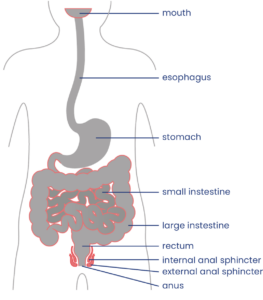
Your bowel function and symptoms may change over time. Most health providers recommend people with SCI to have a check-up once every two to three years or if there are changes to bowel function to keep track of changes to your bowel function and care.

Bowel care is an essential part of staying healthy after an SCI. For most people, a bowel routine is their main method of caring for their bowel health following SCI.

A bowel routine is a regular routine of bowel care techniques that is done every day or every other day to empty the bowel. There is a wide range of different components that may make up a bowel routine, such as hands-on emptying techniques, diet and lifestyle changes, and the use of suppositories, mini-enemas and medications.


Bowel routines are usually done at the same time each day, so the body gets used to the program. The goal of bowel routines is usually to be able to complete the routine within one hour on a regular basis to empty stool from the bowel and prevent accidents.

Every person’s bowel routine is different and often involves some trial and error to find the methods that best meet the person’s unique symptoms, physical abilities, preferences, and lifestyle. Although bowel routines can be complicated and time-consuming, they are a very important part of bowel care to allow for regular emptying and to prevent complications. Poorly managed bowel problems can lead to severe constipation, fecal impaction and other serious bowel complications.

Digital stimulation is a common spastic bowel emptying technique.11

Most people will need to use “hands-on” (manual) techniques to empty the bowel. Some people may need assistance from a care provider to use these techniques. They are usually done while sitting on a toilet or commode or while lying on your left side with the knees bent on an underpad.

Digital stimulation

People with spastic bowel typically use a technique called digital stimulationdigital rectal stimulation, or rectal touch to empty the bowel. Digital stimulation is a technique in which a gloved and lubricated finger (known in anatomy as a “digit”) is gently inserted into the anus and moved in circular motions along the walls of the rectum. This triggers a reflex that causes rectal contractions and loosening of the anal sphincters to allow emptying.

Digital stimulation is one of the most common techniques used for bowel emptying after SCI, however very little research has been done to determine if it is effective.

Watch for signs of autonomic dysreflexia

Cartoon lightning boltsPeople with injuries at T6 and above may trigger autonomic dysreflexia when performing digital stimulation and other bowel techniques. Keep an eye out for signs including sweating, headache, heart rate changes, goose bumps and increasing muscle spasms, and stop any techniques right away. Autonomic dysreflexia can also be triggered by bowel problems such as constipation or hemorrhoids.

People who experience autonomic dysreflexia during digital stimulation may use topical anesthetic medications like Lidocaine (Xylocaine) applied to their finger to numb the nerve endings during digital stimulation. There is moderate evidence that Lidocaine helps to reduce the symptoms of autonomic dysreflexia during digital stimulation.

Refer to our article on Autonomic Dysreflexia for more information! 

Manual removal of stool

People with flaccid bowel do not have bowel reflexes, so digital stimulation is not effective. Instead, the stool needs to be removed manually. This is called manual evacuation, digital disimpaction, or a rectal clear. This technique involves the use of a gloved and lubricated finger, which is inserted into the anus and used to remove stool. It may be done in conjunction with use of a lubricating suppository.

There is limited evidence that manual evacuation helps to reduce the number of bowel accidents, and it is unclear whether it helps to reduce time spent on bowel care.

Abdominal massageDigestive system with a clock for direction reference

Abdominal massage is a technique where a person massages the abdomen by stroking or kneading the lower torso in a clockwise motion to increase movement within the colon. However, research results on whether abdominal massage is effective for helping with bowel care after SCI have been conflicting. Use caution when using abdominal massage after a recent surgery or if you have an abdominal stoma.

Diet and fluid intake play a very important role in maintaining stools that can be moved easily through the bowel. Altering diet and fluid intake is often one of the first changes that are made to try to improve the consistency of stool and manage problems like constipation and diarrhea.

Fibre

The amount and type of fibre consumed can affect stool consistency.14

Fibre plays an important role in maintaining stool consistency so the stool can be moved easily through the bowel. Different types of fibre have different effects in the body. Soluble fibre, such as oatmeal and psyllium fibre, helps to trap waste and water along the digestive tract. It is important to increase water intake together with soluble fibre because it needs to absorb water to work. Soluble fibre may help improve stool consistency if stools are too loose or too hard. Insoluble fibre, such as whole grains, helps propel and move the contents through the colon more quickly.

There is weak evidence that diets high in fibre may actually slow down digestion after SCI. The optimal amount of fibre intake for people with SCI has not yet been studied and is highly variable between individuals, however, most health providers recommend moderate fibre intake to help with bowel care after SCI.

Refer to our article on Fibre to learn more about its role in your diet. 

Fluids

Water is important to bulk up the stool to allow it to move more easily. Health providers often recommend drinking at least 2 litres of healthy fluid per day, however, the optimal amount of fluids for improving bowel movement after SCI has not yet been studied.

It may take trial and error to find the right amount of fibre and fluidBalance scale with question marks on each opposing plate

The amount of fibre and fluids for optimal bowel function is different for everyone. It usually takes some trial and error to find the right amount of fibre for you. Keeping track of your fibre and fluid intake over a period of time can be helpful in figuring out how much you need to maintain your desired stool consistency. Fibre intake should be gradually adjusted to achieve the desired consistency of stool. Speak to your health providers for more information.

Foods contributing to other symptoms

Some people find that certain foods and drinks affect their bowel function in different ways. For example, some foods may cause loose stools or constipation. Every person is different, and it may take some trial and error to find the best diet for you.

Timing of food and drink

The gastrocolic reflex is when food or drink in the stomach triggers movement in the bowel. Some people eat food or drink warm fluids at least 30 minutes before their bowel routine to try to take advantage of this reflex, which is thought to be strongest in the morning. Many people use this technique as part of their bowel care; however the evidence is conflicting about whether the gastrocolic reflex is effective after SCI.

Find out how Charlie manages his fiber and fluid intake depending on the season.

Medications and suppositories may be added to your bowel routine if hands-on techniques and diet changes are not enough.

Stool softeners

Stool softeners are a type of laxative that increases the moisture in the stool to make it easier to pass. Stool softeners are usually taken once or twice per day or as needed. Some common stool softeners include Polyethylene glycol 3350 (PEG) and Docusate sodium (Colace).

Suppositories and Micro-enemas

A suppository is a solid (usually cone or cylinder-shaped) form of medication or other substance that is inserted into the rectum to absorb. Suppositories must make contact with the bowel walls to work, so care should be taken that they are not placed into a stool.

Alternatively, a micro-enema is a liquid form of a drug administered into the rectum before a planned emptying. These methods are used to allow for quick and direct action on the bowel walls.

Stimulant laxatives (including Stimulant suppositories)

Micro-enemas are administered into the rectum before a planned emptying.18

Stimulant laxatives increase the movements in the intestinal wall that help move the stool along. There are many different stimulant laxatives that may be taken by mouth or as a suppository or micro-enema. Some common stimulant laxatives include Bisacodyl (Dulcolax) suppositories and Sennosides (Senokot).

Stimulant suppositories contain medications (such as Dulcolax) which stimulate the bowel reflex. Suppositories are usually inserted 15 to 30 minutes before planned bowel emptying. However, the medication used and even the base that the medication is dissolved in can affect how quickly the medication is absorbed. For example, Dulcolax in a water-soluble polyethylene glycol base (such as a Magic Bullet) allows shorter times to emptying than Dulcolax in a vegetable oil base.

Lubricating suppositories

Lubricating suppositories contain non-medicated substances (such as glycerin) which holds water in the bowel to make the stool softer so it is easier to pass.

Prokinetic agents

Similar to stimulant laxatives, prokinetic agents are medications that increase digestive tract muscle activity to move the stool through digestion. However, prokinetic agents also speed up movement through the upper digestive tract as well as the bowel. Prokinetic agents are not routinely used and considered as a last resort for constipation or in preparation for a bowel procedure, such as a colonoscopy.

There is moderate evidence that the prokinetic agents fampridine, neostigmine, and metoclopramide help provide relief from constipation in people with SCI. There is also some evidence that prucalopride can help SCI individuals with impaired bowel emptying but may cause abdominal pain.

Standing and Exercise

Standing and exercise have long been thought to assist with bowel management after SCI. Standing and exercise are thought to help move abdominal contents toward the rectum. However, there is not enough evidence at this time to determine whether standing and exercise are effective to help with managing bowel changes after SCI.

Assistive devices

A number of devices also exist to help with bowel management. These devices are typically used together with regular bowel care techniques, and may include:

  • Commodes, raised or padded toilet seat, or automated toilet seat
  • Suppository inserter or digital stimulator
  • Mirrors
  • Anal plugs
  • Footstools
  • Bowel irrigation systems

However, limited research has been done on assistive devices for bowel care, so we do not yet know if these treatments are useful to help manage bowel changes after SCI.

Colostomy and ileostomy surgery

Most people are able to manage their bowel function effectively with a bowel routine made up of conservative techniques and medications. However, some people may require the use of additional treatments to achieve optimal bowel care.

Colostomy and ileostomy are surgical procedures that may be used if conservative techniques do not work well or as a way to streamline bowel care. Both of these procedures involve a surgically-created hole between the intestine and the outside of the abdomen. Once the opening is formed, a pouch (collection bag) is attached to allow stool to be collected outside of the body, bypassing the rectum and anus entirely.

A colostomy is when the hole (or stoma) is created between the colon (large intestine) and the outside of the body. An ileostomy is when the hole is created between the ileum (last part of the small intestine) and the outside of the body.

Image showing a person's abdomen with the lower gastrointestinal tract. The large intestine has a hole in the colon labelled as 'stoma' and a bag attached to the stoma labelled 'colostomy bag'.

A colostomy is a surgically created opening between the large intestine (colon) and an external collection device (colostomy bag).19

Although these types of surgeries are invasive, a number of different studies have shown that there are many benefits for bowel care. There is weak evidence that colostomy surgery:

  • Simplifies bowel routines and reduces the amount of time spent on bowel care
  • Reduces the need for use of laxatives and dietary changes in bowel care
  • Reduces the number of hospitalizations related to bowel problems
  • Improves independence and quality of life

These studies showed that, while there were some complications related to stoma surgeries, such as opening of the surgical wound, these were uncommon, and the majority of people were satisfied with the results of their surgery.

Bowel irrigation

Bowel irrigation is the use of water or other fluids (including medications) to stimulate the bowel muscles and flush stool from the bowel. This is done to help loosen stools and treat constipation. Bowel irrigation is sometimes referred to as an enema, although it typically uses more fluid than an enema. The fluid may be introduced into the bowel through the anus (called transanal irrigation) or through a surgically created stoma (called antegrade irrigation).

In MACE, the appendix is made into a stoma.20

Transanal irrigation is done through a catheter that is inserted into the rectum through the anus. The catheter may be surrounded by a balloon that can be inflated to hold the catheter in place and prevent leaking of water. The catheter is connected by a tube to a hand pump or electrical pump system to push water into the bowel. Pulsed water transanal irrigation involves quick pulses of water into the rectum to help loosen stools. There is weak evidence that transanal irrigation and pulsed water transanal irrigation helps improve bowel function after SCI.

When fluid is introduced through a stoma (antegrade irrigation), it is usually done through a small stoma into the beginning of the large intestine (the cecum) called a cecostomy or as part of a surgical technique called the Malone Antegrade Continence Enema (MACE) procedure, where the appendix is made into a stoma. Irrigation is not usually done through an ileostomy because this can cause dehydration. There are several studies which provide weak evidence that MACE is a safe and useful alternative for bowel irrigation after SCI.


Electrical and magnetic stimulation treatments (not in regular use)

Electrical and magnetic stimulation techniques are available to assist with bowel care. However, these treatments are not usually available in most healthcare settings.

Functional magnetic stimulation

Functional magnetic stimulation is a non-invasive procedure in which a magnet is placed over the spinal cord to administer magnetic waves which can stimulate nerve cells to fire. There is weak evidence that functional magnetic stimulation help improve bowel transit time after SCI.

Abdominal muscle stimulation

Abdominal muscle stimulation is another non-invasive procedure in which electrodes are placed on the abdominal muscles to stimulated muscle activity. It has been proposed that activating the abdominal muscles might help to increase pressure within the abdominal cavity, which may assist with bowel emptying for people who do not have abdominal control (injuries above T7).

Sacral anterior root stimulation

Sacral anterior root stimulation is an invasive procedure in which electrodes and a controlling device are implanted onto the sacral nerves during surgery. The device is controlled by an external transmitter and can stimulate the nerves which help empty the bowel.

There is moderate evidence that sacral anterior root stimulation may help improve constipation after SCI. Sacral root stimulation is an emerging area of research and further research in the future may provide more information about whether this treatment works.

Watch SCIRE’s video to learn more about bowel management as you age.21

All people age, and as they do they may notice changes in how they digest certain foods or that they need to go to the bathroom more frequently, or they may not go to the bathroom as successfully. As the body ages, nerve signals to the bowel may decrease and weaken bowel contractions and cause stool to move more slowly through the bowel. As a result, more water gets reabsorbed from the stool which often leads to more constipation. Some may experience more stool leaking.

Aging with SCI

For people aging with SCI, the changes in the bowel from typical aging may combine with the effects of neurogenic bowel dysfunction to worsen problems like constipation. Generally, as people age with SCI, symptoms of constipation increase, the frequency of bowel movements decrease, and the time required to complete a bowel movement increases. Medications and antibiotics taken to treat infections can also have significant effects on bowel function. They can increase constipation, increase leaks/accidents, or make the bowels less predictable.

Bowel changes that people aging with SCI may experience:

Other aging-related problems like pain, osteoarthritis, decreased strength and mobility, and skin issues, can affect the ability to carry out bowel routines. Bowel changes like constipation, hemorrhoids, and spending more time sitting on the toilet can trigger pain, spasticity, and autonomic dysreflexia. All these changes from aging could lead to a need to reassess bowel management strategies.

Refer to the “What bowel problems can happen after SCI?” section above for more information on bowel complications.

Managing bowel changes with age

To manage changes to bowel function from aging with SCI, consult regularly with a health care provider about your bowel management. Strategies to manage bowel changes may include:

  • Modifying your diet and fluid intake
  • Medications for bowel movements like suppositories and laxatives
  • Surgical procedures
  • Reviewing your current medications and supplements (some medications can cause constipation or diarrhea as a side effect)
  • Reviewing toilet and transfer equipment for injury to skin and joints
  • Additional caregiver help

Changing diet and water intake needs

Drinking water and the types of foods you eat both play an important part in how easily stool moves through the bowel. Needing to adjust what you eat and drink as you age is common since people often feel less appetite and thirst, and your body will not be as good at maintaining water and nutrient levels.

The optimal diet and daily water intake are different for each person, but finding the right balance can make a big difference for your bowels. You might also find that adjusting when in the day you eat and drink can affect your bowel movements.

Many people with SCI change their bowel management as they age. In one study, 63% of participants changed their bowel management methods over 20 years. People who used only medication and/or straining to manage their bowels were most likely to change their methods. Use of colostomy and bowel irrigation for management increased during this time.

For some people with long bowel routines (1-2 hours spent on bowel care per day), undergoing a colostomy was able to reduce time spent on bowel care to around 10-20 minutes per day. When considering surgical options for bowel management, people with SCI find it helpful to talk to stoma nurses, SCI doctors, and peers.

There is also research looking at the potential for nerve stimulation to improve bowel function and reduce time needed for bowel management. This may be available depending on location.

Refer to the “Diet and fluids”, “Medications and suppositories”, and “Other treatments and techniques” sections above for more information.

When do I need to review or change my bowel routine?

  1. Your current routine is not working anymore.
  2. The time and energy you spend on bowel management is limiting time spent with family/friends or doing things you love.

Questions to ask yourself to manage bowel changes

  • What has changed (issues, challenges, barriers)?
  • When did the change start?
  • What is the root cause of the change? (medical illness, injury, surgery, mobility, weakness, cognitive changes, social changes, financial changes)
  • What treatments or recommendations have been tried so far?
  • What has worked and what has not?
  • When was the most recent review of bowel with a family doctor or physiatrist?
  • Are current care team members actively involved to provide guidance and support?

Most people experience bowel changes after SCI. Even when well managed, bowel care can take up a lot of time and energy each day, which may interfere with maintaining a regular schedule. Bowel problems can also interfere with important activities like work, socializing and sexual intimacy.

Caring for bowel changes and establishing a bowel routine after SCI is one of the highest priorities for recovery after injury. Bowel care typically involves developing a conservative bowel management routine in combination with various manual techniques, diet changes, and medications. Several other procedures exist that are not as commonly used, such as cecostomy, colostomy and ileostomy surgeries and bowel irrigation.

For a review of how we assess evidence at SCIRE Community and advice on making decisions, please see SCIRE Community Evidence.

Parts of this page have been adapted from the SCIRE Professional “Bowel Dysfunction and Management” and “Autonomic Dysreflexia” Module:

Coggrave M, Mills P, Willms R, Eng JJ, (2014). Bowel Dysfunction and Management Following Spinal Cord Injury. In Eng JJ, Teasell RW, Miller WC, Wolfe DL, Townson AF, Hsieh JTC, Connolly SJ, Noonan VK, Loh E, McIntyre A, editors. Spinal Cord Injury Rehabilitation Evidence. Version 5.0. Vancouver: p 1- 48.
Available from: https://scireproject.com/evidence/bowel-dysfunction-and-management/

Krassioukov A, Blackmer J, Teasell RW, Eng JJ (2014). Autonomic Dysreflexia Following Spinal Cord Injury. In Eng JJ, Teasell RW, Miller WC, Wolfe DL, Townson AF, Hsieh JTC, Connolly SJ, Noonan VK, Loh E, McIntyre A, editors. Spinal Cord Injury Rehabilitation Evidence. Version 5.0. Vancouver: p 1- 35.
Available from: https://scireproject.com/evidence/autonomic-dysreflexia/

Mortenson WB, Sakakibara BM, Miller WC, Wilms R, Hitzig S, Eng JJ (2014). Aging Following Spinal Cord Injury. In Eng JJ, Teasell RW, Miller WC, Wolfe DL, Townson AF, Hsieh JTC, Connolly SJ, Noonan VK, Loh E, McIntyre A, editors. Spinal Cord Injury Rehabilitation Evidence. Version 5.0. Vancouver: p 1- 90.
Available from: scireproject.com/evidence/aging/

Evidence for “Hands-on bowel techniques” is based on the following studies:

[1] Sloots CE, Felt-Bersma RJ, Meuwissen SG, Kuipers EJ. Influence of gender, parity, and caloric load on gastrorectal response in healthy subjects: a barstat study. Dig Dis Sci 2003; 48: 516-521.

[2] Ford MJ, Camilleri MJ, Hanson RB, Wiste JA, Joyner MJ. Hyperventilation, central autonomic control, and colonic tone in humans. Gut 1995; 37: 499-504.

[3] Korsten M, Singal AK, Monga A, Chaparala G, Khan AM, Palmon R, Mendoza JRD, Lirio JP, Rosman AS, Spungen A, Bauman WA. Anorectal stimulation causes increased colonic motor activity in subjects with spinal cord injury. J Spinal Cord Med 2007; 30: 31-35.

[4] Furusawa K, Sugiyama H, Tokuhiro A, Takahashi M, Nakamura T, Tajima F. Topical anesthesia blunts the pressor response induced by bowel manipulation in subjects with cervical spinal cord injury. Spinal Cord 2009;47:144-148.

[5] Solomons J and Woodward S. Digital removal of faeces in the bowel management of patients with spinal cord injury: a review. British Journal of Neuroscience Nursing 2013; 9: 216-222.

[6] Menter R, Weitzenkamp D, Cooper D, Bingley J, Charlifue S, Whiteneck G. Bowel management outcomes in individuals with long-term spinal cord injuries. Spinal Cord 1997; 35: 608-612.

[7] Haas U, Geng V, Evers GC, Knecht H. Bowel management in patients with spinal cord injury — a multicentre study of the German speaking society of paraplegia (DMGP). Spinal Cord. 2005; 43: 724-30.

[8] Ayas S, Leblebici B, Sozay S, Bayramoglu M, Niron EA. The effect of abdominal massage on bowel function in patients with spinal cord injury. Am J Phys Med Rehabil 2006; 85: 951-955.

[9] Hu C, Ye M, Huang Q. Effects of manual therapy on bowel function of patients with spinal cord injury. J Phys Ther Sci 2013; 25: 687-688.

Evidence for “Diet and fluids” is based on the following studies:

[1] Cameron KJ, Nyulasi IB, Collier GR, Brown DJ. Assessment of the effect of increased dietary fibre intake on bowel function in patients with spinal cord injury. Spinal Cord 1996; 34: 277-283.

Evidence for “Medications and suppositories” is based on the following studies:

Stimulant laxatives

[1] House JG, Stiens SA. Pharmacologically initiated defecation for persons with spinal cord injury: effectiveness of three agents. Arch Phys Med Rehabil 1997; 78: 1062-1065.

Prokinetic agents

[1] Cardenas DD, Ditunno J, Graziani V, Jackson AB, Lammertse D, Potter P, Sipski M, Cohen R, Blight AR. Phase 2 trial of sustained-release fampridine in chronic spinal cord injury. Spinal Cord 2007; 45: 158-168.

[2] Korsten MA, Rosman AS, Ng A, Cavusoglu E, Spungen AM, Radulovic M, Wecht J, Bauman WA. Infusion of neostigmine-glycopyrrolate for bowel evacuation in persons with spinal cord injury. Am J Gastroenterol 2005; 100: 1560-1565.

[3] Segal JL, Milne N, Brunnemann SR, Lyons KP. Metoclopramide-induced normalization of impaired gastric emptying in spinal cord injury. Am J Gastroenterol 1987; 82: 1143-1148.

[4] Krogh K, Jensen MB, Gandrup P, Laurberg S, Nilsson J, Kerstens R, De Pauw M. Efficacy and tolerability of prucalopride in patients with constipation due to spinal cord injury. Scand J Gastroenterol 2002; 37: 431-436.

Evidence for “Other treatments and techniques” is based on the following studies:

Assistive Devices

[1] Hoenig H, Murphy T, Galbraith J, Zolkewitz M. Case study to evaluate a standing table for managing constipation. SCI Nursing 2001;18: 74-7.

[2] Uchikawa K, Takahashi H, Deguchi G, Liu M. A washing toilet seat with a CCD camera monitor to stimulate bowel movement in patients with spinal cord injury. Am J Phys Med Rehabil 2007; 86: 200-204.

Colostomy and Ileostomy surgery

[1] Frisbie JH, Tun CG, Nguyen CH. Effect of enterostomy on quality of life in spinal cord injury patients. J Am Paraplegia Soc 1986; 9: 3-5.

[2] Stone JM, Wolfe VA, Nino-Murcia M, Perkash I. Colostomy as treatment for complications of spinal cord injury. Arch Phys Med Rehabil1990b; 71: 514-518.

[3] Kelly SR, Shashidharan M, Borwell B, Tromans AM, Finnis D, Grundy DJ. The role of intestinal stoma in patients with spinal cord injury. Spinal Cord 1999; 37: 211-214.

[4] Rosito O, Nino-Murcia M, Wolfe VA, Kiratli BJ, Perkash I. The effects of colostomy on the quality of life in patients with spinal cord injury: a retrospective analysis. J Spinal Cord Med 2002; 25: 174-183.

[5] Branagan G, Tromans A, Finnis D. Effect of stoma formation on bowel care and quality of life in patients with spinal cord injury. Spinal Cord 2003; 41: 680-683.

[6] Munck J, Simoens Ch, Thill V, Smets D, Debergh N, Fievet F, Mendes da Costa P. Intestinal stoma in patients with spinal cord injury: a retrospective study of 23 patients. Hepatogastroenterology 2008; 55: 2125-2129.

[7] Coggrave MJ, Ingram RM, Gardner BP, Norton C. The impact of stoma for bowel management after spinal cord injury. Spinal Cord 2012; 50: 848-852.

Bowel irrigation

[1] Del Popolo G, Mosiello G, Pilati C, Lamartina M, Battaglino F, Buffa P, Redaelli T, Lamberti G, Menarini M, Di Benedetto P, De Gennaro M. Treatment of neurogenic bowel dysfunction using transanal irrigation: a multicenter Italian study. Spinal Cord 2008; 46: 517-522.

[2] Christensen P, Kvitzau B, Krogh K, Buntzen S, Laurberg S. Neurogenic colorectal dysfunction – use of new antegrade and retrograde wash-out methods. Spinal Cord 2000; 38: 255-261.

[3] Faaborg PM, Christensen P, Kvitsau B, Buntzen S, Laurberg S, Krogh K. Long-term outcome and safety of transanal colonic irrigation for neurogenic bowel dysfunction. Spinal Cord 2009; 47: 545-549.

[4] Kim HR, Lee BS, Lee JE, Shin HI. Application of transanal irrigation for patients with spinal cord injury in South Korea: a 6-month follow-up study. Spinal Cord 2013; 51: 389-394.

[5] Teichman JMH, Harris JM, Currie DM, Barber DB. Malone antegrade continence enema for adults with neurogenic bowel disease. Journal of Urology 1998; 160: 1278-1281.

[6] Christensen P, Kvitzau B, Krogh K, Buntzen S, Laurberg S. Neurogenic colorectal dysfunction – use of new antegrade and retrograde wash-out methods. Spinal Cord 2000; 38: 255-261.

[7] Teichman JMH, Zabihi N, Kraus SR, Harris JM, Barber DB. Long-term results for Malone antegrade continence enema for adults with neurogenic bowel disease. Urology 2003; 61: 502-506.

[8] Worsoe J, Christensen P, Krogh K, Buntzen S, Laurberg S. Long-term results of antegrade colonic enema in adult patients: assessment of functional results. Dis Colon Rectum 2008; 51: 1523-1528.

[9] Puet TA, Jackson H, Amy S. Use of pulsed irrigation evacuation in the management of the neuropathic bowel. Spinal Cord 1997; 35: 694-699.

Electrical and magnetic stimulation

[1] Korsten MA, Fajardo NR, Rosman AS, Creasey GH, Spungen AM, Bauman WA. Difficulty with evacuation after spinal cord injury: Colonic motility during sleep and effects of abdominal wall stimulation. JRRD 2004; 41: 95-99.

[2] Tsai PY. Wang CP, Chiu FY, Tsai YA, Chang YC, Chuang TY. Efficacy of functional magnetic stimulation in neurogenic bowel dysfunction after spinal cord injury. J Rehabil Med 2009; 41: 41-47.

[3] Lin VW, Kim KH, Hsiao I, Brown W. Functional magnetic stimulation facilitates gastric emptying. Arch Phys Med Rehabil 2002; 83: 806-810.

[4] Lin VW, Nino-Murcia M, Frost F, Wolfe V, Hsiao I, Perkash I. Functional magnetic stimulation of the colon in persons with spinal cord injury. Arch Phys Med Rehabil 2001; 82: 167-173.

[5] Binnie NR, Smith AN, Creasey GH, Edmond P. Constipation associated with chronic spinal cord injury: the effect of pelvic parasympathetic stimulation by the Brindley stimulator. Paraplegia 1991; 29: 463-469.

Evidence for “How does aging with SCI affect the bowel?” is based on:

Britton, E., & McLaughlin, J. T. (2013). Ageing and the gut. Proceedings of the Nutrition Society, 72(1), 173–177. https://doi.org/10.1017/S0029665112002807

Cameron, K. J., Nyulasi, I. B., Collier, G. R., & Brown, D. J. (1996). Assessment of the effect of increased dietary fibre intake on bowel function in patients with spinal cord injury. Spinal Cord, 34(5), 277–283. https://doi.org/10.1038/sc.1996.50

Charlifue, S., Jha, A., & Lammertse, D. (2010). Aging with Spinal Cord Injury. Physical Medicine and Rehabilitation Clinics of North America, 21(2), 383–402. https://doi.org/10.1016/j.pmr.2009.12.002

Coggrave, M. J., Ingram, R. M., Gardner, B. P., & Norton, C. S. (2012). The impact of stoma for bowel management after spinal cord injury. Spinal Cord, 50(11), 848–852. https://doi.org/10.1038/sc.2012.66

Fosnes, G. S., Lydersen, S., & Farup, P. G. (2011). Constipation and diarrhoea – common adverse drug reactions? A cross sectional study in the general population. BMC Clinical Pharmacology, 11(1), 2. https://doi.org/10.1186/1472-6904-11-2

Johns, J., Krogh, K., Rodriguez, G. M., Eng, J., Haller, E., Heinen, M., Laredo, R., Longo, W., Montero-Colon, W., Wilson, C. S., & Korsten, M. (2021). Management of Neurogenic Bowel Dysfunction in Adults after Spinal Cord Injury. The Journal of Spinal Cord Medicine, 44(3), 442–510. https://doi.org/10.1080/10790268.2021.1883385

Johns, J. S., Krogh, K., Ethans, K., Chi, J., Querée, M., & Eng, J. J. (2021). Pharmacological Management of Neurogenic Bowel Dysfunction after Spinal Cord Injury and Multiple Sclerosis: A Systematic Review and Clinical Implications. Journal of Clinical Medicine, 10(4), 882. https://doi.org/10.3390/jcm10040882

Nielsen, S. D., Faaborg, P. M., Christensen, P., Krogh, K., & Finnerup, N. B. (2017). Chronic abdominal pain in long-term spinal cord injury: a follow-up study. Spinal Cord, 55(3), 290–293. https://doi.org/10.1038/sc.2016.124

Nielsen, S. D., Faaborg, P. M., Finnerup, N. B., Christensen, P., & Krogh, K. (2017). Ageing with neurogenic bowel dysfunction. Spinal Cord, 55(8), 769–773. https://doi.org/10.1038/sc.2017.22

Parittotokkaporn, S., Varghese, C., O’Grady, G., Svirskis, D., Subramanian, S., & O’Carroll, S. J. (2020). Non-invasive neuromodulation for bowel, bladder and sexual restoration following spinal cord injury: A systematic review. Clinical Neurology and Neurosurgery, 194, 105822. https://doi.org/10.1016/j.clineuro.2020.105822

Rosito, O., Nino-Murcia, M., Wolfe, V. A., Kiratli, B. J., & Perkash, I. (2002). The Effects Of Colostomy On The Quality Of Life In Patients With Spinal Cord Injury: A Retrospective Analysis. The Journal of Spinal Cord Medicine, 25(3), 174–183. https://doi.org/10.1080/10790268.2002.11753619

Savic, G., Frankel, H. L., Jamous, M. A., Soni, B. M., & Charlifue, S. (2018). Long-term bladder and bowel management after spinal cord injury: a 20-year longitudinal study. Spinal Cord, 56(6), 575–581. https://doi.org/10.1038/s41393-018-0072-4

Wahlqvist, M., & Savige, G. (2000). Interventions aimed at dietary and lifestyle changes to promote healthy aging. European Journal of Clinical Nutrition, 54(S3), S148–S156. https://doi.org/10.1038/sj.ejcn.1601037

Wong, S., Santullo, P., Hirani, S. P., Kumar, N., Chowdhury, J. R., García-Forcada, A., Recio, M., Paz, F., Zobina, I., Kolli, S., Kiekens, C., Draulans, N., Roels, E., Martens-Bijlsma, J., O’Driscoll, J., Jamous, A., & Saif, M. (2017). Use of antibiotics and the prevalence of antibiotic-associated diarrhoea in patients with spinal cord injuries: an international, multi-centre study. Journal of Hospital Infection, 97(2), 146–152. https://doi.org/10.1016/j.jhin.2017.06.019

Other references

Anderson KD. Targeting recovery: Priorities of the spinal cord-injured population. Journal of Neurotrauma 2004; 21: 1371-1383.[4] Badiali D, Bracci F, Castellano V, Corazziari E, Fuoco U, Habib FI, Scivoletto G. Sequential treatment of chronic constipation in paraplegic subjects. Spinal Cord 1997; 35: 116-120.

Brading AF, Ramalingam T. Mechanisms controlling normal defecation and the potential effects of spinal cord injury. Prog Brain Res 2006; 152: 345-58.

Byrne CM, Pager CK, Rex J, Roberts R, Solomon MJ (1998): Assessment of Quality of Life in the treatment of patients with neuropathic fecal incontinence. Dis Colon Rectum 2002; 45: 1431-6.

Coggrave M, Burrows D, Durand MA. Progressive protocol in the bowel management of spinal cord injuries. British Journal of Nursing 2006; 15: 1108-1113.

Coggrave M, Norton C, Cody JD. Management of fecal incontinence and constipation in adults with central neurological diseases. Cochrane Database Syst Rev. 2014;1:CD002115.

Coggrave MJ, Norton C, Wilson-Barnett J. Management of neurogenic bowel dysfunction in the community after spinal cord injury: a postal survey in the United Kingdom. Spinal Cord 2009; 47: 323-330.

Coggrave MJ, Norton C. The need for manual evacuation and oral laxatives in the management of neurogenic bowel dysfunction after spinal cord injury: a randomized controlled trial of a stepwise protocol. Spinal Cord. 2010; 48: 504-10.

Correa GI, Rotter KP. Clinical evaluation and management of neurogenic bowel after spinal cord injury. Spinal Cord2000; 38: 301-308.

Cosman BC, Vu TT. Lidocaine anal block limits autonomic dysreflexia during anorectal procedures in spinal cord injury: a randomized, double-blind, placebo-controlled trial. Dis Colon Rectum. 2005;48:1556-61.

Emmanuel A. Review of the efficacy and safety of transanal irrigation for neurogenic bowel dysfunction. Spinal Cord. 2010;48:664-73.

Faaborg PM, Christensen P, Finnerup N, Laurberg S, Krogh K. The pattern of colorectal dysfunction changes with time since spinal cord injury. Spinal Cord 2008; 46: 234-238.

Fajardo NR, Pasiliao RV, Modeste-Duncan R, Creasey G, Bauman WA, Korsten MA. Decreased colonic motility in persons with chronic spinal cord injury. Am J Gastroenterol 2003; 98: 128-34.

Fealey RD, Szurszewski JH, Merrit JL, DiMagno EP. Effect of traumatic spinal cord transection on human upper gastrointestinal motility and gastric emptying. Gastroenterology 1984; 87: 69-75.

Finnerup NB, Faaborg P, Krogh K, Jensen TS. Abdominal pain in long-term spinal cord injury. Spinal Cord 2008; 46: 198-203.

Furusawa K, Sugiyama H, Ikeda A, Tokuhiro A, Koyoshi, H, Takahashi M, Tajima F. Autonomic dysreflexia during a bowel program in patients with cervical spinal cord injury. Acta Med Okayama 2007; 61: 211-227.

Glickman S, Kamm MA. Bowel dysfunction in spinal-cord-injury patients. Lancet 1996; 347: 1651-3.

Gondim FA, Rodrigues CL, da Graça JR, Camurça FD, de Alencar HM, dos Santos AA, Rola FH. Neural mechanisms involved in the delay of gastric emptying and gastrointestinal transit of liquid after thoracic spinal cord transection in awake rats. Auton Neurosci. 2001; 87: 52-8.

Krogh K, Mosdal C, Laurberg S. Gastrointestinal and segmental colonic transit times in patients with acute and chronic spinal cord lesions. Spinal Cord 2000; 38: 615-621.

Leduc BE, Spacek E, Lepage Y. Colonic transit time after spinal cord injury: any clinical significance? J Spinal Cord Med 2002; 25: 161-6.

Lynch AC, Antony A, Dobbs BR, Frizelle FA. Bowel dysfunction following spinal cord injury. Spinal Cord 2001; 39: 193-203.

Lynch AC, Frizelle FA. Colorectal motility and defecation after spinal cord injury in humans. Prog Brain Res 2006; 152: 335-43.

Lynch AC, Wong C, Anthony A, Dobbs BR, Frizelle FA. Bowel dysfunction following spinal cord injury: a description of bowel function in a spinal cord-injured population and comparison with age and gender matched controls. Spinal Cord 2000; 38: 717-723.

Menter R, Weitzenkamp D, Cooper D, Bingley J, Charlifue S, Whiteneck G. Bowel management outcomes in individuals with long-term spinal cord injuries. Spinal Cord 1997; 35: 608-612.

Ng C, Prott G, Rutkowski S, et al. Gastrointestinal symptoms in spinal cord injury: relationships with level of injury and psychologic factors. Dis Colon Rect 2005;48:1562-1568.

Nino-Murcia M, Stone JM, Chang PJ, Perkash I. Colonic transit in spinal cord-injured patients. Invest Radiol 1990; 25: 109-112.

Rajendran SK, Reiser JR, Bauman W, Zhang RL, Gordon SK, Korsten MA. Gastrointestinal transit after spinal cord injury: effect of cisapride. Am J Gastroenterol 1992; 87: 1614-1617.

Stiens SA, Bergman SB, Goetz LL. Neurogenic bowel dysfunction after spinal cord injury: clinical evaluation and rehabilitative management. Arch Phys Med Rehabil 1997;78:S86-S102.

Stone JM, Nino-Marcia M, Wolfe VA, Perkash I. Chronic gastrointestinal problems in spinal cord injury patients: a prospective analysis. Am J Gastroenterol 1990a; 85: 1114-9.

Image credits

  1. The Bowel ©SCIRE, CC BY-SA 3.0
  2. Belly abdominal pain ©Christian Dorn, CC0 1.0
  3. Man toilet bathroom sitting ©Clker-Free-Vector-Images, CC0 1.0
  4. Leak plumbing water drips bathroom ©Clker-Free-Vector-Images, CC0 1.0
  5. Mistake spill slip up accident ©Steve Buissinne, CC0 1.0
  6. Water jump refreshment children ©AxxLC, CC0 1.0
  7. Image ©SCIRE, CC BY-SA 3.0
  8. Bristol Stool Chart ©Cabot Health, CC BY-SA 3.0
  9. Scientist ©H Alberto Gongora, CC BY 3.0 US
  10. Towel napkin dab dry vector ©Isa KARAKUS, CC0 1.0
  11. Modified from: Hand finger pointing ©truthseeker08, CC0 1.0
  12. Electricity ©Artnadhifa, CC BY 3.0 US
  13. Abdomen clockwise clock massage ©bodymybody, CC0 1.0
  14. Breads cereals oats barley wheat ©FotoshopTofs, CC0 1.0
  15. Scale question importance balance ©Arek Socha, CC0 1.0
  16. Alarm clock time of good morning ©congerdesign, CC0 1.0
  17. Suppositories pharmacy ©Andreas Koczwara, CC0 1.0
  18. Microlax Miniklistier ©MedInfo Johnson&Johnson, CC BY-SA 4.0
  19. Cancer Research UK (Original email from CRUK), CC BY-SA 4.0
  20. Reprinted with permission from Malone PSJ. Malone procedure for antegrade continence enemas. In: Spitz L, and Coran AG, eds. Rob & Smith’s Operative Surgery: Pediatric Surgery. 5th ed. London: Chapman & Hall Medical; 1995:459–467.
  21. Aging Bowel Thumbnail ©SCIRE, CC BY-NC 4.0
  22. Clock by Abu Ibrahim Icon, Noun Project
  23. Toilet by Isaac Haq, Noun Project


Disclaimer: This document does not provide medical advice. This information is provided for educational purposes only. Consult a qualified health professional for further information or specific medical advice. The SCIRE Project, its partners and collaborators disclaim any liability to any party for any loss or damage by errors or omissions in this publication.