Breastfeeding Following Spinal Cord Injury: Consumer Guide for Mothers

By | | No Comments

Contributors: Breastfeeding Consumer Guide for Mothers Panel | Published: 11 January 2022 | Updated: ~

Few resources exist that talk about the effects a spinal cord injury (SCI) can have on breastfeeding. It is difficult to find information about the unique questions, challenges, or concerns women with SCI experience when planning or trying to breastfeed.

Our team, which includes women with SCI, health care providers, and SCI researchers, came together to create a guide to help address this information gap.

The purpose of this guide is to share with you the most common issues women with SCI experience during breastfeeding. This guide also provides information, practical suggestions, recommendations, and key resources.

General information about breastfeeding is available on the internet, in books, or from your friends and health care providers. We did not plan to repeat or replace general breastfeeding information or medical advice. Rather, we hope this guide adds to this information by sharing facts, tips, and resources specifically related to breastfeeding after SCI.

Breastfeeding in women with SCI is complex and requires a team of health care providers with complementary expertise. Such a team can include: your family physician, obstetrician, physiatrist, neurologist, occupational therapist, physical therapist, lactation consultant, midwife, psychologist, peer counsellor etc. Your team will vary depending on where you live.

We hope that you find this guide helpful.

Key Points

  • Breastfeeding has many benefits for babies’ physical, developmental, and emotional health regardless of whether the breastfeeding mother has SCI or not.
  • Some women with SCI may experience unique challenges and barriers that can impact how they breastfeed or if they can continue to breastfeed. This can sometimes result in women with SCI feeling alone, lost, or as if they have failed.
  • It is important for every woman to feel supported as she explores how best to balance her own unique needs and abilities with the known benefits and risks of the various ways to feed her child (breastfeeding, supplementing, and/or formula feeding).

It is accepted that breast milk (colostrum, transitional milk and mature milk) is the first choice to help your baby grow and develop. The benefits for your baby include:

  • Providing the ideal nutrition (breast milk has the perfect amount of protein, carbohydrates, fat, vitamins, and minerals, and is easy to digest),
  • Providing antibodies to protect against illness while baby’s immune system develops,
  • Lowering the likelihood of obesity, diabetes, and other diseases developing in childhood,
  • Lowering infant mortality.

The benefits for mothers include:

  • Increasing bonding and relaxation,
  • Reducing costs,
  • Having the convenience of breastmilk available,
  • Lowering the risk of future breast and ovarian cancers.

If breastfeeding is not possible or not enough breast milk is produced by mothers with SCI, then feeding with formula or donated breast milk from a milk bank is both necessary and encouraged.

Refer to the Public Health Agency of Canada’s 10 Great Reasons to Breastfeed Your Baby and the World Health Organization Infant and Young Child Feeding chapter for more information.

Women with SCI, just like women without SCI, are encouraged to give breastfeeding a try. By trying, you can then see how it works for both you and for your baby. If breastfeeding does not work well, there are several safe and healthy options available to you to ensure both your needs and your baby’s needs are met.

Many of the main questions and concerns women with SCI have shared with us are outlined in this guide. The information, recommendations, and resources below are intended to help you to gather information, to help guide your breastfeeding planning, to help you to problem solve common challenges or to help you to explore new ways of doing things as you work to find a feeding plan that is the right fit for you and your baby.

Recommendations:

  • You are encouraged to try to breastfeed and see how it goes for you and your baby.
  • If you have challenges or questions about breastfeeding talk to your health care providers. You may wish to share with them a copy of this resource guide to discuss together.

Everyone is different and it is not possible to predict your ability to produce breast milk and breastfeed your baby. Some challenges during breastfeeding are similar between women with and without SCI. However, it is important to recognize that mothers with SCI face additional concerns during breastfeeding related to SCI.

Depending on their level of injury, women with SCI could have limited milk production, difficulties with positioning and latching their baby, and they may experience episodes of autonomic dysreflexia (AD), an abrupt and often dangerous spike in blood pressure.

Watch SCIRE’s video explaining breastfeeding complications after SCI.

Research shows that the higher and more complete the injury, the more likely difficulties will occur. Yet, with the right supports in place, you can define what successful breastfeeding means to you.


Recommendations:

  • Talk to your health care providers, including a lactation consultant and a physiatrist for more information on how your SCI may be impeding breastfeeding.
  • Review this guide and references for additional information.
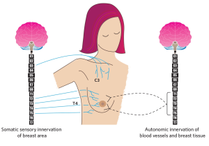
Figure 1. The innervation (control) of the breast by somatic (voluntary) and autonomic (involuntary) nervous systems. The black lines show the autonomic nerves (T1-T5 spinal cord levels) innervating breast tissues and blood vessels. The blue lines show the cervical (C) and thoracic (T) segments of the spinal cord that affect sensory control of the breast and nipple area. Milk production is a result of the coordinated functioning of the somatic and autonomic nervous systems.

The ability to breastfeed could be affected by the level and completeness of your SCI.

First, if your injury is above the first thoracic (T1) spinal cord level the motor functions in your arms/hands could be affected and you could have trouble with positioning and holding your child during breastfeeding. Typically, women with tetraplegia (cervical spinal cord injuries) experience these difficulties (see section on positioning and aids below).

Second, if your injury is above the fifth thoracic (T5 spinal) cord level, this could result in decreased or loss of sensory control of area of the nipple/breast area and affect milk production. These levels of injuries could result also in decrease or loss of body responses that occur during breastfeeding that are important for milk production (physiological reflexes known as “let down reflex”) (See Figure 1)

Watch SCIRE’s video highlighting Sheryl’s experience breastfeeding with a T2-T7 Incomplete SCI.

Recommendation:

Discuss your level of SCI and potential for breastfeeding with medical professionals (such as a physiatrist) and support team before planning a pregnancy or before delivery.
Like all mothers, women with SCI may experience common breastfeeding challenges such as:

  • babies who have difficulty latching/feeding due to issues like prematurity, tongue tie, or jaundice
  • mothers with inverted nipples, infection (clogged ducts, mastitis) or an overabundance of milk

There are also additional considerations specific to SCI that can cause some challenges in latching such as sensory changes, motor effects, and fatigue.

Be aware that sensory changes in your body caused by SCI could affect your ability to breastfeed. If you have decreased nipple sensation, it may lead to altered let-down reflex and impaired milk production as well as limit the ability to recognize poor latch. Visual signs during latching, feeding, and immediately after the feeding can help mothers identify a good latch. Use a mirror or have someone check the latch during the feeding and the breast for abrasions after feeding. During feeding, you should notice that your baby is changing the rate of sucking and appears to be swallowing.  After feeding, you should check the condition of your nipples and if your baby appears to be satisfied.

Be aware that strength and ability to move your upper extremities could impact your ability to breastfeed.   Lack of mobility in your arms/hands, spasms, and impaired trunk strength or balance can affect how well the baby is positioned to access the breast, to get and keep a good latch that does not injure your nipples, and to reposition when needed. If you have poor hand function it may be difficult to express your milk for drainage to prevent infection (clogged ducts and mastitis).  Ineffective or infrequent feeding can also be a cause of inadequate milk production. See positioning and aids section below for ideas during breastfeeding.

Be aware that fatigue is common in people with SCI and could impact your ability to breastfeed successfully.  Having a new baby is intense and requires around the clock attention and care for everyone. For some mothers with SCI, your own requirements for care may conflict with or delay the feeding times of your baby. You may want to create a feeding plan with support from your spouse, partner, family, or aide to assist in the feeding schedule. Make sure you continue to assess the latching, feeding, and breast health for as long as you decide to breastfeed. Remember that there is education and support out there for you if you need it. See section below on community supports.

Recommendations:

  • Pay attention to your baby’s quality of latching and swallowing during breastfeeding, particularly if you have decreased sensation in your nipples and breast area.
  • Examine your nipples (either on your own or ask a care provider to assist you) before and after breastfeeding to look for nipple distortion.
  • Consult with your health care team if you are having a hard time positioning your baby to breastfeed. They may be able to assist or can refer you to a knowledgeable health care provider such as an occupational therapist or physiotherapist to help you explore assistive aids.
  • If you are experiencing fatigue that interferes with your activities and breastfeeding, ask for the support of health care provider (e.g., occupational therapist) for suggestions and energy saving recommendations.

Refer to the Toronto Public Health Division and the Toronto East Health Network, Baby Friendly Initiative Strategy for Ontario: Breastfeeding Protocol: Positioning and Latching for more information.

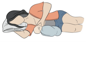
There are many things to consider in choosing a position for breastfeeding and skin-to-skin contact with baby. Your level of injury, fatigue, spasticity, hand function, trunk stability, and time of day can influence if you choose to be in bed, in your wheelchair, or in a different chair to breastfeed. It may also be necessary to have someone else pick up your baby and position them for feeding. The figure below shows the most used positions during breastfeeding by women with SCI.

Figure 2. Alternative breastfeeding positions that may be adapted for women with SCI.

Watch SCIRE’s video explaining ways you can breastfeed with assistive aids.

Recommendations:

  • If you are having difficulties with positioning your baby during breastfeeding ask for the support from your health care providers (e.g., occupational therapists, physical therapists).
  • Wheelchair modifications may be required to provide additional trunk support to increase stability while lifting and holding your baby. Adjusting the seat slope or backrest angle can change centre of gravity and improve stability. A backrest with deeper lateral trunk support or the use of a chest strap may also add stability. Changing the position of sideguards or widening the chair can allow for more room for baby’s body and legs when held in football or cross cradle positions.
  • Wedges can provide support for sitting upright in a bed (providing support for the back, belly or the infant after birth), or be used as leg support to reduce spasms and improve blood pressure.
  • Carriers (harnesses, wraps, ring slings) can be used during breastfeeding and thereafter. They may be useful in positioning your baby when bottle-feeding or using a supplementary nursing system and can help to keep them upright after a feed to reduce spit-up/reflux.
  • Breastfeeding pillows are available in a variety of shapes, weights and stiffness (firm versus lighter and more flexible). Some mothers find that their own regular pillows work well if the breastfeeding pillows do not provide enough support.

Adding a long waist strap to a breastfeeding pillow or infant carrier that also wraps around the wheelchair backrest, can give added security, and reduce the tendency for the pillow to slide forward or for you to lean forward while holding the weight of the infant in front.

Watch SCIRE’s video highlighting Nicole’s breastfeeding experience.

Many mothers worry about low breastmilk supply and that their baby will not get enough milk.  If you have low milk supply, there are a few different options you could consider. You may wish to supplement your baby with breast milk (your own or donated), or formula by bottle or by using supplementary nursing system (SNS). This system works by placing one end of a very thin flexible tube into a formula bottle, taping the tube to your breast, and slipping the other end of the tube into the baby’s mouth once they have latched.

Refer to the Sikana Health video covering “How to use a Supplemental Nursing System” for more information.

Recommendations:

  • Talk to your health care providers, including lactation consultant and paediatrician about the most safe and effective options for feeding your baby, as well as medications for you and your baby.
  • If you have enough hand function, you can express milk directly into your baby’s mouth. Additionally, a breast pump can be used to express the milk into a bottle and then to feed your baby.
  • If you have low milk supply, you can start by feeding your baby from your breast then switching to a bottle after a specific amount of time or once the milk is gone from the breast.
  • You can feed your baby at the breast with additional formula by using a supplementary nursing system.

Many women experience clogged milk ducts or even lactation mastitis (an inflammation of breast tissue that may or may not involve an infection, but causes breast pain, swelling, warmth, fever, or chills) during their breastfeeding experience. Breast lumps are not uncommon.

Depending on the level of injury and hand function women with SCI could be more likely to experience clogged ducts during breastfeeding. This happens when the breast is not fully emptied of milk at each feeding due to incomplete latching and suckling.

Breast health may also be compromised if the baby is not held well to the breast due to limited hand and arm function or muscle fatigue. Limited hand function may also lead to poor milk expression and limit the ability to perform self-lymphatic drainage and massage, which can contribute to the development of clogged ducts and mastitis.

Recommendations:

  • Wear a supportive bra (even at night) but avoid restrictive clothing.
  • Change damp or soiled breast pads often.
  • Shower daily and wash nipples with clear water only (no soap).
  • Eat a healthy diet and stay well hydrated.
  • Use gentle breast massage (If hand function is limited, a massaging device could be used, or assistance requested from partner or caregiver).
  • Alternate use of hot and cold compresses may be helpful if your breasts are engorged.
  • If you have mastitis, check with your health care provider how you can safely continue to breastfeed your baby.
  • Be aware that pain from breastfeeding and mastitis could cause AD.

Refer to the Penn Medicine article on General Breast Health as well as the Cleveland Clinic article on Care for Mastitis for more information!

The common medications used in women with SCI could go through the blood milk barrier into the milk, and many effects of medication on breastfeeding babies are not known. Speak with your health care team as each drug will need to be assessed of its risks compared to its benefits for you and for your baby.

The American Pediatric Association has suggested some medication tips around breastfeeding for all women, which we have quoted below:

Breastfeeding and Medication Tips

  • Many effects of medications on breastfeeding babies simply are not known. Due to this, only take a medication when absolutely needed, at the lowest dose, and for the shortest time possible.
  • When possible, take medications that are given only once a day right after a feeding when your baby will have the longest period without nursing; for many women this is the last feeding of the night before the baby’s bedtime.
  • Watch your baby for side effects such as sleepiness, irritability, other potential or known reactions of the medication.
  • Avoid long-acting, extended-release, and combination forms of medications when possible. Shorter-acting medications are removed from your body more quickly, and single medications give you greater flexibility in dosing.
  • Only water-soluble cream or gel products should be applied to the breast because ointments may expose the baby to high levels of mineral paraffins via licking.
  • Special precautions may be needed in preterm (premature) babies, due to their size and organ systems that are even less developed than a regular term baby.
  • Ask your doctor about the risks and benefits of any medication prescribed while you are breastfeeding, or any medication you choose from over-the-counter options that do not require a prescription.
  • When more than one medication or a combination medication is used, follow the breastfeeding recommendations for the most problematic medication.

Recommendation:

Consult your health care team for advice about what medications to use, adjust, or discontinue while breastfeeding.

Refer to LactMed, a database containing information on drugs and other chemicals which breastfeeding mothers may be exposed to, as well as Drugs.com for information on safe medications during breastfeeding.

Watch SCIRE’s video explaining Kristen’s experience of blood pressure drops while breastfeeding her daughter.

AD is identified by uncontrolled and potentially life-threatening spikes in arterial blood pressure, which may reach as high as 300 mmHg systolic blood pressure. It is common for people with SCI at or above the T6 spinal cord segment to experience episodes of AD that can be caused by painful or non-painful stimuli below the injury level. Episodes of AD have been documented in several studies of breastfeeding by women with higher level injuries. In women with SCI at or above the fourth thoracic segment (T4, level of sensory innervation of nipples),
episodes of AD could be specifically triggered by stimuli from the nipples/breast area during breastfeeding. You should be aware that an episode of AD could be accompanied by severe headaches, sweating, and heart palpitations or you could experience AD without any symptoms at all.

Breastfeeding-related AD may be caused by breastfeeding itself as well as mastitis, nipple fissures or other painful processes originating from the breast. Episodes of AD caused by breastfeeding may be much more common than we know. Recent research shows that one quarter of women with SCI experience AD while breastfeeding, and this number is higher in women with high level SCI (almost 40%).
Frequent and uncontrolled episodes of AD could result not only in discomfort, but also significant negative health consequences, including damage to blood vessels and heart, stroke, seizures, and even death. It is vital to prevent and manage AD in a timely manner. Unfortunately, this topic is not commonly discussed in postpartum care, but it is vital to recognize and address.

Recommendations:

  • Be sure to discuss with your medical practitioner (physiatrist/ family physician/nurse) your blood pressure management and potential for development of AD.
  • Be aware that women with SCI at or above T6 are at higher risk for the development of AD triggered by breastfeeding, clogged ducts/mastitis.
  • If you experience symptoms of AD, you should stop breastfeeding and measure your blood pressure if you can. Be sure that you are in seated position with your legs lowered. Wait until symptoms subside and your blood pressure goes down close to normal before you resume breastfeeding.
  • Be aware that prolong seating while breastfeeding may cause a decrease in your blood pressure (orthostatic hypotension), that could result in light-headedness and dizziness. If this will occur, elevate your legs, or change your position to horizontal.

  • Have a wallet information card on AD in case of admission to an emergency room.
  • Refer to the ABC’s of Autonomic Dysreflexia and the Fact Sheet on AD by Spinal Cord Model System for more information.

    To download a Automatic Dysreflexia wallet card, click here.

Bonding, or what is often called ‘attachment’ or the ‘caring connection’ between a child and parent, is an important part of a child’s physical and emotional development. Some parents report immediately feeling a strong connection with their child during pregnancy or soon after the birth. Other parents report they did not feel an instant connection. Both feelings are okay and normal.
Bonding and attachment can sometimes happen right away and can sometimes take weeks or months of getting to know and understand your baby to find what works best for you and your child and each of your unique needs.
Breastfeeding is not the only way to nurture or bond with your child. There are many other important ways to build connection and attachment between you and your baby.

Recommendations:

  • Find times to hold your baby against your bare skin. There are many positive benefits including body temperature regulation, breathing and heart rate regulation, hormone release for both you and the child, calming, and improved sleep.
  • Cuddle with your child. Ensure you have the chance to hold, rock, or carry your child during times that are not only related to feeding, bathing, diaper changes or consoling. Sometimes those quiet moments are really important moments of connection and affection.
  • Respond to your baby when they cry. This may be by gently touching their skin, picking them up or by using your voice. Let your baby know you are nearby and aware of their needs.
  • Talk to your baby throughout the day. It might feel awkward at first but tell stories, describe people and the world around them, talk about what you are doing, or sing to your child.
  • Look into your newborn’s eyes while you feed or care for your child. This helps your newborn to connect your face with your sounds and actions.
  • Find ways to soothe yourself as well as your child. Take a few deep breaths. Listen to music you enjoy. Visit with friends you feel comfortable with. Look at a picture of a place where you like to spend time. Babies pick up on adults’ feelings of tension and anxiety but also soak up their feelings of calm and comfort.

Why am I feeling so sad or anxious?
The first few weeks after birth are exciting and challenging. It is a time of big changes as you bring your infant home, get to know them, learn how to feed, bathe, and carry them. You also need to adjust to disrupted sleep patterns, manage your physical recovery after the birth, and try to find time for your own daily living and self-care needs (such as eating, dressing, toileting, bathing, etc.). Breastfeeding when you have a SCI can sometimes make some, or all, of these
activities feel more challenging.

You may feel joy and excitement, but also feel sad, worried, or just be overwhelmed. This is understandable in the first few weeks after birth for any mother. This roller coaster of emotions is sometimes called “baby blues” and typically lasts a few days or up to a week or two. However, if the difficult feelings last longer and begin to impact your ability to participate in day-to-day activities, this can be a sign of what is called Postpartum Depression. Postpartum Depression can involve feeling sad, down, or crying easily but it can also sometimes feel like being worried, agitated, anxious, or an overwhelming fear that something awful is about to happen. Women with SCI are at higher risk for Postpartum Depression and anxiety due to several factors including:

  • Mental health concerns prior to pregnancy,
  • The stress of coping with multiple physical challenges during pregnancy and/or after the birth,
  • Worry about other peoples’ opinions or judgments,
  • Struggling with pre-existing fatigue related to SCI and the extra energy required to complete routine tasks as well as new onset fatigue due to baby caregiving needs,
  • Limited access to knowledgeable caregivers or peers who understand the unique joys and challenges of parenting with a SCI,
  • Possible other factors we do not yet know or understand.
    Whatever you are feeling, you are not alone. You are important and valued. Help is available. Talk to your partner, loved ones, or health care providers about your feelings and worries. If needed, immediate help is available 24/7 from local crisis phone lines, Urgent Care Centres or Emergency Departments. With support, many women soon begin to feel their mood improve, their anxiety lessen, feel more attached to their baby, and begin to experience many more positive moments of parenting as they learn tools to help cope with the hard times.

Recommendations:

  • Learn about the signs, symptoms, and resources related to postpartum depression and anxiety, so you, your partner, your family, and your care aides will recognize when extra assistance may be necessary.
  • Talk to your health care providers, seek out information from trusted websites online or connect with local community groups that support new parents.
  • Share this information with your partner, a friend or a family member, or a health care provider during your pregnancy or after baby has arrived. Sometimes we need a bit of help to reach out for the services we need when we are feeling sad or anxious.
  • Watch your mood during pregnancy and after birth and notice any changes in feelings of sadness or worry. Are these feelings improving or getting worse? Are they lasting longer than they did in the past?
  • Know that feelings of sadness or anxiety happen to lots of people. You are not a failure and you are not alone.
  • Help is available 24/7. Accessing help can relieve pressure and often make a big difference in how you are feeling.

There are plenty of books on pregnancy, birth, and all the challenges with motherhood, but there is much less information for pregnant women and new mothers that have a SCI. Just like experiencing and learning about changes to your body during pregnancy, it is important to educate and advocate for yourself about being a mother and breastfeeding with a SCI.

Recommendations:

  • Look to your healthcare team: Your healthcare team involved in your pregnancy and birth is your primary trusted resource. Physiatrists have the medical knowledge to address concerns related to your SCI. Lactation consultants, midwives and staff of public health agencies have knowledge about pregnancy, birth and breastfeeding. These professionals, working together with you can address your specific concerns.
  • Look online for resources: In addition to information provided in this guide and if you don’t have services locally, look for reputable and accredited sources. Check your local or national Spinal Cord Injury organizations. There are many dedicated websites in the medical community. Do an online internet search with some key words like “mom SCI and breastfeeding”. Online videos, articles and peers’ stories provide opportunities for learning and connections among a growing population of moms with a SCI all around the world.
  • Connect with other moms: Peer support is important for all new moms, including moms with SCI. Social media platforms like Facebook have many groups you could join and are a popular place to share questions and concerns and learn about equipment or adaptations ideas.
  • Stay connected: We also know there is a risk of isolation. Currently, there is no way to fully prevent depression or anxiety postpartum but knowing what signs to watch for can be helpful. It’s important to stay connected and not isolated. First, you’re a new mom and that means you share a lot of the same emotions, concerns, and challenges as every new mother. Reach out to other moms in your community or online for advice, support and joint activities, especially nature and outdoor; there are more things in common with able-bodied moms once issues around your SCI are addressed.

Image credits

  1. Smiling Mother Playing With Her Baby Lying On The Floor ©Jacob Lund Photography from NounProject.com
  2. Newborn Being Breast-fed At Home ©Paulo Sousa from NounProject.com
  3. Complications ©SCIRE
  4. Kristen’s breastfeeding story ©SCIRE
  5. Breastfeeding innervation © The Krassioukov Lab
  6. Sheryl’s breastfeeding story ©SCIRE
  7. Breastfeeding Lying down ©SCIRE
  8. Breastfeeding positions ©The Krassioukov Lab
  9. Assistive Aids ©SCIRE
  10. Nicole’s breastfeeding story ©SCIRE
  11. Woman Bottle Feeds A Newborn ©Paulo Sousa from NounProject.com
  12. Medication © Kiran Shastry from Noun Project
  13. Close Up Of Smiling Mother And Baby Daughter ©Noun Project from NounProject.com
  14. Mother Taking Rest Sleeping On A Couch With Her Baby On Her Chest ©Jacob Lund Photography from NounProject.com

Panel Members

Andrei Krassioukov, MD, PhD, FRCPC
Physiatrist
GF Strong Rehabilitation Centre
Professor of Medicine
Division of Physical Medicine & Rehabilitation
University of British Columbia
Vancouver, British Columbia, Canada

Stacy Elliott, MD
Sexual Medicine Physician
Clinical Professor
Departments of Psychiatry and Urologic Sciences
University of British Columbia
Vancouver, British Columbia, Canada

Shea Hocaloski, RN, BTechN
Sexual Health Clinician
GF Strong Rehabilitation Centre
Vancouver, British Columbia, Canada

Olga Krassioukov-Enns, MD
Independent Living Consultant
Principal Researcher, “Axioms of Inclusion”
Winnipeg, Manitoba, Canada

Karen Hodge, MSW, RCSW
Clinical Social Worker, Mother with SCI
Sunny Hill Health Centre/
Adaptability Counselling and Consultation
Vancouver, British Columbia, Canada

Stephanie Gillespie, RN, IBCLC
Lactation Consultant
BC Women’s Hospital
Vancouver, British Columbia, Canada

Sherry Caves
Consumer Consultant
Vancouver, British Columbia, Canada

Teri Thorson
Consumer Consultant
Vancouver, British Columbia, Canada

Marina Green, RN, MSN, IBCLC
Lactation Consultant
BC Children’s and Women’s Health Centre
Vancouver, British Columbia, Canada

Lindsay Alford, BSc OT
Occupational Therapist
GF Strong Rehabilitation Centre
Vancouver, British Columbia, Canada

Melanie Basso, RN MSN PNC(C)
Senior Practice Leader Perinatal at BC Women’s
Vancouver, British Columbia, Canada

Laura McCracken, MSc
Clinical Research Coordinator
International Collaboration on Repair Discoveries
The University of British Columbia
Vancouver, British Columbia, Canada

International Contributors

Kim Anderson, MD
Director
Northeast Ohio Regional SCI Model System, MetroHealth Rehabilitation Institute Professor Department of Physical Medicine and Rehabilitation, MetroHealth Medical Center and Case Western Reserve University School of Medicine, Cleveland, United States

Elena Andretta, MD
Urologist – highly specialized in neuro-urology and bladder dysfunctions
Department of Urology, Dolo Hospital, Venice, Italy

Harvinder Singh Chhabra, MD
Chief of Spine Service & Medical Director
Indian Spinal Injuries Centre, New Delhi, India

Claes Hultling, MD, PhD
Associate Professor, Karolinska Institutet
CEO, Spinalis Foundation, Stockholm, Sweden

Christina-Anastasia Rapidi, MD, PhD
Head of Physical & Rehabilitation Medicine Department
General Hospital “G.Gennimatas”, Athens, Greece

Fin-Biering Sørensen, MD
Clinical Professor, University of Copenhagen Senior consultant, Clinic for Spinal Cord Injuries, Rigshospitalet, Denmark

Ineta Zobina, MD
Specialty doctor in spinal rehabilitation, Welsh Spinal Cord Injury Rehabilitation Centre (WSCIRC), Cardiff, United Kingdom

Francois Theron, MD
Medical Director, Muelmed Rehabilitation Centre: Orthopedic surgeon and lecturer, Department of Orthopedic Surgery, University of Pretoria, Pretoria, South Africa

International Reviewers

Allison Kessler, MD, MSc
Assistant Professor Department of PM&R Northwestern University Feinberg School of Medicine and Section Chief, Renée Crown Center for Spinal Cord Innovation, Chicago, IL, USA

Frederique Courtois PhD
Professor, Department of Sexology, Universite du Québec à Montréal, Montreal, QC, Canada

Maryam Berri, MD
Clinical Assistant Professor in the Department of Physical Medicine and Rehabilitation at the University of Michigan Medical School, Ann Arbor, MI, USA

This work was supported by the Craig Neilson Foundation Creating Opportunity and Independence Grant through “Breastfeeding and Spinal Cord Injury: Establishing Guidelines for Health professionals and Consumers” project lead by Dr. Andrei Krassioukov, ICORD, UBC.

We also would like to recognize the expertise and assistance of the panel members, reviewers, project participants and staff, whose contribution was integral to the development of this document.


Disclaimer: This document does not provide medical advice. This information is provided for educational purposes only. Consult a qualified health professional for further information or specific medical advice. The SCIRE Project, its partners and collaborators disclaim any liability to any party for any loss or damage by errors or omissions in this publication.

Community Stories: John Cobb on Occupational Therapy

By | | No Comments

Authors: Sarah Yada Seto, Dominik Zbogar | Published: 30 November 2021


Insights and Experiences of an Occupational Therapist

We spoke to John Cobb, Occupational Therapist (OT) in the Acute Spine Unit at Vancouver General Hospital. John has been an OT for 28 years and provides his advice and insights on his role, and how work in this field has evolved over the years. 

Can you describe your role as a healthcare provider? 

I work in acute care, so I primarily look after people with new injuries – they tend to be traumatic injuries from car accidents, falls, and sports. We also admit patients with spinal cord injury (SCI) from cancer as well as infections. The length of stay for patients varies from about 3 weeks and, in rare cases, up to a year. It’s about taking care of people and doing much more than just applying your knowledge and skills. People with SCI are in a tough spot, and don’t know what to do next. You need to connect with the person, help them be empowered and regain control of their life. 

What changes have you seen in rehab, treatment and outcomes for people with SCI over time? How has your rehab practice changed over time? 

There has been a big shift to evidence-based practice and standardization. Nowadays, the work is based on both clinical experience and knowledge, but also by integrating research outcomes and taking things from ‘bench to bedside’. In terms of those first hours and days, and how people are diagnosed … all of that has improved. The surgical management they receive has also improved. With continued revisions to the International Standards for Neurological Classification of Spinal Cord Injury (ISNCSCI) – the diagnosis tool we use – more and more patients are being diagnosed as incomplete. It’s interesting. It may be that more incomplete injuries occur these days, but the increase could be the result of being more accurately diagnosed. The diagnosis sets the trajectory of care. We can now say to people – an incomplete injury is more ‘open’ in terms of the possibility for improvements. There are now different expectations and different hopes.

What are some of the greatest challenges you have seen in your field?

One of the biggest challenges relates to the complexity of the injury. On a medical and physical level it’s managed pretty well in the acute and rehabilitation phases of care. With some long-term issues like spasticity and pain, a lot of work has been done but I still think that the spinal cord injury community would say its not good enough yet. They would say, “If I have to live with a SCI, could I at least be pain free?” There’s a certain kind of complexity, acuity, and dependency that are thrust upon these individuals in the beginning, and then there is the ‘push and pull’ of the system that is trying to meet those needs. Challenges related to having the time, equipment, space, technology that you need to do your best job arise. It’s not all bad… but I do feel that people are discharged out of the formal health care system quickly. 

What inspires you most about your role? 

First, it’s the staff who are willing to go the extra mile and do whatever they need to do. They give their patients every chance to succeed. Second, it’s the people who are newly living with SCI and have every reason to give up, and complain, and be mad… but they just find a new and unique way to dig deep, face the challenge, and have a good life! 

How has technology in rehab advanced over time?

Innovative technology is constantly being created and developed. Matching the right tech with the right person is key. I hope the next big step will be to make all these innovations universally available – quickly, easily, and affordably. If a piece of technology is awesome but a person cannot have it, it does them no good. Apple products tend to be disability friendly and starting with that can simply mean, “Hey, let’s turn on your voice control so you can control your iPhone or iPad.” SCI is so complex though – you can have tech like voice control to access your iPhone, but it doesn’t mean you are completely physically independent. In acute care – if you don’t have somebody to set you up but you need it, then it doesn’t even matter if the tech is in the room… Sometimes it feels like the system does not want to deal with that level of detail, but living with SCI is in the details. 

What are some of the best resources you recommend for people with SCI? 

For those who are in acute care and rehab, I think one of the best resources is the knowledgeable staff; there are many professionals who are deeply dedicated to this unique population. Also, there are lots of community-based organizations that are there to provide ongoing support including SCI-BC and SCIRE Community. Once the patient returns to the community – it’s invaluable to connect with other people with SCI who have lived it and know it. It’s really big. My hope that is that everybody that goes into the community will connect with someone.

What keeps you sane?

Sometimes I like being by myself and getting in some quiet time, but usually I’m pretty active. Vancouver is great for staying active – and I have a close network of family and friends. I enjoy hiking, cross-country skiing, going out on the seawall, and going to restaurants when I can! 

What advice do you have for those who will be entering your field?

This work is not easy but it’s important – and people will truly rely on you to be excellent and for that reason, it’s completely worth it.

Wheelchair Maintenance

By | | No Comments

Author: Sharon Jang | Reviewer: Ian Denison | Published: 3 January 2025 | Updated: ~

Maintaining your wheelchair is important to expanding its lifespan and injury prevention. This page provides an overview on how you can keep your wheelchair in good shape.

Key Points

  • Maintaining your wheelchair on a regular basis can help save money on repairs, extend the life of your wheelchair, and prevent injuries.
  • Maintenance involves two aspects: checking to make sure your wheelchair is in good shape (inspections) and minor tune-ups.
  • Maintaining your wheelchair is beneficial, but does not replace an annual wheelchair inspection by your wheelchair provider.

Regularly maintaining your wheelchair can help you save money from repairs, extend the life of your wheelchair, and prevent injuries. Some weak evidence research has found that individuals who did not regularly maintain their wheelchairs are over 10 times more likely to have a wheelchair-related accident. Knowing what to do when your wheelchair malfunctions may provide you with more independence and reassurance when travelling. Although more complicated and technical maintenance should be left to a wheelchair maintenance expert, there are many things you can do at home yourself.

Maintenance scheduling

The numerous maintenance tasks listed in this article may seem overwhelming. This article goes over many aspects of your wheelchair to inspect and covers multiple maintenance tasks to be completed. Completing all of these tasks in one go may require a lot of time and energy. If this is not feasible for you, try breaking up the monthly maintenance items into different weeks. For example:

  • Week 1: Check tires– inspect tires/casters, check wheel lock.
  • Week 2: Frame maintenance – inspect frame, check nuts and bolts, wipe down frame.
  • Week 3: Wheel Maintenance – clean axles, lubricate if needed, inspect wheel bearing, check spokes.
  • Week 4: Supports – check back rest, foot support, clean upholstery.
ToolImagePurpose
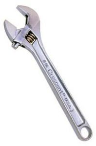
WrenchTo turn nuts and bolts or to prevent them from turning when loosening or tightening.
Lubricant (e.g., graphite, PTFE/Teflon)Lubricates moving parts and prevents them from corroding.
Screwdrivers (with various heads)
Slotted
Philips
Torx
Robertson
Pozidrive
Used to undo or tighten screws on a wheelchair.
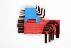
Hex keys (Allen keys)Used to turn sockets with a hexagonal head. You will either need metric or imperial hex keys, depending on your wheelchair.
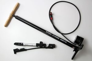
Tire pumpsStanding tire pumps are recommended as they are needed to pump tires up to over 50 psi.
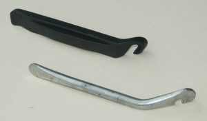
Tire leversUsed to lift the tire off and to access the inner tube of a tire.

Several parts of a manual wheelchair may require maintenance. We discuss these maintenance checks below. If you find anything wrong with your wheelchair, contact your wheelchair service provider.

Weekly maintenance

Tire inflation

To maximize pushing efficiency, it is important that your tires are always properly inflated. Weak research evidence suggests that tires deflated by more than 50% result in using more energy when pushing. However, it must also be noted that softer tires perform better on soft surfaces, such as grass or gravel. For general daily use, tires should always be inflated to the recommended values indicated on the side of the tire.

There are two ways to check tire pressure:

  1. For a more accurate reading, use a standing bicycle tire pump with a gauge.
  2. If you do not have a pump with a gauge, press down firmly on your tire with your thumb. If it presses in at all, it requires inflation.

Types of valves: Presta (left), Schraeder (right)

The amount of air required for your specific tire is indicated on the side of your tire. To inflate your tire, a standing pump, a gas station pump, or a hand pump may be used. Note that most hand pumps are not able to inflate tires over 50 psi; a high-pressure hand pump is required for tires. In addition, gas station pumps are only able to pump up Schraeder valves as Presta valves require an adaptor.

Refer to our article on Manual Wheelchairs for more information!

Cushion

Your cushion is essential to maintaining a good seated posture and for skin health. To ensure that your cushion is in its best shape, inspect the cushion and cover on a weekly basis. When inspecting the cushion cover, look for any holes, signs of wear, or flaking on the underside of the cushion, and make sure the zipper is working properly.

Maintaining your cushion depends on what kind of cushion you have:

  • For gel cushions: knead the gel from outside to inside. Ensure that the gel is redistributed, and that gel is present under areas of high pressure (e.g., in the area of your sit bones). In addition, ensure that there are no leaks in your cushion.
  • For foam cushions: check that the foam is not breaking down or crumbling anywhere.
  • For air cushions: ensure that your cushion is properly inflated. Check for leaks. If you think your cushion may have a leak, submerge it underwater and check for bubbles.

Monthly maintenance

Cushion and cushion cover

Keeping your cushion clean is important, as dirt on the cushion may lead to skin breakdown, and may leave a smell on your cushion. Once a month, wipe down the cushion with a clean damp cloth and soap. Wash the cushion cover in a washing machine, and follow the instructions in the cushion guide. Make sure to hang dry the cover, as placing the cushion cover in the dryer may result in shrinkage which may result in the cover being too small for your cushion.

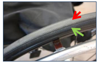
Intact (green) and worn out (red) tread on a wheelchair tire.15

Tires

The tires are a key component of the wheelchair subjected to daily wear. Inspect your tires once a month to make sure they are in good shape. Look for any signs of wear, cracks, bulges, looseness, damage, or flat spots.

Wheel bearing

The wheel bearings are located within the hub of the rear tire, and help to allow the wheel to turn freely and smoothly. Bearings normally wear out over time with use. You will know it is time to replace a bearing if you start hearing a knocking, or more infrequently, a squeaking sound as you wheel. If you suspect that you need your wheel bearing replaced, contact your local wheelchair service provider.

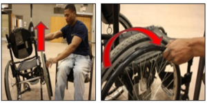
The wheel bearing should be tightened to a happy medium: a wheel bearing that is too loose may result in side to side movement of a wheel, while a wheel bearing that is too tight may result in additional resistance, resulting in an increased amount of energy spent while using your wheelchair. To check that the wheel bearings are not too tight or too loose, lift one side of the wheelchair up and spin the wheel. The tire should spin easily and should not slow and stop quickly after being spun. After the wheel stops spinning, it should spin backwards a little and should not wiggle side to side too much.

Large wheel axle

Rear wheels may be fixed (i.e., not removable) or quick release (i.e., removable). To ensure that the wheels are in place and are not loose, wiggle the wheel in all directions. If you have a fixed axle, there should be no play in the wheel. If you have a quick-release axle, some play is acceptable.

If you have a quick-release axle, test the release mechanism and ensure that the wheel securely locks back in place. A wheel that does not latch back in securely may result in an accident and should be addressed as soon as possible.

Wheel alignment – wheelchair tracking

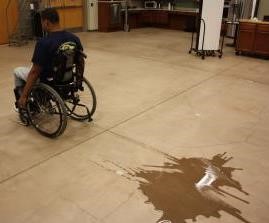
When looking at your wheelchair from above, the two rear wheels should be parallel to each other. Having wheels that are misaligned may result in greater energy expenditure and veering of the wheelchair when pushing. Needing to constantly correct for a veer when pushing may result in reduced control over the direction that a wheelchair is moving in, a strain on one arm and/or an increased use of energy.

To check whether your tires are aligned, roll through a puddle of water and allow the wheelchair to coast. The wheelchair should maintain its direction, and the tracks of the chair should be straight.

Spokes

Spokes are attached from the wheel rim (outer part of the wheel) to the hub (center part of the wheel), and help to distribute the forces of wheeling, such as the weight of the user, wheeling over surfaces, and braking. The spokes on a wheel act to prevent the tire from collapsing and adds stiffness to a wheel by acting as an anchor for the hub of the wheel.
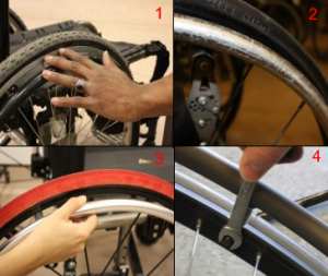
When inspecting your spokes, you want to check that none of the spokes are bent and that there is enough tension in the spokes. Having enough tension in each of the spokes is particularly important, as having one loose spoke will lead to others becoming loose. Signs of loose spokes include a faint metallic snapping sound as you move. To check tension in the spokes, you have two options:

  1. Squeeze the spokes in pairs around the entire wheel. If a spoke gives when being squeezed gently, it may be loose.
  2. The ping test: spin the wheel and hold a pencil against each spoke. You should hear a normal pinging sound. Any spoke that sounds off indicates a loose spoke.
Wheel locks
Casters

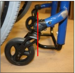
Caster stem that is not aligned with the caster wheel.19

Casters are the small wheels found on the front of the wheelchair that help to stabilize the wheelchair. Begin by inspecting your casters for wear, cracks, looseness, tears, and bulges.

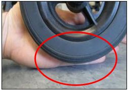
An example of a floating caster. Note the space between the bottom of the wheel and the ground.18

Secondly, to ensure that the casters are effectively stabilizing the wheelchair, check that both caster wheels are in contact with the floor and that the caster stem is aligned vertically. As the casters are required for maneuvering, it is important to ensure that they are able to turn freely around the axle. Check the casters for fluttering, or a shimmy/rapid vibration of the casters when moving.

Clean the caster wheel. Remove any dirt, lint, or hair that may have been collected in the caster axle using scissors, pliers, or tweezers. Further, clean the caster using a clean damp cloth or with a toothbrush.

Caster bearing

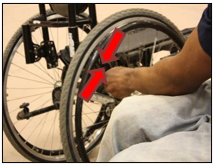
1. Inspect for wear. 2. An example of a handrim with scratches. 3.Inspect the tightness by pulling out on the hand rim. 4. Tighten as necessary.21

Like the rear wheel, casters consist of wheel bearings to ensure smooth rolling. To check that the bearings are at a happy medium in tension, spin the caster wheels and the caster assembly, and push the caster side to side. Grinding and excessive play in the caster bearings are indicative of a problem.
Handrim

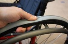
The handrims are rings connected to the rear wheel by bolts, and are used to propel the wheelchair. Most handrims are made out of plastic or metal (e.g., aluminum, steel), and may be coated in vinyl for extra grip. It is recommended to inspect your handrims monthly for wear, dents, cracks, or bends – they should be smooth all around. In addition, make sure that the hand rims are not loose. If loose, try tightening the bolts that connect the handrims to the wheel.

Frame

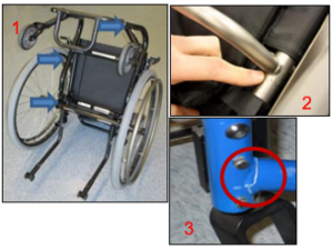
1. Common weld points.
2. Inspect the weld points.
3. Example of a cracked weld.22

Wheelchair frames consist of a series of metal tubes that have been welded together. Each month, check the welds to make sure that the tubes are held together. Inspect the frame for cracks or fractures. In addition, wipe down the frame each month with a clean damp rag. A toothbrush may be used to remove more difficult dirt. Avoid using a hose, power washer, or washing your wheelchair in the shower as it may cause the bearings to rust.

Nuts and Bolts

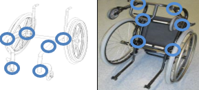
Common sites of nuts and bolts on manual wheelchairs.23

There are many nuts and bolts used on a wheelchair to hold various parts together. Loose nuts and bolts on your wheelchair may not only lead to rattling noises, but may not hold the part correctly and may fall out. Check the nuts and bolts on your wheelchair, and tighten them if loose. Make sure not to over tighten the nut or bolt, as it could damage the part or increase wheeling resistance.

Backrest

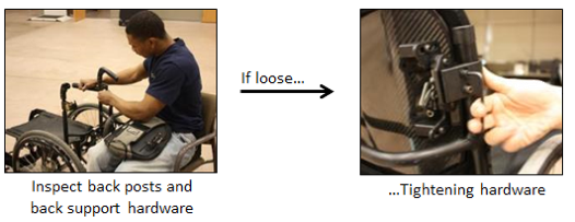
As the backrest is used to support your sitting posture and can impact your skin health, it is important to check that it is in good shape. To do so, check the upholstery for tears, wear, stretching, or metal parts that have poked through. If you have a rigid back, check that the backrest does not wiggle and is tightly secured. In addition, make sure that the backrest height is level. It is possible for a backrest bracket to become loose, resulting in one side of the backrest being higher than the other.
If you have an adjustable sling back, observe the tension of the backrest as it may stretch over time. Adjust the back as needed.

Foot support

The foot support is often the first part of the wheelchair that comes into contact with obstacles. For example, it may be used to help open doors, act as bumpers, and may be scraped along the ground. As the foot support is used to help maintain posture, it is important to keep it intact. Inspect the footrest to ensure that it is not loose. If you have swivelling foot rests, ensure that they swing away with ease, and can latch back properly. Also be sure to check the footrests for wear on the pins, bolts, and bushing, and tighten these parts if necessary.

Maintaining a power wheelchair may seem intimidating given the integration of electronics, but most activities are fairly simple. Below we discuss the tasks you should complete with your wheelchair.
Refer to our article on Power Wheelchairs for more information!

Daily

Battery

Properly charging your battery is important in maintaining its health. Do not charge the battery too frequently with little use, or let your battery completely die. If you are using your wheelchair every day, charge your batteries every night. Batteries should be charged for 8-12 hours, even though the charging lamp has gone off.

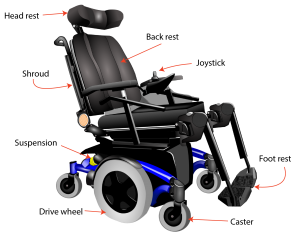
Plastic shrouds

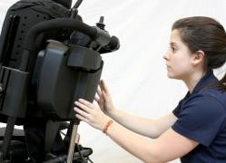
Shrouds are the plastic coverings that protect the electronics and the battery of the wheelchair from dirt and moisture. To check them, make sure that they are present and intact. Try to jiggle the shrouds around to ensure they are not loose.

Brakes

The brakes are essential to safe use of your wheelchair. On a power wheelchair, the brakes are connected to the motor. When you drive, they automatically disengage, and when you stop, they automatically re-engage with an audible clicking sound. If you suspect something wrong with your brakes, try the following:

    1. Turn down the speed of your wheelchair
    2. Push the joystick forward and then stop. Upon stopping, you should hear a clicking sound. This indicates that your brakes are working.

Weekly

Tire inflation, Cushion inspection

Similar to manual wheelchairs, pneumatic tires and cushions should be inspected on a weekly basis to optimize their performance. Refer to the tire inflation and cushion inspection instructions in the manual wheelchair maintenance section above.

Motor

The motor is an integral part of the wheelchair, as its job is to convert power from the battery into energy to move the chair. It is normal for the motor to make some noise when it is being used. Try to become accustomed to what your motor sounds like so you are able to detect any changes. Over time, it is normal for the motor to become a bit louder; however, excessive noise may be indicative of an issue. If you notice any sounds that you are unable to recognize, contact your wheelchair provider.

Controller and joystick

The controller of a wheelchair often consists of a power button, a screen, and a joystick. It is the interface used to control the driving, speed, and positioning of your wheelchair. Before inspecting your controller, make sure that it is switched off. There are two main aspects of inspecting the controller:

    1. Check the joystick and the rubber connection between the joystick and the control for any cracks or wear. This protective covering acts to keep dust, dirt, and moisture out of the electronics. And so damage to the covering may eventually lead to failure of the control.
    2. Check the wiring of the joystick. Ensure that none of the connecting cables are frayed or showing through the insulation.

Monthly

Cushion, cushion cover, and foot plates.

Cushions, cushion covers, and footplates should be maintained and inspected at least once a month. The maintenance and checking process for these items are similar to manual wheelchairs. For more information, refer to the section “What maintenance and checks should I do for my manual wheelchair?” above.

Tire

The treads on your tire play a key role in maintaining traction and maintaining the stability and maneuverability of the wheelchair. Some tires may have less tread than others; note how much tread your tire starts off with. Check the tire treads monthly to ensure that they are not worn.
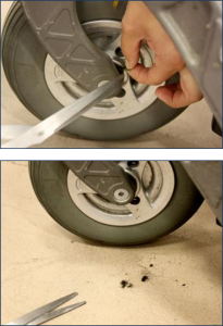
Caster

The axles of the front caster wheels of the wheelchair are the lowest to the ground, and thus are susceptible to picking up hair, dust, lint, and dirt. Buildup on your axles can lead to premature wearing and increased rolling resistance. For example, hair wrapped around the caster wheel can lead to breakage. Using a pair of scissors, tweezers, a toothbrush, or pliers, remove debris from the caster.

Frame

Inspect the frame and weld points on the wheelchair and ensure there are no cracks. Make sure all fasteners are appropriately tight.

Backrest

The backrest of your wheelchair is important for support and your posture. Maintaining the backrest of a power wheelchair is similar to a manual wheelchair. Refer to the manual wheelchair section for further details.

Wiring and electronics

There are many wires throughout a powered wheelchair that are essential to making the wheelchair move. To safeguard the use of your wheelchair, make sure that all of the wires are in place and free from dirt and corrosion. If you notice any exposed wires or corrosion on the wires, take it to a dealer. If wires are hanging out or are in the way of your day-to-day use, it may be beneficial to connect the wires to a support (e.g., armrest, frame, etc) as shown below.

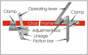
Diagram of the wheel locks and the bolts that can be adjusted.31

Wheel lock not locking wheels

If your wheelchair is still moving despite your wheel locks being on, first check to see if your wheels may be underinflated. Try inflating your tires to the recommended pressure. If the wheel locks are still ineffective, try adjusting the position of the wheel lock by loosening the bolts securing the clamp on the frame of the wheelchair. Slide the adjustment bar as required, and tighten up the bolts. If your brakes look like they are worn, contact your local wheelchair service provider for a replacement.

Wheelchair Keeps veering to one side

One way to check tracking is by rolling through a puddle of water. Take note of the water trail made from the wheels. Are they parallel and straight?32

As you push your wheelchair, it should travel straight forward. However, your wheelchair may sometimes veer, or pull to one side, when you intend to go forward in a straight line. Before trying to resolve this issue, ensure that your chair is actually pulling one way, and that it isn’t related to an uneven surface or unequal strength. To do this, propel your wheelchair forward as far as you can with one push. Note any deviations to a side. Turn around, and perform the same action in the opposite direction. This is to cancel out the effect of an uneven surface. If you have identified a pulling of your wheelchair in one direction, something is causing poor tracking. There are multiple reasons why your wheelchair may veer to one side when you are pushing.

  • Caster:
    • Vertical alignment of the caster may be off.
    • Caster fork may be misaligned.
    • Hair may be wrapped around one of the casters.
  • Tire:
    • Check to ensure that the tire pressure on both sides are equal.
    • Make sure that axles are mounted symmetrically on the frame.
  • Frame:
    • Make sure the frame is sitting evenly. Check that the footplates are sitting at an equal height.

Patching a flat inner tube

If you are using a wheelchair with air-filled tires, chances are you may encounter a flat tire. If you only have a patch kit on you, follow the instructions below on how to fix a tire with a patch kit. Depending on the extent of the damage done on your inner tube, a patch may suffice. Patch kits, tires, and inner tubes may be purchased from bicycle shops or wheelchair vendors.

  1. Deflate the tire as much as possible.
  2. Remove the inner tube from the tire. To do this, insert a tire lever under the edge of the tire above a spoke. Secure the hooked end of the tire lever around a spoke. Insert a second lever a few inches away from the first, and push down on it until that area of the tire flips over the rim. Slide this lever around the tire in a clockwise direction until the tire is removed.
  3. Remove the inner tube under the tire.
  4. Determine where the leak is by over-inflating the tire and listening/feeling for the air escaping. If you are unable to successfully locate the leak, submerge the air-filled tire underwater and watch for bubbles.
  5. Once you have identified the hole, mark it with a pen or marker.
  6. Use the sandpaper in the patch kit to sand the area around the hole. This will help the patch adhere to the tube better.
  7. Let the air out and apply a thin layer of rubber cement over the hole. Make sure you spread the cement over an area large enough to encompass the patch. Wait for the cement to dry.
  8. When the cement is dry, apply the patch firmly to the inner tube. Now we are ready to put the tire back together.
  9. Inflate the tube until it holds its shape.
  10. Find the valve and align it with the valve hole on the rim. Use your hands to knead the tire back onto the rim. You may need to use your tire levers to help put the last bit of the tire back onto the rim, but be careful not to pinch the inner tube.
  11. Re-inflate the tube to the recommended value on the tire wall.

To replace an inner tube instead of patching it, skip steps 4-8.

Fixing a leaky air cushion

If you notice a leak in your air cushion, it can be easily repaired with a patch. While ROHO cushions come with a patch, other brands may require you to order some from the manufacturer.

  1. Determine where the leak is. To do so, inflate your cushion and submerge it underwater. Where you see bubbles is indicative of the leak spot.
  2. Mark the hole by placing a toothpick into the hole.
  3. Allow the cushion to completely dry by laying it out on a towel.
  4. Clean the area around the hole using the alcohol wipe provided. Let it dry.
  5. Peel the backing off of the patch and place it over the hole. Firmly press on the patch until there is a good seal.
  6. Reinflate the cushion

Maintaining your wheelchair is important to its longevity and its performance. Completing various inspections and simple maintenance tasks on a regular basis is fairly simple, and can be done by yourself or a family, friend, or caregiver.

It is best to discuss all wheelchair modifications and big maintenance with your wheelchair provider should you find any major issues. This article is not intended to replace yearly professional wheelchair maintenance/tune-ups.

For a review of how we assess evidence at SCIRE Community and advice on making decisions, please see SCIRE Community Evidence.

Evidence for “Why is wheelchair maintenance important” is based on:

Chen WY, Jang Y, Wang JD, Huang WN, Chang CC, Mao HF, Wang YH. Wheelchair-related accidents: relationship with wheelchair-using behavior in active community wheelchair users. Archives of physical medicine and rehabilitation 2011;92(6):892-898.

Evidence for “What maintenance should be done for a manual wheelchair?” is based on:

Boninger, M., Kirby, R.L., Oyster, M., Pearlman, J. Cooper, R.A.,…Toro, M. (2017). Wheelchair maintenance training program: Clinician’s reference manual. Retrieved from: http://www.upmc-sci.pitt.edu/node/924

Golden, J., Colescott, D. (2017). Manual wheelchair maintenance checklist [PDF]. Retreived from: http://sci.washington.edu/summit2017/MANUAL_WHEELCHAIR_MAINTENANCE_CHECKLIST-SCI_Summit2017.pdf

Manual Wheelchair Maintenance Checklist.(n.d.). Retreived from: http://sci.washington.edu/summit2017/MANUAL_WHEELCHAIR_MAINTENANCE_CHECKLIST-SCI_Summit2017.pdf

Model systems knowledge translation center (2018). Maintenance guide for users of manual and power wheelchairs [PDF]. Retrieved from: https://msktc.org/sci/factsheets/maintenance-guide-users-manual-and-power-wheelchairs

Sawatzky BJ, Miller WC, Denison I. Measuring energy expenditure using heart rate to assess the effects of wheelchair tire pressure. Clinical Rehabilitation 2005;19(2):182-7.

Evidence for “What maintenance should be done for a power wheelchair?” is based on:

Model systems knowledge translation center (2018). Maintenance guide for users of manual and power wheelchairs [PDF]. Retrieved from: https://msktc.org/sci/factsheets/maintenance-guide-users-manual-and-power-wheelchairs

Boninger, M., Kirby, R.L., Oyster, M., Pearlman, J. Cooper, R.A.,…Toro, M. (2017). Wheelchair maintenance training program: Clinician’s reference manual. Retrieved from: http://www.upmc-sci.pitt.edu/node/924

Evidence for “What are some simple repairs I can do?” is based on:

Denison, I. (2006). Wheelchair maintenance series [PDF]. Retrieved from: http://www.vch.ca/Documents/Equipment-Wheelchair-Maintenance-Series.pdf

Image credits

  1. Crescent brand 8-inch adjustable wrench. ©Rico402. CC0 1.0
  2. WD-40 Specialist Dirt & Dust resistant dry lube PTFE spray. ©WD-40 2020
  3. Screw head – slotted. ©Inductive load. Public domain.
  4. Diagram of a screw head – Phillips. ©Inductive load. Public domain
  5. Diagram of a screw head – Pozidrive. ©Inductive load. Public domain
  6. Diagram of a screw head – Robertson square drive. ©Inductive load. Public domain
  7. Diagram of a screw head – Torx. ©Inductive load. Public domain
  8. Construction tool hardware construct allen key. ©Max Pixel. CC0 1.0
  9. Lezyne and Topeak Road Morph bike pumps. © CC-BY-2.0
  10. Different kinds of handicapped equipment. © Modified by the SCIRE Community Team
  11. Tyre Levers ©Ian Harvey. CC0
  12. Checking tire pressure. ©Manual Wheelchair Maintenance Program. CC-BY-NC-ND-3.0.
  13. Types of tire valves. ©Manual Wheelchair Maintenance Program. CC-BY-NC-ND-3.0.
  14. Checking cushion cover. ©Manual Wheelchair Maintenance Program. CC-BY-NC-ND-3.0.
  15. Tire treads. ©Manual Wheelchair Maintenance Program. CC-BY-NC-ND-3.0.
  16. Checking the wheel bearing. ©Manual Wheelchair Maintenance Program. CC-BY-NC-ND-3.0.
  17. Inspecting spokes. ©Manual Wheelchair Maintenance Program. CC-BY-NC-ND-3.0.
  18. Floating caster. ©Manual Wheelchair Maintenance Program. CC-BY-NC-ND-3.0.
  19. Misaligned caster stem. ©Manual Wheelchair Maintenance Program. CC-BY-NC-ND-3.0.
  20. Cleaning caster. ©Manual Wheelchair Maintenance Program. CC-BY-NC-ND-3.0.
  21. Inspecting handrim. ©Manual Wheelchair Maintenance Program. CC-BY-NC-ND-3.0.
  22. Inspecting wheelchair frame. ©Manual Wheelchair Maintenance Program. CC-BY-NC-ND-3.0.
  23. Nuts and bolts locations. ©Manual Wheelchair Maintenance Program. CC-BY-NC-ND-3.0.
  24. Checking backrest. ©Manual Wheelchair Maintenance Program. CC-BY-NC-ND-3.0.
  25. Modified from: Wheel isolated ©MBGX2, Pixabay License
  26. Inspecting shrouds. ©Power Wheelchair Maintenance Program. CC-BY-NC-ND-3.0.
  27. Checking the braking mechanism. ©Power Wheelchair Maintenance Program. CC-BY-NC-ND-3.0.
  28. Tire treads. ©Power Wheelchair Maintenance Program. CC-BY-NC-ND-3.0.
  29. Caster wheels. ©Power Wheelchair Maintenance Program. CC-BY-NC-ND-3.0.
  30. Tying up wires. ©Power Wheelchair Maintenance Program. CC-BY-NC-ND-3.0.
  31. Wheel lock. ©Ian Denison. Used with permission
  32. Wheelchair tracking. ©Manual Wheelchair Maintenance Program. CC-BY-NC-ND-3.0.


Disclaimer: This document does not provide medical advice. This information is provided for educational purposes only. Consult a qualified health professional for further information or specific medical advice. The SCIRE Project, its partners and collaborators disclaim any liability to any party for any loss or damage by errors or omissions in this publication.

Robotic Exoskeletons

By | | No Comments

Author: Sharon Jang | Reviewer: Riley Louie | Published: 7 April 2021 | Updated: ~

New technology has led to the creation of wearable robotic devices to improve leg movement for activities such as standing and walking. This page discusses the use of robotic exoskeletons in people after spinal cord injury (SCI).

Key Points

  • Robotic exoskeletons are electromechanical devices that are worn around the limbs to support activities such as standing and walking in SCI people.
  • In addition to improved mobility, robotic exoskeletons allow increased levels of activity for general health benefits related to regular exercise.
  • Limitations of using exoskeletons include their high costs, limited availability, and restricted use in real-world settings.
  • Moderate evidence shows that exoskeletons increase safety and decrease energy requirements during walking for people with thoracic SCI.

Robotic exoskeletons (also known as powered gait orthoses) are wearable electromechanical devices that enhance movement of weak or paralyzed legs. They are electrically powered at the joints, allowing the hips, knees, and ankles to move. The greatest advantage exoskeletons have over passive orthotics or braces is that they are programmed to enable coordinated movement without much effort from the user. This is especially so since exoskeletons carry their own weight as well as that of the user. Movements that most exoskeletons assist include sit-to-stand and walking.

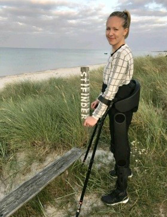
As of 2019, many different models of exoskeletons exist, and this number continues to grow as research and technology progresses. However, it is important to note that only three have been approved by the FDA for sale in North America and only two have been approved for home use, while the other is only approved for research and rehabilitation purposes. The two that are approved for home use are the ReWalk and the Indego, while the Ekso has been approved for research and rehabilitation purposes. The current models of exoskeletons have the ability to move from 0.2-2.6 km/hr, and weigh between 12-38 kg.
Exoskeletons are slowly moving into the community. Currently, most models can only move over flat, smooth surfaces. However, some can go up inclines. Moreover, newer models have the added function of enabling the user to sit in or wheel a wheelchair without having to take off the device. These features can increase the independence of people with SCI and enable/enhance the performance of activities such as standing, walking, and climbing stairs. However, these devices are still expensive, ranging from $70,000-120,000 USD.

In deciding if robotic exoskeletons are an appropriate option for you, a health care provider will perform an assessment where they account for factors such as your level of injury, risk for falls and fractures, and range of movement. Once exoskeletons are deemed appropriate, training will be required to learn how to properly use the device. A physiotherapist or caregiver may help you put on and take off the device. Generally, the feet are placed on footplates and the torso, hips, and legs are strapped into the exoskeleton. Some models extend upwards to include a backpack-like structure, which offers more trunk support and contains the computer and battery. Other models are secured only at the waist and below. In addition, some models can be programmed externally using a tablet. A pair of forearm crutches or a walker is often used to maintain balance. As someone with SCI walks, the built-in sensors and motorized joints (hip, knee, and/or ankle) constantly accommodate to encourage a rhythmic walking pattern. When the individual improves their own walking function and requires less assistance, the exoskeleton can be adjusted to provide less support.


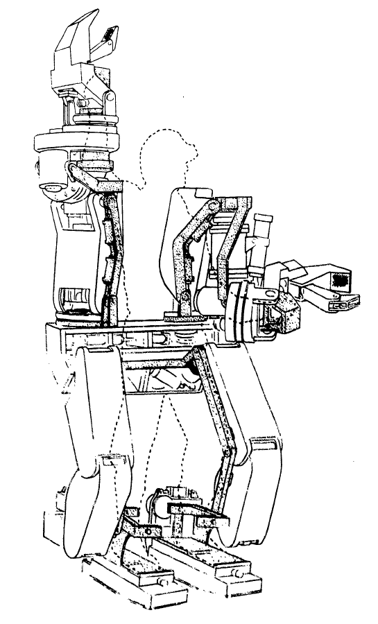
A conceptual diagram of the Hardiman.

The term “exoskeleton” was originally borrowed from animal biology. An exoskeleton (such as a shell) is an outer cover that protects and supports the animal. In a similar way, robotic exoskeletons have been designed to help externally support and enhance human movement.

The earliest exoskeletons were developed with a military focus, aimed to help soldiers carry heavier loads, run faster, and jump higher. In 1968, the first exoskeleton, dubbed “the Hardiman” was developed in partnership with the US military. This exoskeleton was originally designed to help amplify soldiers’ strength by 25 times (i.e., lifting a 1500 item would feel like you are lifting a 60 lb item). Although the Hardiman was created and worked, it was not without limitations. First of all, the exoskeleton itself weighed 1500 lb. It was hydraulically-powered and required pumps and bladders that could fill a room. Despite its abilities, it was not very functional.

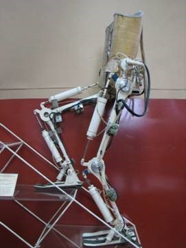
In 1972, a team from Yugoslavia developed the first functional exoskeleton: the kinematic walker. This device was the first of its kind; a powered robot consisting of a single hydraulic actuator (motor), which reduced its size. The kinematic walker was designed as a walking orthotic, and allowed for smooth movements to be made. Some movements the exoskeleton was able to perform included flexion and extension of the hip, knee, and ankle, in addition to abduction and adduction of the legs. Although this exoskeleton only weighed 12 kg, it required a separate power source and a computer, which were externally located from the exoskeleton.
For more information on treadmill-based robotics, refer to our articles on Body Weight Supported Treadmill Training and Functional Electrical Stimulation for more information on treadmill based robotics.

In 2001, the first exoskeleton products started being sold. The Lokomat, a robotic exoskeleton that was suspended over a treadmill, was one of the first exoskeletons used for rehabilitation. Meanwhile, the US Defense Advanced Research Projects Agency (DARPA) had announced their Human Performance Augmentation program which offered funding to develop exoskeletons for military use. From this funding, two separate groups developed exoskeletons – the Berkeley Lower Extremity Exoskeleton (BLEEX) and the Raytheon XOS suit. Both suits were created to help soldiers carry extra weight (up to 200 lb!) without feeling it.

Starting in 2010, more rehabilitation-based exoskeletons started entering the market. These include gait-assistive devices commonly seen in the media today, such as the Ekso, the ReWalk, and the Indego exoskeletons. These models are described in the section below


There are many types of exoskeletons that are currently available for use for individuals with SCI. While some exoskeletons have only been cleared for rehabilitation purposes, others have separate models for personal or home use. Below we discuss the general differences between rehabilitation and personal exoskeletons.

Rehabilitation Exoskeletons

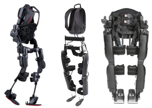
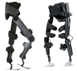
The Ekso (left), the ReWalk (middle), and the REX (right) exoskeletons.4-6

Exoskeletons used for rehabilitation are generally heavier than personal exoskeletons and are often controlled by a computer and battery located in a backpack worn by the user. However, rehabilitation exoskeletons often come with more customizable abilities. For example, the Ekso and the ReWalk allow the clinician to modify the amount of power (i.e., support from the robot) for each leg, depending on the ability of the user. Similarly, the therapy version of the Indego allows the clinician to control how much weight is supported by the exoskeleton and how much movement assistance is provided by the exoskeleton. Moreover, how steps are triggered can be programmed in different ways: e.g., by the press of a button, by shifting your body weight, or by initiating a step using your own muscles. These bulkier clinical models are usually one-size-fits-all, allowing clinicians to adjust the dimensions of the device (i.e., leg length, hip width, etc.) to treat their clients of varying body sizes.

The REX differs from all of the aforementioned exoskeletons in that it is controlled with a joystick and is able to self-balance, making it essentially “hands-free”. This feature allows individuals to perform exercises while standing, including squats, lunges, and upper body exercises using both hands.

The Indego (left) and the ReWalk (right).

Personal Exoskeletons

Exoskeletons that have been designed for home use are generally lighter and provide limited support to the torso. Personal exoskeletons have a battery pack that is connected to the waist, but otherwise operates similarly to the rehabilitation models in that a weight shift initiates a step. Furthermore, some exoskeleton companies have developed apps that accompany the exoskeleton, allowing the user to independently access performance data. These personal exoskeletons are usually custom-developed to fit the user’s unique body dimensions.

While there are many purported benefits of using robotic exoskeletons among individuals with SCI, strong research evidence is lacking. Exoskeletons are largely used to enhance mobility in SCI, where they can be used for walk (gait) training during rehabilitation or in the community (at home) to perform simple daily activities. Although further research is required, some research has suggested that walking with an exoskeleton may have additional health benefits:

Spasticity

There are mixed findings regarding the effects of exoskeletal walking on spasticity. Many (weak evidence) studies have found that using exoskeletons can result in decreased spasticity. Despite these noted benefits, one weak evidence study reported mixed findings on spasticity as 26.7% of their study sample saw a decrease in spasticity, while 62.2% of participants saw no change and 11.1% saw an increase in spasticity. The impact of exoskeletal walking on spasticity may be related to the user’s baseline level of spasticity. In a weak evidence study, users who had low levels of spasticity prior to using an exoskeleton experienced an increase in spasticity; however, this increase in spasticity ultimately decreased over 12 weeks back to near zero. Individuals who already had high levels of spasticity prior to walking in an exoskeleton saw no changes in spasticity.

Bowel function

There is some weak research evidence that suggests walking with an exoskeleton can help with various bowel functions, including improved regularity of bowel movements, less time required for bowel management, and decreased enema dose. However, two (weak evidence) studies have found no effects on bowel function.


Bone Health

After SCI, bone mineral density in the legs declines at rapid rates due to inactivity and weight bearing activities have the potential to help restore bone mineral density. One study (weak evidence) found that walking in an exoskeleton may increase bone mineral density up to 14% with 6 weeks of training.

Fitness

Some research (weak evidence) suggests that walking in an exoskeleton can provide good exercise for the heart and upper and lower limb strength in those with incomplete SCI. More details about the effect of exoskeletons on fitness are discussed below in the section: “Can I get exercise benefits from walking in an exoskeleton?”

Pain

A few studies (weak evidence) report a decrease in pain with exoskeleton use, with one noting a reduction in pain, but not enough to significantly impact everyday life (i.e., clinical significance). On the other hand, some weak evidence studies have found no effect of exoskeleton walking on pain.
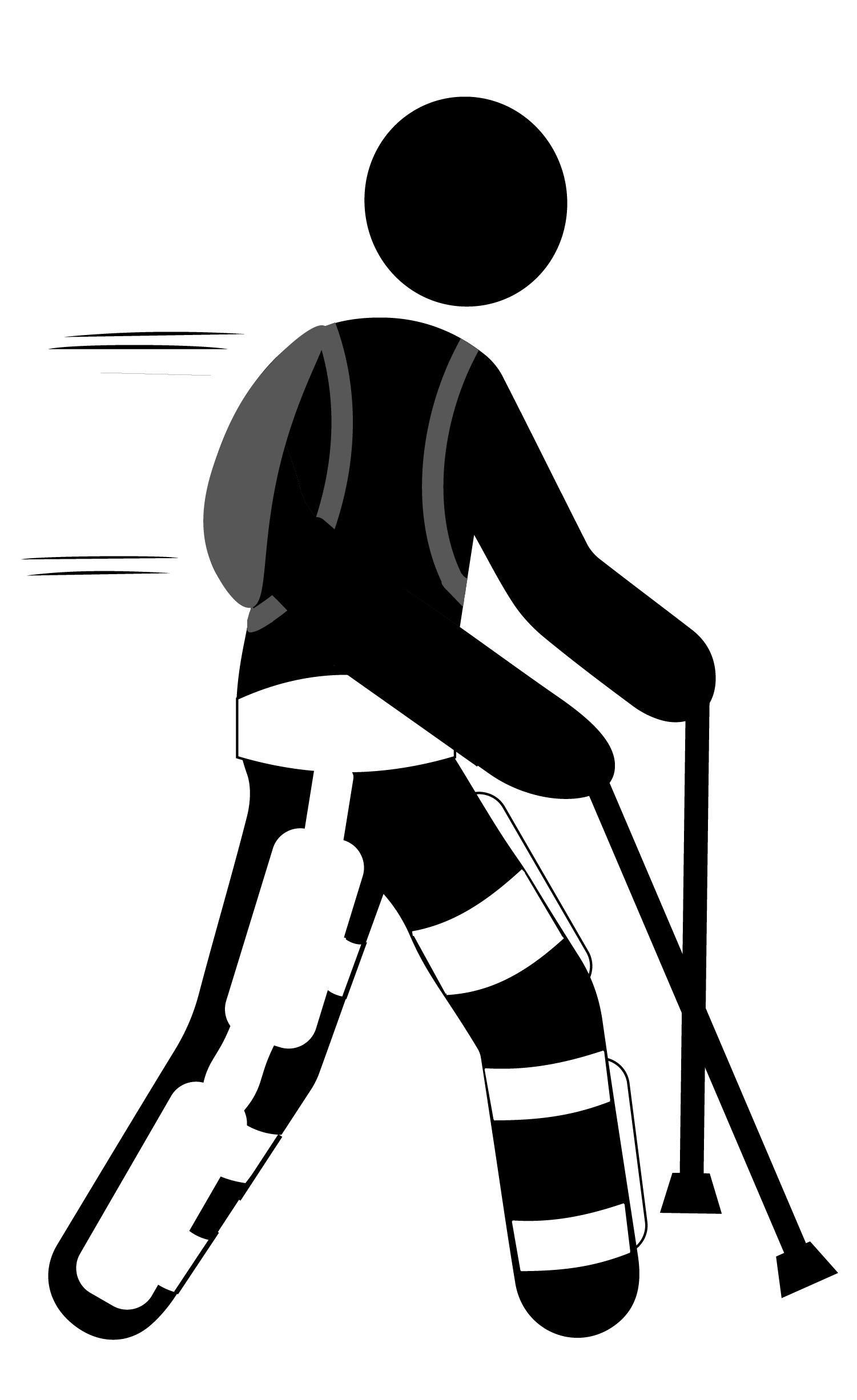
Pressure Sores

There is some weak research evidence that walking in an exoskeleton may help avoid the negative effects of prolonged standing or sitting  (e.g., pressure ulcers).


Refer to our articles on Pressure Sores, Pain and Spasticity for more information!

While a number of benefits are associated with exoskeleton use, there are also factors to consider prior to using the device in therapy or for the long term.

Risks of using an exoskeleton

Robotic exoskeletons are generally safe when used with discretion. However, there are some risks associated with its use. Mild adverse effects that have been reported in research include: skin redness, small abrasions (i.e., scrapes), mild joint swelling, and mild bruising. Additionally, like other simple orthotics and braces, falls and fractures have been identified as a risk. It is suggested (weak evidence) that family and friends of exoskeleton users should be trained to deal with emergency situations, such as falls or the exoskeleton shutting off unexpectedly.

Considerations of using an exoskeleton in the community

While exoskeleton home use may seem promising, there are some limitations. These include:

  • Slow walking speeds, which may not be ideal for everyday activities.
  • High costs to purchase the device.
  • Lack of availability of community-based exoskeletons.
  • Limited capacity or poor efficiency in moving on uneven surfaces (e.g., hills, steps) or complex movements (turning, side-stepping, backwards walking).
  • Being prone to water damage (they are not waterproof).

There are certain situations where extra attention is needed to determine whether robotic exoskeletons are appropriate and safe. Consult a qualified health provider for further safety information. Robotic exoskeletons are not recommended for individuals:

Extreme contractures are a contraindication for using exoskeletons.

  • Who are unable to tolerate standing, even with an assistive device (walking frame, bracing), due to pain or other complications (autonomic dysreflexia, orthostatic hypotension).
  • With severe neurological injuries (apart from SCI).
  • With severe or uncontrolled spasticity.
  • With osteoporosis.
  • With fractures.
  • With severe contractures (deformities that cause joint and muscle stiffness and limit normal or functional movement of the limbs).

Using Functional Electrical Stimulation (FES) to counter spasticity

Researchers built a novel device that integrates FES into an exoskeleton to address the issue of severe spasticity affecting exoskeleton use. In this FES-exoskeleton hybrid, FES complemented the exoskeleton by stimulating tight extensor muscles to facilitate walking. The authors found that spasticity was temporarily reduced when FES was used when walking with an exoskeleton. Furthermore, it was found that the knee was more easily extended when moving from a sit-to-stand position, and the forces applied on the knee during sit-to-stand were reduced. While this (weak evidence) study provides some promise for individuals with severe spasticity to also use exoskeletons, further research is required.

Using an exoskeleton requires practice. Although walking in an exoskeleton may seem daunting at first, research suggests that walking proficiency improves over time. In both newly injured (i.e., less than 6 months since injury) and chronically injured individuals with SCI, weak evidence shows that walking in the exoskeleton improves over time.

You get faster

Among newly injured individuals, weak evidence from one study suggests that walking speed in an exoskeleton becomes 3.2x faster after 25 1-hour training sessions. Moreover, these individuals were able to walk further in an exoskeleton after their training sessions. Among chronically injured individuals, similar trends are seen with increases in exoskeleton walking speed in two weak evidence studies. In another weak evidence study, it was found that 21 sessions were required to achieve the near-maximal walking speed at the end of a 12-week period, while 62 sessions were required to achieve near-maximal walking distance.

Less effort is Required

There is both weak and moderate evidence suggesting that the amount of effort it takes to walk using an exoskeleton decreases over time. This has been evaluated both subjectively (i.e., people feel like walking in an exoskeleton is not as hard over time) and physiologically (i.e., less demand on your body). This suggests that individuals are able to walk longer distances with lower effort after training to use an exoskeleton.

Is using an exoskeleton different for those with acute injuries versus chronic injuries?

Researchers have noted differences in exoskeleton use among newly injured (e.g., in patients) and chronically injured individuals with respect to adverse effects and benefits received. Among people who have recently sustained an SCI, weak evidence indicates that the most common adverse effect was orthostatic hypotension (a sudden drop in blood pressure). One study found that orthostatic hypotension commonly occurred after the first stand or after pauses (e.g., to take vitals, or to turn). However, the frequency of orthostatic hypotension episodes tapered off after a couple of sessions. Furthermore, another study (weak evidence) suggests that newly injured individuals may see improvements in their independence and quality of life, whereas those with chronic injuries do not. More research is required to determine the significance of differences between acutely injured and chronically injured individuals who use exoskeletons.

What factors influence walking speed?

Among individuals with SCI who use current exoskeleton devices, the average walking speed is 0.26 meters per second. This speed is fairly slow, and is lower than the average speed required to walk proficiently in the community (0.8 meters per second) and to cross the street safely (1.06 meters per second). However, walking speed in an exoskeleton is subject to improvement, depending on various factors.

Some factors that influence walking speed include age, level and type of injury, and the amount of training one receives. There is some (weak) evidence suggesting that those with incomplete, lower level SCI are more likely to exhibit faster walking speeds. In particular, one (weak evidence) study found that those with lower level paraplegia (i.e., T9-L1) were able to walk at significantly higher speeds. Moreover, there was a weak correlation between older age and faster walking speeds (i.e., older adults walk slightly faster than younger adults), though this could be related to the age-related difference in injury severity (that is, older individuals had lower levels of injury). No correlation was found with a greater time since injury.

What factors influence skill acquisition?

The time it takes to acquire proficient skills to walk in an exoskeleton varies greatly, ranging from 6-23 sessions. A variety of factors influence skill acquisition, including lifestyle, age, age at injury, and body mass index (BMI). Weak evidence suggests that an active lifestyle is the most important predictor of skill performance, although being younger and having a lower BMI are also associated with higher skill level. Additionally, the authors note that while having a lower level of injury was a positive predictor of skill between 2-4 weeks of using an exoskeleton, it did not predict final skill levels.

The type of exoskeleton being used may also influence skill acquisition. For example, weak evidence notes that a device with more support to the torso may facilitate skill acquisition as it provides more stability. Although we have summarized the research on factors influencing skill acquisition, the type of exoskeleton used in each study was not accounted for. As a result, we are unable to tease apart the effects of the aforementioned factors (e.g., lifestyle, age, BMI) and of the exoskeleton type on acquiring skills. The time it takes to acquire proficient skills to walk in an exoskeleton varies greatly, ranging from 6-23 sessions. A variety of factors influence skill acquisition, including lifestyle, age, age at injury, and body mass index (BMI). Weak evidence suggests that an active lifestyle is the most important predictor of skill performance, although being younger and having a lower BMI are also associated with higher skill level. Additionally, the authors note that while having a lower level of injury was a positive predictor of skill between 2-4 weeks of using an exoskeleton, it did not predict final skill levels.

Although walking in an exoskeleton is used primarily for rehabilitation purposes, the effort required to use the device is strenuous enough to be considered exercise. For example, one (weak evidence) study found that walking in an exoskeleton requires 3.34 times more effort than pushing a wheelchair, and 1.9 times more effort compared to walking without impairment, despite walking 7.4 times slower. Not surprisingly, participants also perceive themselves to be working harder. Participants from three (weak evidence) studies exercised at a moderate intensity, which is enough to get cardiovascular benefits, while walking in a robotic exoskeleton. Although some participants from a (weak evidence) study reported working at a low intensity, the authors noted that based on their heart rate and oxygen consumption, they were actually working at a moderate intensity. This suggests that some people may actually be working their bodies harder than they feel they are!

So why is walking in an exoskeleton so much work? Research suggests that using an exoskeleton requires a lot of work from the arms and torso to support an upright posture and to shift weight to initiate stepping. However, relative to other walking orthotics (e.g., robotic gait orthoses, hip-knee-ankle-foot orthoses), FES, and bracing, walking in an exoskeleton is considerably less effort. So then why would you use an exoskeleton for exercise if using other walking orthotics is harder work? The important consideration is stamina. You might work harder walking with rigid braces, but you may tire out quickly. With an exoskeleton, the understanding is that the assisted walking could allow you to exercise at moderate intensity for much longer.

Currently, there are only two exoskeleton models that have been approved for community use in North America. As such, there is limited evidence for using an exoskeleton in the community. In order to use an exoskeleton independently, individuals should be able to put on and take off the exoskeleton without professional help. Weak evidence has shown that individuals with paraplegia are able to independently put on and take off these devices, although those with tetraplegia are not. Additionally, weak evidence has shown that the time it takes to put on and take off an exoskeleton can be reduced with practice. Regarding walking speed in indoor versus outdoor environments, one weak evidence study has found that there are no significant differences in speed.

Some studies have looked at home- and community-based skills that can be completed using an exoskeleton. One weak evidence study found that the majority of participants could walk independently without a trainer in an exoskeleton, and perform tasks such as reaching high cupboards, using a stove, and using a sink, but were not able to walk on carpet and ramps. However, the authors note that some tasks were more difficult to complete in an exoskeleton, including reaching low cupboards and opening a fridge and getting items. Other (weak evidence) studies have found that a small proportion of people were able to do more advanced community-based tasks, such as entering/exiting elevators, operating automatic doors, navigating revolving doors, and ordering at a café. More research is required to determine how quickly individuals can pick up these skills.

There are currently many models of robotic exoskeletons continuing to be developed and refined. Exoskeletons are primarily used for rehabilitation purposes, although some models are available for community use. Using an exoskeleton has been shown to be relatively safe and easy to learn. Many benefits have been reported, including being able to ambulate, improvements in bone health, heart health, spasticity, bowel functioning, fitness, and pressure sores. While there are a lot of positive findings for robotic exoskeletons, this is an emerging field and stronger research is required to support these beneficial claims. A strong consideration is to weigh these benefits against cost, and also compare how these benefits compare to other to achieve similar gains.

For a review of how we assess evidence at SCIRE Community and advice on making decisions, please see SCIRE Community Evidence.

Parts of this page have been adapted from the SCIRE Project (Professional) “Lower Limb” Chapter:

Lam T, Wolfe DL, Domingo A, Eng JJ, Sproule S (2014). Lower Limb Rehabilitation Following Spinal Cord Injury. In: Eng JJ, Teasell RW, Miller WC, Wolfe DL, Townson AF, Hsieh JTC, Connolly SJ, Noonan VK, Loh E, McIntyre A, editors. Spinal Cord Injury Rehabilitation Evidence. Version 5.0. Vancouver: p 1-74.

Available from: scireproject.com/evidence/rehabilitation-evidence/lower-limb/ 

Evidence for “What are robotic exoskeletons?” is based on the following studies:

Khan, A. S., Livingstone, D. C., Hurd, C. L., Duchcherer, J., Misiaszek, J. E., Gorassini, M. A., … Yang, J. F. (2019). Retraining walking over ground in a powered exoskeleton after spinal cord injury: a prospective cohort study to examine functional gains and neuroplasticity, 1–17. Retrieved from https://doi.org/10.1186/s12984-019-0585-x

Ali, H. (2014). Bionic Exoskeleton: History, Development and the Future. IOSR Journal of Mechanical and Civil Engineering (IOSR-JMCE), 2014, 58–62. Retrieved from http://iosrjournals.org/iosr-jmce/papers/ICAET-2014/me/volume-5/12.pdf?id=7622

Gardner, A. D., Potgieter, J., & Noble, F. K. (2017). A review of commercially available exoskeletons’ capabilities. 2017 24th International Conference on Mechatronics and Machine Vision in Practice, M2VIP 2017, 2017Decem, 1–5.

He, Y., Eguren, D., Luu, T. P., & Contreras-Vidal, J. L. (2017). Risk management and regulations for lower limb medical exoskeletons: A review. Medical Devices: Evidence and Research, 10, 89–107.

Evidence for “What is the history behind robotic exoskeletons?” is based on the following studies:

Yang, C. J., Zhang, J. F., Chen, Y., Dong, Y. M., & Zhang, Y. (2008). A review of exoskeleton-type systems and their key technologies. Proceedings of the Institution of Mechanical Engineers, Part C: Journal of Mechanical Engineering Science, 222(8), 1599–1612.

Ali, H. (2014). Bionic Exoskeleton: History, Development and the Future. IOSR Journal of Mechanical and Civil Engineering (IOSR-JMCE), 2014, 58–62. Retrieved from http://iosrjournals.org/iosr-jmce/papers/ICAET-2014/me/volume-5/12.pdf?id=7622

Evidence for “What are exoskeletons used for?” is based on the following studies:

Esquenazi, A., Talaty, M., Packel, A., & Saulino, M. (2012). The Rewalk powered exoskeleton to restore ambulatory function to individuals with thoracic-level motor-complete spinal cord injury. American Journal of Physical Medicine and Rehabilitation, 91(11), 911–921.

Kolakowsky-Hayner, S. A. (2013). Safety and Feasibility of using the EksoTM Bionic Exoskeleton to Aid Ambulation after Spinal Cord Injury. Journal of Spine.

Kozlowski, A. J., Bryce, T. N., & Dijkers, M. P. (2015). Time and effort required by persons with spinal cord injury to learn to use a powered exoskeleton for assisted walking. Topics in Spinal Cord Injury Rehabilitation, 21(2), 110–121.

Kressler, J., Thomas, C. K., Field-Fote, E. C., Sanchez, J., Widerström-Noga, E., Cilien, D. C., … Nash, M. S. (2014). Understanding therapeutic benefits of overground bionic ambulation: Exploratory case series in persons with chronic, complete spinal cord injury. Archives of Physical Medicine and Rehabilitation, 95(10), 1878-1887.e4.

Zeilig, G., Weingarden, H., Zwecker, M., Dudkiewicz, I., Bloch, A., & Esquenazi, A. (2012). Safety and tolerance of the ReWalkTM exoskeleton suit for ambulation by people with complete spinal cord injury: A pilot study. Journal of Spinal Cord Medicine, 35(2), 96–101.

Juszczak, M., Gallo, E., & Bushnik, T. (2018). Examining the effects of a powered exoskeleton on quality of life and secondary impairments in people living with spinal cord injury. Topics in Spinal Cord Injury Rehabilitation, 24(4), 336–342.

Khan, A. S., Livingstone, D. C., Hurd, C. L., Duchcherer, J., Misiaszek, J. E., Gorassini, M. A., … Yang, J. F. (2019). Retraining walking over ground in a powered exoskeleton after spinal cord injury: a prospective cohort study to examine functional gains and neuroplasticity, 1–17. Retrieved from https://doi.org/10.1186/s12984-019-0585-x

Karelis, A. D., Carvalho, L. P., Castillo, M. J. E., Gagnon, D. H., & Aubertin-Leheudre, M. (2017). Effect on body composition and bone mineral density of walking with a robotic exoskeleton in adults with chronic spinal cord injury. Journal of Rehabilitation Medicine, 49(1), 84–87.

Escalona, M. J., Brosseau, R., Vermette, M., Comtois, A. S., Duclos, C., Aubertin-Leheudre, M., & Gagnon, D. H. (2018). Cardiorespiratory demand and rate of perceived exertion during overground walking with a robotic exoskeleton in long-term manual wheelchair users with chronic spinal cord injury: A cross-sectional study. Annals of Physical and Rehabilitation Medicine, 61(4), 215–223.

Mcintosh, K., Charbonneau, R., Bensaada, Y., Bhatiya, U., & Ho, C. (2019). The Safety and Feasibility of Exoskeletal-Assisted Walking in Acute Rehabilitation After Spinal Cord Injury. Archives of Physical Medicine and Rehabilitation. Retrieved from https://doi.org/10.1016/j.apmr.2019.09.005

Stampacchia, G., Rustici, A., Bigazzi, S., Gerini, A., Tombini, T., & Mazzoleni, S. (2016). Walking with a powered robotic exoskeleton: Subjective experience, spasticity and pain in spinal cord injured persons. NeuroRehabilitation, 39(2), 277–283.

Baunsgaard, C. B., Nissen, U. V., Brust, A. K., Frotzler, A., Ribeill, C., Kalke, Y. B., … Benito Penalva, J. (2018). Exoskeleton gait training after spinal cord injury: An exploratory study on secondary health conditions. Journal of Rehabilitation Medicine, 50(9), 806–813.

Evidence for “What are the risks and considerations for using an exoskeleton?” is based on the following studies:

Tefertiller, C., Hays, K., Jones, J., Jayaraman, A., Hartigan, C., Bushnik, T., & Forrest, G. F. (2018). Initial outcomes from a multicenter study utilizing the indego powered exoskeleton in spinal cord injury. Topics in Spinal Cord Injury Rehabilitation, 24(1), 78–85.

Mcintosh, K., Charbonneau, R., Bensaada, Y., Bhatiya, U., & Ho, C. (2019). The Safety and Feasibility of Exoskeletal-Assisted Walking in Acute Rehabilitation After Spinal Cord Injury. Archives of Physical Medicine and Rehabilitation. Retrieved from https://doi.org/10.1016/j.apmr.2019.09.005

Mekki, M., Delgado, A. D., Fry, A., Putrino, D., & Huang, V. (2018). Robotic Rehabilitation and Spinal Cord Injury: a Narrative Review. Neurotherapeutics, 15(3), 604–617.

Miller, L. E., Zimmermann, A. K., & Herbert, W. G. (2016). [Miller, 2016] Clinical effectiveness and safety of powered exoskeleton-assisted walking on SCI patients, 455–466.

Van Herpen, F. H. M., Van Dijsseldonk, • R B, Rijken, • H, Keijsers, • N L W, Louwerens, J. W. K., & Van Nes, • I J W. (2019). Spinal Cord Series and Cases Case Report: Description of two fractures during the use of a powered exoskeleton. Retrieved from https://doi.org/10.1038/s41394-019-0244-2

Kandilakis, C., & Sasso-Lance, E. (n.d.). Exoskeletons for Personal Use After Spinal Cord Injury. Retrieved from https://doi.org/10.1016/j.apmr.2019.05.028

Evidence for “Are there restrictions or precautions for using robotic exoskeletons?” is based on the following studies:

Miller, L. E., Zimmermann, A. K., & Herbert, W. G. (2016). [Miller, 2016] Clinical effectiveness and safety of powered exoskeleton-assisted walking on SCI patients, 455–466.

Murray, S. A., Farris, R. J., Golfarb, M., Hartigan, C., Kandilakis, C., & Truex, D. (2018). FES Coupled with A Powered Exoskeleton for Cooperative Muscle Contribution in Persons with Paraplegia. Proceedings of the Annual International Conference of the IEEE Engineering in Medicine and Biology Society, EMBS, 2018July, 2788–2792.

Ekelem, A., & Goldfarb, M. (2018). Supplemental stimulation improves swing phase kinematics during exoskeleton assisted gait of SCI subjects with severe muscle spasticity. Frontiers in Neuroscience, 12(JUN).

Evidence for “How does walking change over time?” is based on the following studies:

Mcintosh, K., Charbonneau, R., Bensaada, Y., Bhatiya, U., & Ho, C. (2019). The Safety and Feasibility of Exoskeletal-Assisted Walking in Acute Rehabilitation After Spinal Cord Injury. Archives of Physical Medicine and Rehabilitation. Retrieved from https://doi.org/10.1016/j.apmr.2019.09.005

Ramanujam, A., Momeni, K., Husain, S. R., Augustine, J., Garbarini, E., Barrance, P., … Forrest, G. F. (2018). Mechanisms for improving walking speed after longitudinal powered robotic exoskeleton training for individuals with spinal cord injury. Proceedings of the Annual International Conference of the IEEE Engineering in Medicine and Biology Society, EMBS, 2018July, 2805–2808.

Tefertiller, C., Hays, K., Jones, J., Jayaraman, A., Hartigan, C., Bushnik, T., & Forrest, G. F. (2018). Initial outcomes from a multicenter study utilizing the indego powered exoskeleton in spinal cord injury. Topics in Spinal Cord Injury Rehabilitation, 24(1), 78–85.

Khan, A. S., Livingstone, D. C., Hurd, C. L., Duchcherer, J., Misiaszek, J. E., Gorassini, M. A., … Yang, J. F. (2019). Retraining walking over ground in a powered exoskeleton after spinal cord injury: a prospective cohort study to examine functional gains and neuroplasticity, 1–17. Retrieved from https://doi.org/10.1186/s12984-019-0585-x

Escalona, M. J., Brosseau, R., Vermette, M., Comtois, A. S., Duclos, C., Aubertin-Leheudre, M., & Gagnon, D. H. (2018). Cardiorespiratory demand and rate of perceived exertion during overground walking with a robotic exoskeleton in long-term manual wheelchair users with chronic spinal cord injury: A cross-sectional study. Annals of Physical and Rehabilitation Medicine, 61(4), 215–223.

Delgado, A. D., Escalon, M. X., Bryce, T. N., Weinrauch, W., Suarez, S. J., & Kozlowski, A. J. (2019). Safety and feasibility of exoskeleton-assisted walking during acute/sub-acute SCI in an inpatient rehabilitation facility: A single-group preliminary study. Journal of Spinal Cord Medicine. Retrieved from https://www.tandfonline.com/action/journalInformation?journalCode=yscm20

Baunsgaard, C. B., Nissen, U. V., Brust, A. K., Frotzler, A., Ribeill, C., Kalke, Y. B., … Benito Penalva, J. (2018). Exoskeleton gait training after spinal cord injury: An exploratory study on secondary health conditions. Journal of Rehabilitation Medicine, 50(9), 806–813.

Evidence for “What influences walking speed and skill acquisition when using an exoskeleton?” is based on the following studies:

Louie, D. R., Eng, J. J., & Lam, T. (2015). Gait speed using powered robotic exoskeletons after spinal cord injury: A systematic review and correlational study. Journal of NeuroEngineering and Rehabilitation, 12(1), 1–10. Retrieved from http://dx.doi.org/10.1186/s12984-015-0074-9

Hartigan, C., Kandilakis, C., Dalley, S., Clausen, M., Wilson, E., Morrison, S., … Farris, R. (2015). Mobility outcomes following five training sessions with a powered exoskeleton. Topics in Spinal Cord Injury Rehabilitation, 21(2), 93–99.

van Dijsseldonk, R. B., Rijken, H., W van Nes, I. J., van de Meent, H., W Keijsers, N. L., & W Keijsers,  el L. (2019). Predictors of exoskeleton motor learning in spinal cord injured patients. Disability and Rehabilitation, 1-7.

Khan, A. S., Livingstone, D. C., Hurd, C. L., Duchcherer, J., Misiaszek, J. E., Gorassini, M. A., … Yang, J. F. (2019). Retraining walking over ground in a powered exoskeleton after spinal cord injury: a prospective cohort study to examine functional gains and neuroplasticity, 1–17. Retrieved from https://doi.org/10.1186/s12984-019-0585-x

Evidence for “Can I get exercise benefits from walking in an exoskeleton?” is based on the following studies:

Khan, A. S., Livingstone, D. C., Hurd, C. L., Duchcherer, J., Misiaszek, J. E., Gorassini, M. A., … Yang, J. F. (2019). Retraining walking over ground in a powered exoskeleton after spinal cord injury: a prospective cohort study to examine functional gains and neuroplasticity, 1–17. Retrieved from https://doi.org/10.1186/s12984-019-0585-x

Escalona, M. J., Brosseau, R., Vermette, M., Comtois, A. S., Duclos, C., Aubertin-Leheudre, M., & Gagnon, D. H. (2018). Cardiorespiratory demand and rate of perceived exertion during overground walking with a robotic exoskeleton in long-term manual wheelchair users with chronic spinal cord injury: A cross-sectional study. Annals of Physical and Rehabilitation Medicine, 61(4), 215–223.

Kozlowski, A. J., Bryce, T. N., & Dijkers, M. P. (2015). Time and effort required by persons with spinal cord injury to learn to use a powered exoskeleton for assisted walking. Topics in Spinal Cord Injury Rehabilitation, 21(2), 110–121.

Mcintosh, K., Charbonneau, R., Bensaada, Y., Bhatiya, U., & Ho, C. (2019). The Safety and Feasibility of Exoskeletal-Assisted Walking in Acute Rehabilitation After Spinal Cord Injury. Archives of Physical Medicine and Rehabilitation. Retrieved from https://doi.org/10.1016/j.apmr.2019.09.005

Evidence for “What evidence is there for using an exoskeleton in the community?” is based on the following studies:

Tefertiller, C., Hays, K., Jones, J., Jayaraman, A., Hartigan, C., Bushnik, T., & Forrest, G. F. (2018). Initial outcomes from a multicenter study utilizing the indego powered exoskeleton in spinal cord injury. Topics in Spinal Cord Injury Rehabilitation, 24(1), 78–85.

Hartigan, C., Kandilakis, C., Dalley, S., Clausen, M., Wilson, E., Morrison, S., … Farris, R. (2015). Mobility outcomes following five training sessions with a powered exoskeleton. Topics in Spinal Cord Injury Rehabilitation, 21(2), 93–99.

Khan, A. S., Livingstone, D. C., Hurd, C. L., Duchcherer, J., Misiaszek, J. E., Gorassini, M. A., … Yang, J. F. (2019). Retraining walking over ground in a powered exoskeleton after spinal cord injury: a prospective cohort study to examine functional gains and neuroplasticity, 1–17. Retrieved from https://doi.org/10.1186/s12984-019-0585-x

Spungen, A. M., Asselin, P. K., Fineberg, D. B., Kornfeld, S. D., & Harel, N. Y. (2012). Exoskeletal-Assisted Walking for Persons with Motor-Complete Paraplegia. VA Rehabilitation Research and Development National Center of Excellence for the Medical Consequences of Spinal Cord Injury. Retreived from: http://www.ryzur.com.cn/uploadfile/2016/0830/20160830115519272.pdf

Miller, L. E., Zimmermann, A. K., & Herbert, W. G. (2016). Clinical effectiveness and safety of powered exoskeleton-assisted walking on SCI patients. Medical Devices: Evidence and Research, 9:455–466.

Image credits

  1. Walking with a Clinician ©The SCIRE Community Team
  2. Hardiman I ©Bruce R. Fick and John B. Makinson, General Elerctric Co., Public Domain
  3. Active Suit ©Robotics Laboratory, Mihailo Pupin Institute
  4. Ekso Exoskeleton ©Ekso Bionics 2020
  5. ReWalk Exoskeleton ©ReWalk Robotics 2020
  6. REX Exoskeleton ©REX Bionics Ltd 2020
  7. Indego Exoskeleton ©Parker Hannifin Corp 2020
  8. ReWalk Exoskeleton ©ReWalk Robotics 2020
  9. Spasticity ©The SCIRE Community Team
  10. Colon ©Servier Medical Art, CC BY 3.0
  11. Femur ©Servier Medical Art, CC BY 3.0
  12. Modified from: Beating heart ©Lillit Kalachyan, CC BY 3.0
  13. Lightning ©FLPLF, CC BY 3.0
  14. ModifiedfromSpasticity ©The SCIRE Community Team
  15. Ankle sprain ©Servier Medical Art, CC BY 3.0
  16. Exoskeleton Icon ©The SCIRE Community Team
  17. Downtown New York City streets ©Free-photos, Pixabay License
  18. Candles ©The SCIRE Community Team
  19. Modified from: Weight Scale ©Sandra, CC BY 3.0
  20. Wheelchair Tennis ©Gan Khoon Lay, CC BY 3.0
  21. Personal user photo courtesy of Parker Hannifin Corporation, USA


Disclaimer: This document does not provide medical advice. This information is provided for educational purposes only. Consult a qualified health professional for further information or specific medical advice. The SCIRE Project, its partners and collaborators disclaim any liability to any party for any loss or damage by errors or omissions in this publication.

Community Stories: Sherry Caves on Motherhood

By | | No Comments

Authors: Sarah Yada Seto, Dominik Zbogar | Published: 4 March 2021


Resilience Has No Bounds –
Sherry’s Journey as a Mother Living With SCI

When you first meet Sherry Caves, you can immediately sense she has a strong will, and is full of determination. Hit by a drunk driver when she was just seven years old, Sherry is paraplegic and has undergone 13 or 14 spinal surgeries over the years. Despite this accident, Sherry continues to live an active life, full of adventure – she’s gone sailing, hiking, zip lining and even bungee jumping. She and her husband, Darryl, also have a son, Aidan, now 26-years old.

“I never grew up feeling like I had much of a disability,” Sherry mused. “As a teenager, when I travelled from my hometown to compete in sports, I had met one or two girls my age with an SCI. As an adult, though, I really didn’t know many girls or women with an SCI. “When I became pregnant with my son, Aidan, I tried to seek out peers that had experienced pregnancy and parenting but with no luck. So, I went through it with a bit of ignorant bliss; with the same angst as any other mom-to-be but not knowing how my body was going to respond as I grew. I soon realized that doing this while living with an SCI … that your disability could be magnified and at times be at the forefront.” When Aidan was born, Sherry and Darryl’s lives changed like any new parents’ lives would. Sherry quickly found ways to adapt to her new life as a mom. Back then it was less about technological help, and more about the mental strength and fortitude to persevere during the ups and downs of raising a child.
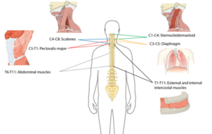
What years were the most challenging for Sherry as a mother? “I’d have to say when your child is under 2 years old, and is not mobile. For me, when he was a baby to toddler, it was more of a physical challenge; it was a little harder for me, with balance and with being fused. I learned that when I dressed him in overalls, it made it easier to grab hold of this crawling baby from the floor. Over time, a certain dynamic develops – children discover quickly that you aren’t able to pick them up the same way as others perhaps and they figure out how to crawl up using your chair to get on your lap. They’re always motivated to be with you. After that, it’s normal parenting.”

“I lived in the West End and would have Aidan walk beside me from when he was about 2 or 3. My rule was that he would have to have his hand on my lap for safety if just he and I were out for a walk. If he took his hand off my lap, he knew he’d be back on my lap or we’d go home. He learned it was all about safety.”

The family’s passion for the outdoors continued through the years raising Aidan. She needed to find adaptable ways to cope with wheeling across grass, parks, and beaches. During those times she would often use a scooter for a day outing to English Bay or Second Beach pool back when Aidan was 4 or 5 years old. In recent years, using a Freewheel attachment has been helpful.

“You want to keep up with them. You don’t want to limit them. There has been an explosion in wheelchair technology recently which I didn’t have access to when my son was younger. Nowadays there are power assist wheels or e-front wheel drive attachments that are quite affordable that enable a level of independence depending on your level of SCI. I’ve also had the same handcycle since my son was about 9 years old – I wished I had a Bionx (e-bike motor add-on) back then! They came out with them years later and it allowed me to cycle easier and more enjoyably while keeping up with the family.” Fostering a love for cycling would propel Aidan to eventually become a member of the Canadian Men’s National Track Cycling Team, medaling at several international World Cups, Commonwealth Games and multi-Pan-American games, retiring just this year.

Sherry Caves, pictured here with husband, Darryl and son, Aidan, on the Access Challenge overnight hike in Manning Park over 20 years ago.

Does Sherry have any final words of advice for people living with SCI – looking to become new parents in the future – particularly when the world is facing a global pandemic? “It’s wonderful, rewarding, and challenging. Focus on your health. If you’re not healthy, it complicates everything. COVID-19 has added that extra layer of risk. But I feel people with SCI have already been down for the count with issues like AD, wounds or UTI’s. You just have to stop, recalculate and wait until you’re healthy, and start again. I guarantee this isn’t the first time we’ve all had to stop and restart.”

“Join a support network. SCI-BC and BCMOS (BC Mobility Opportunities Society) are great resources and have awesome support networks. A friend and I started a ‘Women with SCI’ support group years ago and to this day, there are about 10 of us that still go out for lunch and dinner (pre-COVID-19 times).” Sherry stops for a moment and adds one more word of advice. “Take things in stride – life is ever changing. No one phase will ever stay the same.”

These are words that all of us are sure to agree with and can take to heart.

Respiratory Changes After Spinal Cord Injury

By | | No Comments

Author: Sharon Jang | Reviewer: Tova Plashkes | Published: 24 September 2020 | Updated: 7 December 2021

This page provides an overview of how spinal cord injury (SCI) affects breathing and coughing, and the acute treatments used to address these issues.

Key Points

  • Spinal cord injury can damage the muscles of breathing, affecting the ability to take a deep breath, cough and clear mucus, and maintain adequate oxygen levels. The extent of these changes depends on the level and completeness of the SCI with higher cervical injuries being more affected.
  • A wide range of management options may be used to assist or improve the effectiveness of breathing and coughing in acute and chronic SCI, including tracheostomy and intubation in severe cases, non-invasive ventilation, along with assisted coughing techniques.
  • A number of secondary respiratory complications can affect people with SCI long after injury, including lung infections like pneumonia.
  • Preventative strategies such as flu shots, smoking cessation, and healthy living are an important component of respiratory care.

The breathing process: During inhalation, the diaphragm (dark pink) moves down and the ribs expand. During exhalation, the diaphragm moves up and the ribs contract.1

The respiratory system is responsible for helping you breathe in (inhaling) and out (exhaling). Breathing is done through the nose and mouth, although the nose is more often used. One reason the nose is used more often is because it acts as a filter for debris, which protects the lungs. Coughing is another important part of the respiratory system, as it helps to clear mucus from your lungs and airways.

Many muscles help with respiratory functions. The muscles used for inhaling are controlled from the spinal nerves of the neck (C3-C5 primarily), with some help from the nerves of the lower neck and thorax (C6-T12). Inhaling is mostly facilitated by your diaphragm, which is a large dome shaped muscle underneath the lungs. When you breathe in, your diaphragm lowers and the space in your chest increases, pulling air into the lungs. When you exhale, your ribs move back in and the diaphragm moves back up.

Breathing during exercise and coughing requires extra effort. To help, the abdominal muscles (over your belly) and the intercostal muscles (which help to squeeze the ribs) are activated, allowing for a stronger inhalation and a forceful cough.

When someone is unable to breathe or cough by themselves, their function, independence, and health are affected. These respiratory complications arise in 36-83% of individuals after SCI. This is due to disruption of the breathing nerves after an injury, and to secondary complications of an SCI, such as spasticity. After an SCI, breathing muscles may be partially or completely affected, depending on the completeness of the injury. However, breathing ability may improve over time.

Many muscles are required for breathing. The diagram above shows the main muscles of breathing, and the sections of the spinal cord that innervate them.2

The chances of experiencing respiratory complications depend on a variety of factors, including:

  • The level of injury (dictates which muscles are spared)
  • The completeness of an injury
  • Timing of a tracheostomy*
  • The cause of injury
  • Age*

* Conflicting evidence

Refer to our page on Evidence Rating for more information on conflicting evidence.

Changes in breathing

Changes in amount of air getting into the lungs

After injury, the amount of air that can be inhaled and exhaled are significantly reduced for people with cervical and higher thoracic (neck and upper back) level injuries. More specifically, the amount of air that can fill the lungs (known as the total lung capacity) is reduced to 60-80% of normal values. Additionally, the amount of air that can be exhaled after the biggest breath in (known as the vital capacity) is reduced to 50-80% of normal values. This contributes to inefficient breathing that may be tiresome and difficult. In addition, a lower vital capacity can impact voice volume, making it difficult to speak at louder volumes.
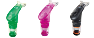
Changes to the lung

An SCI can affect the lung itself. The main change is a reduction in lung compliance, or the lung’s ability to stretch and expand. As a result, the lung does not spring back “closed” after being open. In addition, the compliance of the rib cage (chest wall) may also decrease, causing the chest to become rigid in individuals with tetraplegia. Reduced compliance results in a decreased ability to take a deep breath independently or with the help of a breathing bag or ventilator.

Changes in coughing

Coughing is important to keep the airway and lungs clear from mucus. This is because a build up of mucus can collapse the lungs, and mucus in the airways can result in infection. In order to perform a cough, one needs to inhale deeply then have a forceful exhale while a structure called the glottis closes the entrance to the windpipe. The intercostal muscles and the abdominal muscles assist with the ability to increase the force of exhaled air. As these muscles are innervated by nerves in the chest region, individuals with spinal cord injuries may have an impaired coughing function. Cough function may be completely absent in some individuals, while others may have limited or ineffective coughing abilities.

Changes in lung irritability and mucous production

Soon after injury, it is common for individuals with high-level SCI to produce a lot of mucus in their lungs and have smaller airways deep in the lung. The lungs are also very irritable to stimuli like too much suctioning of mucus, or smoking.  This may be due to the increased influence of the parasympathetic nervous system after SCI. In people with acute tetraplegia, it has been reported that an excess of up to 1 liter of mucus is produced each day. In combination with an inability to cough, this excess production of mucus can result in a buildup of fluid in the lungs and airway.

Changes in swallowing

Although swallowing is important for eating, it is also important for clearing the throat to prevent food, drink, stomach contents, or saliva from entering the lungs (also known as aspiration). After SCI, the risk of aspiration increases as:

    • Your ability to cough may be limited by medical conditions and weakness due to your injury
    • Surgical procedures on the spine may compress your throat
    • You may feel less alert due to sedative medications
    • Some medications you may be on can lead to dry mouth
    • Your sensation may be impaired, which prevents you from feeling food or liquid in the spaces at the back of your throat

The lack of effective swallowing can cause mucus to collect in your airway. Over time, the stagnant mucus can encourage the growth of bacteria, which may travel down to your lungs and potentially result in pneumonia.

Secretion Removal Techniques

Efficient removal of mucus from the airways is important to prevent choking and lung infections, especially when independent coughing is difficult. Although research on the topic of secretion removal techniques is scarce, one study with moderate evidence showed that manual removal techniques combined with mechanical removal techniques are effective in SCI early after injury. Different techniques are outlined below:

Postural drainage

Certain body positions can use gravity to drain mucus towards the throat to be excreted easier. For example, laying on your side with your feet elevated can help drain the lower lung. In order for these positions to be effective, your body must be positioned in specific angles. Refer to your healthcare professional for more information. To facilitate breakup and movement of the mucus buildup in the lungs, postural drainage can be paired with applying pressure to the chest (chest percussion) or shaking the chest (vibration).

Manual assisted coughing

Physical pressure is applied to the chest or abdomen right before expiration to help the individual breathe out. This can be done on yourself or by a trained family member or caregiver.

Mechanically assisted coughing (insufflation-exsufflation)

There are machines that help loosen secretions, clear mucus, and can trigger a cough. They work by delivering a deep breath by pushing air into the lungs, then facilitate exhalation by sucking the air out.

Suction

A tube can be inserted through the mouth or tracheostomy site to suction mucus that is stuck in the upper airways. Suctioning may also reflexively trigger a cough.

For a review of what we mean by “strong”, “moderate”, and “weak” evidence, refer to the SCIRE Community Evidence Ratings.

Respiratory Muscle Training

Weak inspiratory muscles can result in breathlessness. Like exercise training, inspiratory muscle strength and endurance can increase with training and decrease bouts of shortness of breath (dyspnea) and coughing. Inspiratory muscle training involves using devices that create resistance when breathing in.

Resistive trainers have adjustable settings that allow individualized training programs.5

Refer to our article on Inspiratory Muscle Training for more information!

Drug Treatments

Bronchodilators

People with tetraplegia have increased sensitivity of their airways, resulting in more frequent narrowing. To treat this, a family of drugs called bronchodilators can be used to enlarge the airways for air to pass through with more ease. The use of bronchodilators is supported by multiple (weak evidence) studies, which have found that bronchodilators can help improve expiration among individuals with tetraplegia. There is also one strong evidence study that indicates that the bronchodilator salmeterol can improve both respiratory functioning and the strength of breathing muscles. While bronchodilators can help positively influence respiratory functioning, their use carries a potential negative side effect of thickening mucus.

Mechanical ventilation, or machine assisted breathing, is becoming more common as there has been an increase in the number of people who survive cervical level injuries over the past 40 years. Mechanical ventilation is used by people who are unable to breathe independently, often right after injury. The machine works by pushing air into the lungs until a pre-set volume or pressure is reached. Once the pre-set value has been met, the machine stops pushing air in and the air is exhaled by the person.

In general, there are two forms of mechanical ventilation: a non-invasive approach where a mask is placed over the mouth and nose (known as a Bilevel Positive Airway Pressure (BiPAP) or Continuous Positive Airway Pressure (or CPAP)), or an invasive approach where a tube is inserted into the windpipe via the mouth and throat (intubation) or directly into the windpipe through a surgical incision (tracheostomy). Intubation or tracheostomy is used in more severe cases to ensure air gets to the lungs and that mucus is filtered out of the lung. Factors that increase the chances of requiring invasive mechanical ventilation include having a complete injury, having a higher level of injury, or having a compound injury Whenever possible, the healthcare team tries to help people breathe on their own and “weans” the person off the ventilator if possible.

Intubation

The process of intubation consists of running a tube into the trachea either through the nose or mouth. This process is completed as soon as someone is in respiratory distress, which is normally at the scene of the accident or upon admission to the hospital. Intubation is often used for a short term (i.e., less than 10 days), as prolonged use can lead to severe weakness of breathing muscles, pneumonia, more difficulty in breathing, mobility limitations, prolonged ventilator weaning, and can make lung and mouth hygiene difficult.

Tracheostomy

A tracheostomy is a surgical procedure that involves placing a tube through an opening in the throat and windpipe. This creates a pathway for air delivery from a ventilator and to facilitate secretion removal. However, after the tracheostomy tube is taken out, speaking and eating may be difficult as the throat muscles become weakened and uncoordinated.

A tracheostomy tube is inserted into the throat through a surgical hole.6

A tracheostomy is performed in the event that breathing support is required for a minimum of 3 weeks. Individuals who may require a tracheostomy (weak to moderate evidence) include: having a complete or higher level of injury, having a complete injury or a lower AIS motor grade, and old age.

Once an individual is able to independently breathe, the tube is removed from the windpipe. Weak evidence suggests that tube removal is more successful in individuals with certain characteristics:

  • Those who have lower level spinal cord injuries.
  • Those who have not had a tracheostomy but have only been intubated.

Continuous Positive Airway Pressure (CPAP) Ventilation

Continuous Positive Airway Pressure (CPAP) is a form of mechanical ventilation commonly used to address obstructive sleep apnea. Sleep apnea occurs when breathing ceases in short bouts during sleep, and can result in feeling tired during the day. CPAP machines are used to manage this condition by acting as a “pneumatic splint”, keeping airways open during sleep.

Loss of independent breathing and cough function can lead to secondary respiratory issues. These issues need to be medically addressed, as they may be life-threatening if left untreated.

Common secondary respiratory issues following SCI

Atelectasis

A collapsed lung in comparison to a healthy lung.7

A condition where a part of the lung becomes partially or fully collapsed due to a lack of air. This results in a reduced ability to exchange oxygen and carbon dioxide. When the body does not get enough oxygen, organs will start to shut down. Atelectasis can result from anything that prevents the lungs from fully expanding, including:

    • Weak or paralyzed muscles, which can prevent being able to take in a deep full breath. This is the most important cause in SCI.
    • A buildup of mucus, which may block an area of the lung from fully expanding.
    • Shallow breathing due to surgery or pain, which can result in poor inflation of the lungs.
Pneumonia

Pressure from outside the lungs, which can result in the inability to fully inflate. This external pressure may stem from fluid or air, abdominal organs, or external hardware such as a brace.

Pneumonia is a medical name for a lung infection. After an SCI, several factors make pneumonia very common:

Requiring a ventilator, suctioning (removal of secretions with a special machine), or a tracheostomy may often be necessary, but tends to introduce bacteria despite best efforts at hygiene and air filtration. The risk of getting pneumonia increases if someone:

  • is unable to cough and clear mucus,
  • is reliant on mechanical ventilation to assist with breathing,
  • has a severe injury,
  • has a traumatic higher-level injury involving fractures, or,
  • has had a surgical tracheostomy.

Refer to our article on Infectious Respiratory Conditions for more information!

Pulmonary embolism

A pulmonary embolism occurs when a clot (red groups) gets caught in the lungs.8

A pulmonary embolism is a blockage of an artery in the lungs by a blood clot that has moved from elsewhere in the body through the bloodstream (embolism). As a result of paralysis or immobility, a blockage may develop in a vein, often in the lower leg. This is called a blood clot, or deep vein thrombosis. The clot may travel to the lung and block blood vessels, resulting in sudden shortness of breath. The prevalence of this condition is highest within the first three months of injury. Weak evidence suggests that pulmonary embolisms occur in a range of 1.25% to 4.5% of people with SCI in the first 90 days. However, pulmonary embolisms have been considered to occur rarely after the first three months of SCI, and have decreased significantly due to preventative measures (e.g., blood thinners). That said, weak evidence suggests that pulmonary embolisms may still be an issue in chronic SCI, but may not be severe enough to cause any symptoms.

Pulmonary edema

Pulmonary edema is a build-up of fluid in the lung. This often occurs in early stages following injury. It can affect as much as 50% of individuals with acute tetraplegia. There are several causes, with the most common being excess fluids given to people with SCI. After an SCI, blood pressure may drop to very low levels. Depending on the cause and type of injury, this may be due to blood loss from a traumatic injury, or impairment of nerves that keep blood pressure at its normal level with a cervical or high thoracic injury. As a result, a lot of fluids are given to patients to help their blood pressure recover.

Respiratory failure

Respiratory failure occurs when the respiratory system is damaged to the extent where the body does not get enough oxygen and is unable to get rid of carbon dioxide. Oxygen levels in the body may drop to critically low values and carbon dioxide, which is poisonous at very high levels, builds up. The risk of a respiratory failure increases with higher levels of injury, and most commonly occurs in acute SCI. This usually results in the need for mechanical ventilation.

Getting vaccinated is one of the ways to prevent secondary complications such as pneumonia.9

Prevention is important to avoid getting respiratory illnesses when you have an SCI. Some things you can do to stay as healthy as possible include:

  • Avoiding smoking any substances and taking in second hand smoke. The lungs of people with SCI are easily irritated, and those who smoke are more susceptible to lung infections.
  • Staying hydrated – drink plenty of water. This helps to keep mucus in the lungs from being too thick.
  • Ensuring proper nutrition to help maintain a healthy weight and ensure the body has enough vitamins, minerals and protein to heal well when sick.
  • Exercising, as it can help by:
    • Helping you maintain a healthy weight, as lung complications become more prevalent in people who are overweight or obese,
    • Strengthening your breathing muscles.
  • Getting vaccinated for influenza (the flu) and pneumonia. This can help decrease your odds of getting these illnesses.
  • Coughing on a regular basis. Coughing is important for keeping your airways clear of secretions. If you have difficulties coughing by yourself, have someone help you perform manual assist coughs, or use a cough assist machine.
  • Maintaining mobility and proper posture. In order to prevent build up in the lungs, try to sit up everyday and turn when laying in bed.

Secretion removal techniques

Equipment used for lung volume recruitment.10

Upon returning to the community, common secretion removal techniques include glossopharyngeal breathing and the use of lung volume recruitment (LVR) bags with an assisted cough. Glossopharyngeal breathing (or frog breathing) is a technique that is used to get a deeper breath. This is done by rapidly taking “gulps” of breaths one after the other, followed by exhaling. This can help create a cough, or facilitate assisted coughing.

LVR, or “breath stacking” is done with an LVR kit which consists of a resuscitation bag connected with a flexible tube to a mouth piece with a one-way valve. The individual will inhale the most they can, and once this point is reached, a clinician (or second person) will squeeze the bag to “stack” breaths to fully inflate the lungs. This allows the individual to breathe more air than they are able to themselves, and to exhale more air more quickly to produce an improved cough. This also can help with maintaining chest mobility and flexibility.

Exercise Training

Exercise training involving arm and leg movements can improve muscle strength and cardiovascular endurance. Breathing muscles are also challenged with exercise and may become stronger with exercise. This increase in strength can help decrease the effort of breathing at rest and with functional activity, like transfers. An example of a method of exercise training for individuals with higher levels of injuries include the use of a Functional Electrical Stimulation (FES) bike. Other exercises like arm cycling or strengthening exercises are commonly prescribed by a physiotherapist or health care professional. While exercise can help strengthen respiratory muscles, low-moderate evidence studies debate whether lung volumes are impacted. This is to say, exercising may help make breathing feel easier, but it is unknown whether the amount of air you can take into your lungs is affected. High intensity exercise three times per week for six weeks has shown to significantly improve respiratory function. However, standard guidelines for high intensity exercise have not yet been established.
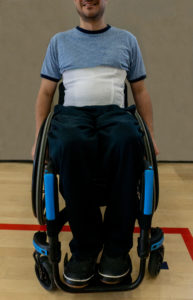
An abdominal binder wrapped around the abdomen to correct the positioning of the diaphragm.12

Refer to our article on Functional Electrical Stimulation for more information!

Girdle/Abdominal Binder

Girdles or abdominal binders are garments that apply pressure around the abdominal area to help keep the diaphragm in an optimal position. Abdominal binders are also used for managing orthostatic hypotension and blood pooling. Although there may be short-term improvements when using a girdle or binder, more research is needed in determining their long-term utility.

Refer to our article on Abdominal binders for more information!

Electrical Stimulation

For people who are ventilator dependent, various electrical stimulation techniques are available to assist with breathing. This includes phrenic nerve stimulation/diaphragm pacing, abdominal electrical stimulation, and epidural stimulation.

Phrenic nerve stimulation/Diaphragm pacing

The diaphragm is the main muscle responsible for breathing and is activated by the phrenic nerve to contract. After SCI, the connection between the brain and the phrenic nerve is disrupted, which contributes to impaired breathing. Researchers have been looking at ways we can stimulate this nerve to reactivate the diaphragm through a process called phrenic nerve stimulation. This process involves surgically implanting electrodes and a receiver close to the phrenic nerve, either in the chest or the neck, and a receiver in the chest wall. This device is controlled with an external remote and antenna (which is used to connect to the electrode receiver).

For phrenic nerve stimulation to work, the diaphragm must have normal function, and the phrenic nerve needs to be intact (i.e., sends a signal when stimulated). As a result, individuals who have a C3, C4, or C5 level injury may not be eligible as they often have impaired phrenic nerve function. It is important to note that this procedure can only facilitate inspiratory functions, but not expiratory. As a result, an individual who receives phrenic nerve stimulation may not require mechanical ventilation, but will still require assistance with coughing and clearing secretions. Tracheostomies and mechanical ventilation are often still used in combination with phrenic nerve stimulation as a back-up.

Some weak evidence supports the use of phrenic nerve stimulation. One study found that diaphragm pacemakers have better results with long term implantation (i.e., 6.3 years in the study). Another study showed that diaphragmatic paces can improve survival rates, decrease the cost of care, improve the quality of speech, increase rates of social participation, and improve management of using a powered wheelchair. Many complications have been reported in the research in regards to using a phrenic nerve stimulator. These complications include wires breaking or getting displaced, device failure, inhaling food when eating, shoulder or abdominal pain, and infections.

Abdominal electrical stimulation

As diaphragm pacing only helps with inhalation, limited research suggests that electrically stimulating the abdominal muscles helps with expiration and coughing. Ideally, the abdominal muscles would be used to support voluntary efforts to cough. There have been mixed findings on the effectiveness of stimulating the abdominal muscles to enhance cough. While some weak studies have found abdominal stimulation to improve cough, other weak evidence studies have found no noticeable changes. More research is required to determine the efficacy of stimulating the abdominal muscles to enhance cough in SCI.

Epidural Stimulation

Epidural stimulation is conducted through surgically implanting an electrode over the spinal cord. Once done, the electrode, which is controlled with a remote outside of the body, stimulates various parts of the spinal cord. Emerging research on epidural stimulation suggests that it may benefit respiratory function after SCI. By directly stimulating nerve cells in the spinal cord, weak evidence suggests that breathing muscles can be activated. The muscles are activated in a pattern that resembles normal breathing, while reducing fatigue. Additionally, weak evidence suggests that epidural stimulation can improve other respiratory functions including coughing and speaking.

Respiratory problems are common after SCI. The extent and type of these problems depend on the level of injury and completeness of injury. Both conservative and invasive options for managing respiratory health following an SCI are available. Due to impaired respiratory function, a variety of secondary complications to the lungs frequently occur after SCI. While prevention using proper respiratory hygiene is best, should you experience a secondary respiratory complication, a variety of management techniques can be applied. Some techniques are more common in the acute stages of SCI, while others are more suited to chronic SCI. It is best to discuss all treatment options with your health providers to find out which treatments are suitable for you.

For a review of what we mean by “strong”, “moderate”, and “weak” evidence, please see SCIRE Community Evidence Ratings.

Parts of this page has been adapted from SCIRE Project (Professional) “Respiratory Management during the Acute Phase of Spinal Cord Injury” Chapter:

Mullen E, Mirkowski M, Vu V, McIntyre A, Teasell RW. (2015). Respiratory Management during the Acute Phase of Spinal Cord Injury. In Eng JJ, Teasell RW, Miller WC, Wolfe DL, Townson AF, Hsieh JTC, Connolly SJ, Noonan VK, Loh E, McIntyre A, editors. Spinal Cord Injury Research Evidence. Version 5.0: p 1-50.

Available from: https://scireproject.com/evidence/acute-evidence/respiratory-management-during-acute-phase-of-spinal-cord-injury/


Evidence for “How does an SCI affect the respiratory system” is based on

Warren, P. M., Awad, B. I., & Alilain, W. J. (2014). Drawing breath without the command of effectors: The control of respiration following spinal cord injury. Respiratory Physiology & Neurobiology. https://doi.org/10.1016/j.resp.2014.08.005

Lemons, V. R., & Wagner, F. C. (1994). Respiratory Complications After CSCI. In Spine (Vol. 19, Issue 20, pp. 2315–2320).

Romero-Ganuza, J., Gambarrutta, C., Merlo-Gonzalez, V. E., Marin-Ruiz, M. Á., Diez De La Lastra-Buigues, E., & Oliviero, A. (2011). Complications of tracheostomy after anterior cervical spine fixation surgery. American Journal of Otolaryngology – Head and Neck Medicine and Surgery, 32(5), 408–411. https://doi.org/10.1016/j.amjoto.2010.07.020

Romero, J., Vari, A., Gambarrutta, C., & Oliviero, A. (2009). Tracheostomy timing in traumatic spinal cord injury. European Spine Journal, 18(10), 1452–1457. https://doi.org/10.1007/s00586-009-1097-3

Aarabi, B., Harrop, J. S., Tator, C. H., Alexander, M., Dettori, J. R., Grossman, R. G., Fehlings, M. G., Mirvis, S. E., Shanmuganathan, K., Zacherl, K. M., Burau, K. D., Frankowski, R. F., Toups, E., Shaffrey, C. I., Guest, J. D., Harkema, S. J., Habashi, N. M., Andrews, P., Johnson, M. M., & Rosner, M. (2012). Predictors of pulmonary complications in blunt traumatic spinal cord injury. Journal of Neurosurgery: Spine, 17, 38–45.

Jain, N. B., Higgins, L. D., Katz, J. N., & Garshick, E. (2010). Association of shoulder pain with the use of mobility devices in persons with chronic spinal cord injury. PM and R, 2(10), 896–900. https://doi.org/10.1016/j.pmrj.2010.05.004

Anke, A., Aksnes, A. K., Stanghelle, J. K., & Hjeltnes, N. (1993). Lung volumes in tetraplegic patients according to cervical spinal cord injury level. Scandinavian Journal of Rehabilitation Medicine, 25(2), 73–77. http://www.ncbi.nlm.nih.gov/pubmed/8341994

Brown, R., DiMarco, A. F., Hoit, J. D., & Garshick, E. (2006). Respiratory dysfunction and management in spinal cord injury. Respiratory Care, 51(8), 853-68;discussion 869-70. http://www.ncbi.nlm.nih.gov/pubmed/16867197%0Ahttp://www.pubmedcentral.nih.gov/articlerender.fcgi?artid=PMC2495152

Berlly, M., & Shem, K. (2007). Respiratory management during the first five days after spinal cord injury. Journal of Spinal Cord Medicine, 30(4), 309–318. https://doi.org/10.1080/10790268.2007.11753946

Schilero, G. J., Spungen, A. M., Bauman, W. A., Radulovic, M., & Lesser, M. (2009). Pulmonary function and spinal cord injury. Respiratory Physiology and Neurobiology, 166(3), 129–141. https://doi.org/10.1016/j.resp.2009.04.002

Schilero, G.J., Grimm, D.R., Bauman, W.A., Lenner, R., Lesser, M. (2005). Assessment of airway caliber and bronchodilator responsiveness in subjects with spinal cord injury. Chest, 127(1), 149-155. http://dx.doi.org/10.1378/chest.127.1.149

Bhaskar, K. R., Brown, R., O’Sullivan, D. D., Melia, S., Duggan, M., & Reid, L. (1991). Bronchial Mucus Hypersecretion in Acute Quadriplegia: Macromolecular Yields and Glycoconjugate Composition. American Review of Respiratory Disease, 143(3), 640–648. https://doi.org/10.1164/ajrccm/143.3.640

Chaw, E., Shem, K., Castillo, K., Wong, S., & Chang, J. (2012). Dysphagia and associated respiratory considerations in cervical spinal cord injury. Topics in Spinal Cord Injury Rehabilitation, 18(4), 291–299. https://doi.org/10.1310/sci1804-291

Evidence for “How does an SCI affect the respiratory system” is based on

Almenoff PL, Alexander LR, Spungen AM, Lesser MD, Bauman WA. Bronchodilatory effects of ipratropium bromide in patients with tetraplegia. Paraplegia 1995; 33: 274-7.

Spungen AM, Dicpinigaitis PV, Almenoff PL, Bauman WA. Pulmonary obstruction in individuals with cervical spinal cord lesions unmasked by bronchodilator administration. Paraplegia 1993;31:404-7.

Schilero GJ, Grimm D, Spungen AM, Lenner R, Lesser M.  Bronchodilator responses to metaproterenol sulfate among subjects with spinal cord injury. J Rehabil Res Dev 2004; 41: 59-64.

Grimm DR, Schilero GJ, Spungen AM, Bauman  WA, Lesser M. Salmeterol improves pulmonary function in persons with tetraplegia. Lung 2006; 184: 335–339.

Evidence for “How can mechanical ventilation be used to help with breathing” is based on

Devivo, M. J. (2012). Epidemiology of traumatic spinal cord injury: Trends and future implications. Spinal Cord, 50(5), 365–372. https://doi.org/10.1038/sc.2011.178

Gregoretti, C., Olivieri, C., & Navalesi, P. (2005). Physiologic comparison between conventional mechanical ventilation and transtracheal open ventilation in acute traumatic quadriplegic patients. Critical Care Medicine, 33(5), 1114–1118. https://doi.org/10.1097/01.CCM.0000162559.74446.09

Como, J. J., Sutton, E. R. H., McCunn, M., Dutton, R. P., Johnson, S. B., Aarabi, B., & Scalea, T. M. (2005). Characterizing the need for mechanical ventilation following cervical spinal cord injury with neurologic deficit. Journal of Trauma – Injury, Infection and Critical Care, 59(4), 912–916. https://doi.org/10.1097/01.ta.0000187660.03742.a6

Montoto-Marqués, A., Trillo-Dono, N., Ferreiro-Velasco, M. E., Salvador-De La Barrera, S., Rodriguez-Sotillo, A., Mourelo-Fariña, M., Galeiras-Vázquez, R., & Meijide-Failde, R. (2018). Risks factors of mechanical ventilation in acute traumatic cervical spinal cord injured patients. Spinal Cord, 56(3), 206–211. https://doi.org/10.1038/s41393-017-0005-7

Seidl, R. O., Wolf, D., Nusser-Müller-Busch, R., & Niedeggen, A. (2010). Airway management in acute tetraplegics: A retrospective study. European Spine Journal, 19(7), 1073–1078. https://doi.org/10.1007/s00586-010-1328-7

Velmahos, G. C., Toutouzas, K., Chan, L., Tillou, A., Rhee, P., Murray, J., & Demetriades, D. (2003). Intubation after cervical spinal cord injury: to be done selectively or routinely? The American Surgeon, 69, 891–894. https://doi.org/10.1257/0002828041464551

Durbin, C. G., Bell, C. T., & Shilling, A. M. (2014). Elective intubation. Respiratory Care, 59(6), 825–849.

Shirawi, N., & Arabi, Y. (2005). Bench-to-bedside review: Early tracheostomy in critically ill trauma patients. In Critical Care (Vol. 10, Issue 1). BioMed Central Ltd. https://doi.org/10.1186/cc3828

Biering-Sorensen, M. (1992). Tracheostomy in spinal cord injured: frequency and follow up. 30, 656–660.

Ganuza, J. R., Forcada, A. G., Gambarrutta, C., Buigues, E. D. D. L. L., Gonzalez, V. E. M., Fuentes, F. P., & Luciani, A. A. (2011). Effect of technique and timing of tracheostomy in patients with acute traumatic spinal cord injury undergoing mechanical ventilation. Journal of Spinal Cord Medicine, 34(1), 76–84. https://doi.org/10.1179/107902610X12886261091875

McCully, B. H., Fabricant, L., Geraci, T., Greenbaum, A., Schreiber, M. A., & Gordy, S. D. (2014). Complete cervical spinal cord injury above C6 predicts the need for tracheostomy. American Journal of Surgery, 207(5), 668–669. https://doi.org/10.1016/j.amjsurg.2014.01.001

Yugué, I., Okada, S., Ueta, T., Maeda, T., Mori, E., Kawano, O., Takao, T., Sakai, H., Masuda, M., Hayashi, T., Morishita, Y., & Shiba, K. (2012). Analysis of the risk factors for tracheostomy in traumatic cervical spinal cord injury. Spine, 37(26), 1633–1638. https://doi.org/10.1097/BRS.0b013e31827417f1

Leelapattana, P., Fleming, J. C., Gurr, K. R., Bailey, S. I., Parry, N., & Bailey, C. S. (2012). Predicting the need for tracheostomy in patients with cervical spinal cord injury. Journal of Trauma and Acute Care Surgery, 73(4), 880–884. https://doi.org/10.1097/TA.0b013e318251fb34

Menaker, J., Kufera, J. A., Glaser, J., Stein, D. M., & Scalea, T. M. (2013). Admission ASIA motor score predicting the need for tracheostomy after cervical spinal cord injury. Journal of Trauma and Acute Care Surgery, 75(4), 629–634. https://doi.org/10.1097/TA.0b013e3182a12b86

O’Keeffe, T., Goldman, R. K., Mayberry, J. C., Rehm, C. G., & Hart, R. A. (2004). Tracheostomy after anterior cervical spine fixation. Journal of Trauma – Injury, Infection and Critical Care, 57(4), 855–860. https://doi.org/10.1097/01.TA.0000083006.48501.B2

Harrop, J. S., Sharan, A. D., Scheid, E. H., Vaccaro, A. R., & Przybylski, G. J. (2004). Tracheostomy placement in patients with complete cervical spinal cord injuries: American Spinal Injury Association Grade A. Journal of Neurosurgery, 100(Spine 1), 20–23.

Peterson, W., Charlifue, W., Gerhart, A., & Whiteneck, G. (1994). Two methods of weaning persons with quadriplegia from mechanical ventilators. Paraplegia, 32(2), 98–103. https://doi.org/10.1038/sc.1994.17

Kornblith, L. Z., Kutcher, M. E., Callcut, R. A., Redick, B. J., Hu, C. K., Cogbill, T. H., Baker, C. C., Shapiro, M. L., Burlew, C. C., Kaups, K. L., DeMoya, M. A., Haan, J. M., Koontz, C. H., Zolin, S. J., Gordy, S. D., Shatz, D. V, Paul, D. B., Cohen, M. J., & Western Trauma Association Study Group. (2013). Mechanical ventilation weaning and extubation after spinal cord injury: a Western Trauma Association multicenter study. The Journal of Trauma and Acute Care Surgery, 75(6), 1060–1069; discussion 1069-70. https://doi.org/10.1097/TA.0b013e3182a74a5b

Evidence for “What secondary respiratory issues occur with acute SCI?” is based on

Berlly, M., & Shem, K. (2007). Respiratory management during the first five days after spinal cord injury. Journal of Spinal Cord Medicine, 30(4), 309–318. https://doi.org/10.1080/10790268.2007.11753946

Garcia-Arguello, L. Y., O’Horo, J. C., Farrell, A., Blakney, R., Sohail, M. R., Evans, C. T., & Safdar, N. (2017). Infections in the spinal cord-injured population: a systematic review. Spinal Cord, 55(6), 526–534. https://doi.org/https://dx.doi.org/10.1038/sc.2016.173

Alabed, S., De Heredia, L. L., Naidoo, A., Belci, M., Hughes, R. J., & Meagher, T. M. (2015). Incidence of pulmonary embolism after the first 3 months of spinal cord injury. Spinal Cord, 53(11), 835–837. https://doi.org/10.1038/sc.2015.105

Aito, S., Pieri, A., D’Andrea, M., Marcelli, F., & Cominelli, E. (2002). Primary prevention of deep venous thrombosis and pulmonary embolism in acute spinal cord injured patients. Spinal Cord, 40(6), 300–303. https://doi.org/10.1038/sj.sc.3101298

Frisbie, J. H., & Sharma, G. V. R. K. (2012). The prevalence of pulmonary embolism in chronically paralyzed subjects: A review of available evidence. Spinal Cord, 50(6), 400–403. https://doi.org/10.1038/sc.2011.154

Sezer, N., Akkuş, S., & Uğurlu, F. G. (2015). Chronic complications of spinal cord injury. World Journal of Orthopaedics, 6(1), 24–33. https://doi.org/10.5312/wjo.v6.i1.24

Evidence for “What is the emerging research on processes to help with breathing” is based on

DiMarco, A. F., Kowalski, K. E., Hromyak, D. R., & Geertman, R. T. (2014). Long-term follow-up of spinal cord stimulation to restore cough in subjects with spinal cord injury. The Journal of Spinal Cord Medicine, 37(4), 380–388. https://doi.org/10.1179/2045772313Y.0000000152

Carter, R. E. (1993). Experience with ventilator dependent patients ! In Paraplegia (Vol. 31). https://doi.org/10.1038/sc.1993.28

Hirschfeld, S., Exner, G., Luukkaala, T., & Baer, G. A. (2008). Mechanical ventilation or phrenic nerve stimulation for treatment of spinal cord injury-induced respiratory insufficiency. Spinal Cord, 46(11), 738–742. https://doi.org/10.1038/sc.2008.43

Esclarin, A., Bravo, P., Arroyo, O., Mazaira, J., Garrido, H., & Alcaraz, M. A. (1994). Tracheostomy ventilation versus diaphragmatic pacemaker ventilation in high spinal cord injury. Paraplegia, 32(10), 687–693. https://doi.org/10.1038/sc.1994.111

DiMarco, A. F., Kowalski, K. E., Geertman, R. T., & Hromyak, D. R. (2006). Spinal cord stimulation: a new method to produce an effective cough in patients with spinal cord injury. American Journal of Respiratory and Critical Care Medicine, 173(12), 1386–1389. https://doi.org/10.1164/rccm.200601-097CR

Hachmann, J. T., Grahn, P. J., Calvert, J. S., Drubach, D. I., Lee, K. H., & Lavrov, I. A. (2017). Electrical Neuromodulation of the Respiratory System After Spinal Cord Injury. Mayo Clinic Proceedings, 92(9), 1401–1414. https://doi.org/10.1016/j.mayocp.2017.04.011

Harkema, S. J., Wang, S., Angeli, C. A., Chen, Y., Boakye, M., Ugiliweneza, B., & Hirsch, G. A. (2018). Normalization of Blood Pressure With Spinal Cord Epidural Stimulation After Severe Spinal Cord Injury. Frontiers in Human Neuroscience, 12, 83. https://doi.org/10.3389/fnhum.2018.00083

Image Credits:

  1. The breathing process ©The SCIRE Community Team
  2. Modified from: Musculi colli base © Olek Remesz, CC-BY-SA 2.5; Muscles that move the humerus ©OpenStax, CC BY 4.0; Thorax ©OpenStax, CC BY 4.0; Respiratory System ©Theresa Knott, CC BY-SA 3.0, Vertebral Column ©Servier Medical Art, CC BY 3.0; Outline ©Servier Medical Art, CC BY 3.0
  3. Lungs ©Mahmure Alp, CC BY 3.0
  4. Sneezing icon ©j4p4n, CC 0
  5. POWERbreathe Plus, ©POWERbreathe
  6. Tracheostomy NIH ©National Heart Lung and Blood Institute, CC 0
  7. Blausen 0742 Pneumothorax ©Bruce Blaus, CC BY 3.0
  8. Pulmonary embolism ©Servier Medical Art, CC BY 3.0
  9. Cure medical care medication pharmacology vaccination ©Bicanski, CC 0
  10. Lung volume recruitment set ©The SCIRE Community Team
  11. Using the FES ©The SCIRE Community Team
  12. Abdominal Binder ©The SCIRE Community Team
  13. Modified from: Diagram 1 of 3 showing stage 3A lung cancer CRUK 008 ©Cancer Research UK, CC BY 4.0; Breathing ©Servier Medical Art, CC BY 3.0


Disclaimer: This document does not provide medical advice. This information is provided for educational purposes only. Consult a qualified health professional for further information or specific medical advice. The SCIRE Project, its partners and collaborators disclaim any liability to any party for any loss or damage by errors or omissions in this publication.

Infectious Respiratory Conditions After SCI

By | | No Comments

Author: Sharon Jang | Reviewers: Phillip Popovich, Katherine Mifflin | Published: 2 September 2020 | Updated: 10 January 2025

The adverse effects of a spinal cord injury (SCI) on the respiratory and immune systems can increase the risk of getting an infectious respiratory condition. This page reviews the relationship between SCI and infectious respiratory conditions.

Key Points

  • Many respiratory changes occur after SCI, including a weakened ability or a loss of breathing and/or coughing function.
  • Individuals with acute SCI are more susceptible to infectious respiratory conditions due to a weakened immune system and a potentially lessened ability to cough or clear secretions.
  • As individuals with SCI transition from the acute to chronic, they are less likely to catch infectious respiratory conditions. However, if they do, the condition may present more severely.
  • The best thing to do is to try to prevent these conditions! Strategies such as vaccinations and good hand washing practice can help.

The upper respiratory system (red) and lower respiratory system (blue).1

The respiratory system involves the lungs, and is responsible for breathing, coughing, and speaking. It consists of the upper respiratory tract (nose, mouth, and the throat (pharynx)), and the lower respiratory tract (voice box (larynx), airways (windpipe or trachea), and lungs). Breathing and coughing both depend on various muscles in the chest and neck. Changes to respiratory function after SCI depend on the level of injury and completeness of the injury. After SCI, especially high cervical level injury, some muscles required for breathing may be affected. These include:

  • The diaphragm, which is the main muscle that pulls air into the lungs,
  • The abdominal muscles, which helps to expel air from the lungs and produce a forceful cough, and
  • The muscles in between your ribs (intercostal muscles), which help squeeze air out of the lungs.

The muscles used to breathe are mostly controlled by the upper parts of the spine.2

As a result, sustaining a higher-level injury may result in impaired respiratory function. Some of these changes include:

  • A reduction in the amount of air you are able to breathe,
  • A stiffer lung, making it difficult to take a full, deep breath,
  • A weak or ineffective cough,
  • An increased amount of mucus, and
  • Difficulties with swallowing.

Despite these changes, many technologies and techniques are available that can assist with breathing and coughing. Moreover, the greatest amount of air you are able to blow out after taking your biggest breath in increases over time from injury.

For more information, refer to our article on Respiratory Changes After SCI.

The immune system is responsible for fighting infections and preventing illness. To keep our bodies healthy, the immune system does three main things:

  1. Recognizes harmful germs, such as bacteria and viruses, when they enter the body,
  2. Kills germs and removes them from the body, and
  3. Fights changes in the body that may cause illness (e.g., cancer cells)

Who are the key players in the immune system?

The immune system is comprised of two main parts: the innate immune system and the adaptive (or acquired) immune system. The innate immune system refers to a non-specific line of defence (i.e., it acts against all germs in the same manner) that you are born with. It is often the first line of defense, which is made up of parts of your body that prevent germs from entering. This includes:

  • The outer layer of the skin, which acts as a physical barrier to germs,
  • Mucus and hair, which traps germs,
  • Saliva, which rinses out germs from the mouth,
  • Bodily fluids, such as stomach acid, which kills bacteria, and
  • Urination and defecation, which excretes germs from the body.

Many organs throughout the body contribute to producing cells required to keep you healthy.3

If germs manage to invade the body, the innate immune system is the first system to recognize them. The innate immune system will then trigger a general attack, such as inflammation and fever. This attack affects the entire body, and does not directly target the germ. Innate immune cells then enlist the help of the adaptive immune system.

The second line of defense involves the adaptive immune system, which initiates specific defences for each germ that enters your body. For example, the body will react differently to a virus that causes influenza versus one that causes measles. The main players in the second line of defense are various types of white blood cells. This includes: natural killer cells, which kill any cell that is not recognized as part of the body, lymphocytes, which help the body remember the invaders for the future and destroy them, and phagocytes, which help “eat” and break up invading organisms.

The cells that contribute to the first and second line of defence are produced in organs all over the body, including the bone marrow, spleen, lymph nodes, adrenal glands, tonsils, and thymus.

Spinal Cord Injury Immune Depression Syndrome (SCI-IDS) is a condition that weakens the immune system after SCI. Weak evidence suggests that SCI-IDS commonly occurs among those with acute SCI. That said, there is also early evidence that SCI-IDS can persist and be present in those with chronic (>1 year) SCI. Although researchers are unsure why the immune system is weakened after SCI, hypotheses have been made:

  • SCI-IDS may be a self-defence mechanism that lowers the body’s immunity to prevent the body from attacking itself after the damage that has occurred in the spinal cord.
  • Many of the organs associated with the immune system, such as the spleen, thymus, and lymph nodes, are controlled by the sympathetic nervous system. These nerves are impaired when an individual sustains an injury at T6 or above. As a result, the immune system may not be as active.

A natural killer cell. The number of these cells is decreased after SCI.4

Weak evidence suggests that immune system changes may occur regardless of level of injury. For example, the amount of natural killer cells are reduced in adults with SCI, regardless of level of injury, in comparison to an able-bodied population. This reduces the body’s ability to fight off germs, which may cause infections, disease, and illnesses. Moreover, early animal research suggests that individuals with SCI may be more susceptible to viruses, such as the flu due to impairments in the body’s immunity. However, it is important to note that these findings have not yet been replicated in humans.

Although the immune system recovers after acute SCI, some (weak) evidence suggests that lowered immunity may extend into the chronic stage of SCI. One study investigated the genes responsible for programming and developing immune cells. The authors found that the genes that normally create natural killer cells are reduced, resulting in lower amounts of these germ-fighting cells throughout the body. A second study supported these findings, as individuals with SCI were found to have lower amounts of natural killer cells in their blood compared to an able-bodied population.
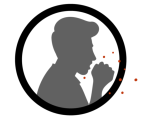
Having an SCI in combination with a weakened immune system has many implications for secondary complications. For example, after SCI many individuals may have difficulties with or be unable to effectively void urine, which encourages the growth of bacteria. This, in combination with a weakened immune system, may explain why urinary tract infections (UTIs) are a common secondary complication of SCI. Although SCI complications and a weakened immune system may contribute to many other secondary complications (e.g., UTI, pressure sores), this article will focus on infectious respiratory conditions that are common with SCI.

For more information, refer to our articles on UTIs and Pressure Injuries!

Respiratory infections can occur in anyone, but people with SCI are at a higher risk for the following reasons:

  • Weakened immune system: After SCI, individuals may have a weakened immune system, some researchers believe that this may make them more prone to infections.
  • Reduced/absent respiratory functioning: As people with SCI may have an inability/weakened ability to cough, mucus begins to build up in the airways and the lung. This accumulation of mucus creates a breeding ground for bacteria and viruses.
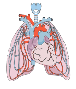
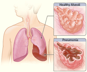
  • Inhaling your food, drinks, or saliva (aspiration) is common after SCI. This results in these substances collecting in your lung, which may result in pneumonia.
  • Use of mechanical respiratory devices: The use of mechanical ventilation can cause ventilator-assisted pneumonia, especially in hospital environments. If parts of the ventilation system (e.g., tubing) are not cleaned properly, bacteria can grow

The heart (in the center) and lungs are complexly interconnected. The lungs help oxygenate the blood, which is pumped by the heart.7

Infectious respiratory diseases can target either the upper respiratory tract (i.e., the nose, mouth, and throat), or the lower respiratory tract (i.e., the voice box, windpipe, airways into the lungs, and the lungs). Oxygen is required for each organ to function properly. When a part of your respiratory system becomes infected, the amount of air you breathe in may be reduced. This reduces the amount of oxygen available for the body, and may quickly affect the function of the brain and other organs. Moreover, these infections can spread all over the body (sepsis). Once it spreads, it becomes more difficult to treat.

As an SCI can negatively affect respiratory and immune function, the rates of respiratory diseases, such as bacterial pneumonia and influenza, among individuals with SCI is high. In fact, respiratory diseases account for just over 80% of all deaths after SCI. In addition, respiratory conditions often present more severely in SCI. This was shown in a (weak evidence) study, where people with SCI who contracted influenza or pneumonia were 37 times more likely to die from the infection compared to an able-bodied population. Two of the most common respiratory infections in people with SCI are pneumonia and the flu. These conditions are particularly infectious, and are caught through tiny droplets of fluid in the air that may be released as a cough or a sneeze. These droplets may fall on surfaces, and can spread if someone touches the surface, then touches their mouth or eyes.

Some (weak evidence) research done to predict who is more likely to experience respiratory illness after SCI indicated that those with a complete injury and those with tetraplegia are at an increased risk of dying from a respiratory related infection. Another study found that during acute care, those with a complete injury were at a greater risk of getting pneumonia. This is related to the lack of/weakened ability to cough and clear mucus from the airways. Secondary complications that may predispose people with SCI to respiratory illnesses include: obesity, heart disease, asthma, chronic obstructive pulmonary disease (COPD), chronic coughing, chronic existence of phlegm, wheezing, and the use of pulmonary medications.

Pneumonia and SCI

Pneumonia is an infection caused by a bacteria or virus, which leads to an infection of the small air sacs in the lungs. It is one of the most common infections in acute SCI with about 30% of individuals with acute SCI experiencing pneumonia (weak evidence), dropping to about 3.5% in the chronic stages of SCI (i.e., 1-20 years post injury). Although the chance of getting pneumonia decreases after acute SCI, it is important to remember that pneumonia manifests more severely in people with SCI. That is to say, while your chances of getting pneumonia may be reduced at longer times after SCI, if you do get it, it is more severe.

Influenza and SCI

Influenza, or the flu, is a respiratory condition caused by a virus. There are multiple types and sub-types of influenza, although type A and B are the strains that most often cause the flu season. The flu virus affects the nose, throat, and sometimes the lungs, and can lead to secondary conditions such as pneumonia. Flu vaccines are recommended, especially for people with SCI as they are a vulnerable population. There is weak evidence that supports the use of flu vaccines for people with SCI, as their immune system responds similarly to the vaccine compared to able-bodied individuals. That said, those with tetraplegia may have a reduced response to the vaccine. Animal studies suggest that vaccines may be less effective with higher levels of SCI. More research is required to determine the response to influenza vaccines after SCI.

Other infectious respiratory conditions and SCI

There are many other infectious respiratory conditions that exist, such as the common cold, tuberculosis, and coronaviruses. However, little to no research has been done on the impact of these conditions in the SCI population. Although the available research is limited, it is important to note that individuals with SCI are still at an increased risk of contracting these conditions. The following section describes various respiratory infections in the able-bodied population unless otherwise noted.

Common Cold

The common cold (or simply, the cold) is a general term for a mild upper respiratory tract condition affecting the nose and throat. Common symptoms include stuffy nose, sneezing, sore throat, and cough. Unlike other conditions, there are multiple types of viruses that can cause a cold. Rhinoviruses, coronaviruses, and influenza viruses account for the majority of cases. The cold occurs most frequently in the fall, and decreases upon the arrival of spring. On average, a person will catch the cold once a year, but it is likely that this rate is underestimated.
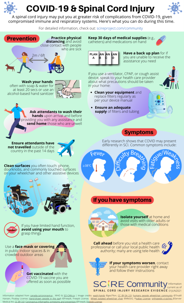
Coronaviruses

A coronavirus (above), which gets its name from the spikes on the outside that resemble a crown, which is “corona” in Latin.10

Coronaviruses are responsible for many health conditions including the common cold, Middle East Respiratory Syndrome (MERS), Severe Acute Respiratory Syndrome (SARS), and novel coronavirus disease 2019 (COVID-19). These are the four strains of coronavirus now known to affect humans, and they are responsible for 10-30% of upper respiratory tract infections. Although these viruses are genetically related, they cause very different conditions. The severity of the conditions they may cause varies from mild (such as the common cold, described above), to severe (such as MERS, SARS, and COVID-19). Though MERS, SARS, and COVID-19 can all lead to pneumonia, each condition affects the body differently: MERS has a greater impact on the digestive system and kidneys, while SARS and COVID-19 most heavily impact the respiratory system, clotting function, and heart activity.

The 2019 Novel Coronavirus (COVID-19)

The COVID-19 outbreak began in 2019 in Wuhan, China, and has since spread around the world in 2020. Currently, little is known about the virus and the infection it causes, though research is on-going. The virus appears to attack the respiratory system, resulting in symptoms such as cough, shortness of breath, and pneumonia, in addition to fever and kidney failure. The extent of the symptoms ranges from mild to severe, and it is possible for someone to be infected without symptoms. This virus has caused a sense of unease among the SCI community. In a survey conducted by a group of researchers, some of the most common concern included increased vulnerability to infection, decreased availability of caretakers, inability to obtain medical supplies, the inability to be appropriately tested, an inability to travel to medical appointments, and an inability to self-quarantine.

To date, few weak evidence studies on COVID-19 and SCI have been completed. This biggest take away is that the typical symptoms of COVID-19 as reported by the World Health Organization (i.e., cough, fever, and shortness of breath) are not necessarily applicable to people with spinal cord injuries. As coughing is often impaired with SCI, it can be absent in reported cases of COVID-19 in people with SCI.

Instead, common symptoms of COVID-19 in people with SCI include a fever and feeling weaker than normal. Other symptoms that have been reported include shortness of breath, body aches/worsening pain, sweating, chest pain, and increased spasticity, a worsened ability to clear secretions, and abnormally fast breathing. Although people with SCI may have fewer of the typical COVID-19 symptoms, one study has found that they are more likely to experience COVID-19 more severely compared to able-bodied individuals.

Tuberculosis

Tuberculosis is an infection of the lungs that can be caused by Mycobacterium tuberculosis bacteria. The presence of tuberculosis is higher in developing countries in comparison to developed countries. This is related to factors such as lower rates of vaccination and higher rates of HIV (an immune compromising condition) in developing countries. Treating tuberculosis is particularly difficult, as many strains of the virus/bacteria are resistant to drugs.

Upper respiratory tract infections

Upper respiratory tract infections are a group of conditions that affect the nose and throat. Some conditions include pharyngitis (sore throat) and laryngitis (inflammation of the voice box; when you lose your voice). These infections are of particular note to those using ventilators, as over 90% of pneumonia and hospitalizations start with an upper respiratory tract infection.

In order to avoid getting infectious respiratory conditions, prevention is key, especially in the community. Here is what you can do to stay healthy:

  • Wash your hands with warm water and soap for 20-30 seconds
  • Get vaccinated for pneumonia and the flu. Vaccinations are especially important, as weak evidence suggest that rates of vaccination are still low.
  • Stay hydrated! Drinking water can help loosen up the mucus in your lungs.
  • Clean surfaces that may have been in contact with a sick person. This includes parts of your wheelchair, including the joystick, pushrims, etc.
  • Avoid smoking. Smoking can damage the lung’s ability to fight infections, which can compound issues with an already weak immune system.
  • Practice good health habits, such as exercising and having a healthy diet.
  • Stay home if you are sick.
  • Let the people around you know you are feeling unwell. This way they can check up on you and know to avoid close contact.

After an SCI, respiratory functions (i.e., breathing and coughing) and the immune system are compromised. Researchers are still unsure about why the immune system is suppressed. While there is some weak evidence for why the immune system changes after SCI, more clinical trials are required to determine the specific effects of SCI on the immune system.

Given the changes to respiratory and immune functioning after SCI, there is a higher risk of getting an infectious respiratory disease. The best thing to do is to work at prevention, which can be done through a variety of ways such as getting vaccinated and staying hydrated. Discuss all treatment options with your health providers to find out which treatments are suitable for you.

For a review of how we assess evidence at SCIRE Community and advice on making decisions, please see SCIRE Community Evidence.

SCIRE Community. “Respiratory Changes After SCI”. Available from: https://community.scireproject.com/topic/respiratory-changes/

SCIRE Community. “COVID-19 & SCI Infographic”. Available from: https://community.scireproject.com/covid-19/infographics/

Parts of this page has been adapted from SCIRE Professional “Respiratory Management Following Spinal Cord Injury” Module:

Sheel AW, Welch JF, Townson AF (2018). Respiratory Management Following Spinal Cord Injury. In: Eng JJ, Teasell RW, Miller WC, Wolfe DL, Townson AF, Hsieh JTC, Connolly SJ, Noonan VK, Loh E, Sproule S, McIntyre A, Querée M, editors. Spinal Cord Injury Rehabilitation Evidence. Version 6.0. Vancouver: p. 1-72.
Available from: https://scireproject.com/evidence/respiratory-management-rehab-phase/ 

Evidence for “What is the immune system” is based on:

Tortora, G.J., and Derrickson .B.(2013).The Lymphatic System and Immunity. In Roesch, B. (Eds.), Principles of Anatomy and Physiology (pp. 366-447). Biologtical Science Textbooks

Evidence for “What happens to the immune system after SCI” is based on:

Allison, D. J., & Ditor, D. S. (2015). Immune dysfunction and chronic inflammation following spinal cord injury. Spinal Cord, 53(1), 14–18. https://doi.org/10.1038/sc.2014.184

Campagnolo, D. I., Dixon, D., Schwartz, J., Bartlett, J. A., & Keller, S. E. (2008). Altered innate immunity following spinal cord injury. Spinal Cord, 46(7), 477–481. https://doi.org/10.1038/sc.2008.4

Popovich, P., & McTigue, D. (2009). Beware the immune system in spinal cord injury. Nature Medicine, 15(7), 736–737. https://doi.org/10.1038/nm0709-736

Schwab, J. M., Zhang, Y., Kopp, M. A., Brommer, B., & Popovich, P. G. (2014). The paradox of chronic neuroinflammation, systemic immune suppression, autoimmunity after traumatic chronic spinal cord injury. Experimental Neurology, 258, 121–129. https://doi.org/10.1016/j.expneurol.2014.04.023

Riegger, T., Conrad, S., Schluesener, H. J., Kaps, H. P., Badke, A., Baron, C., … Schwab, J. M. (2009). Immune depression syndrome following human spinal cord injury (SCI): A pilot study. Neuroscience, 158(3), 1194–1199. https://doi.org/10.1016/j.neuroscience.2008.08.021

Herman, P., Stein, A., Gibbs, K., Korsunsky, I., Gregersen, P., & Bloom, O. (2018). Persons with Chronic Spinal Cord Injury Have Decreased Natural Killer Cell and Increased Toll-Like Receptor/Inflammatory Gene Expression. Journal of Neurotrauma, 35(15), 1819–1829. https://doi.org/10.1089/neu.2017.5519

Evidence for “Why are people with SCI at higher risk for respiratory infections” is based on:

Brommer, B., Engel, O., Kopp, M. A., Watzlawick, R., Müller, S., Prüss, H., … Schwab, J. M. (2016). Spinal cord injury-induced immune deficiency syndrome enhances infection susceptibility dependent on lesion level. Brain, 139(3), 692–707. https://doi.org/10.1093/brain/awv375

Burns, S. P. (2007). Acute Respiratory Infections in Persons with Spinal Cord Injury. Physical Medicine and Rehabilitation Clinics of North America, 18(2), 203–216. https://doi.org/10.1016/j.pmr.2007.02.001

Evidence for “What infectious respiratory conditions should I be aware of?” is based on:

Northwest Regional Spinal Cord Injury System. (2004, October 12). Common respiratory problems in SCI – What you need to know. http://sci.washington.edu/info/forums/reports/common_respiratory.asp

DeVivo, M. J., Black, K. J., & Stover, S. L. (1993). Causes of death during the first 12 years after spinal cord injury. Archives of Physical Medicine and Rehabilitation, 74(3), 248–254. https://doi.org/10.5555/uri:pii:000399939390132T

Stolzmann, K. L., Gagnon, D. R., Brown, R., Tun, C. G., & Garshick, E. (2010). Risk factors for chest illness in chronic spinal cord injury: a prospective study. American Journal of Physical Medicine & Rehabilitation, 89(7), 576–583. https://doi.org/10.1097/PHM.0b013e3181ddca8e

Berlly, M., & Shem, K. (2007). Respiratory management during the first five days after spinal cord injury. Journal of Spinal Cord Medicine, 30(4), 309–318. https://doi.org/10.1080/10790268.2007.11753946

McKinley, W. O., Jackson, A. B., Cardenas, D. D., & DeVivo, M. J. (1999). Long-term medical complications after traumatic spinal cord injury: A Regional Model Systems Analysis. Archives of Physical Medicine and Rehabilitation, 80(11), 1402–1410. https://doi.org/10.1016/S0003-9993(99)90251-4

Trautner, B. W., Atmar, R. L., Hulstrom, A., & Daroiuche, R. O. (2004). Inactivated Influenza Vaccination for People With Spinal Cord Injury. Arch Phys Med Rehabil, 85(11), 1886–1889. https://doi.org/10.1038/jid.2014.371

Centers for Disease Control and Prevention. (2020a). Understanding influenza viruses. https://www.cdc.gov/flu/about/viruses/index.htm

Centers for Disease Control and Prevention. (2020b). Prevent seasonal flu. https://www.cdc.gov/flu/prevent/index.html

Burns, S. P. (2007). Acute Respiratory Infections in Persons with Spinal Cord Injury. Physical Medicine and Rehabilitation Clinics of North America, 18(2), 203–216. https://doi.org/10.1016/j.pmr.2007.02.001

Rao, N. M. (2015). Swine flu the pandemic disease – preventive measures to take. Journal of Medical Science and Technology, 4(1), 1–2.

Heikkinen, T., & Järvinen, A. (2003). The common cold. Lancet, 361(9351), 51–59. https://doi.org/10.1016/S0140-6736(03)12162-9

Paules, Catharine, I., Marston, H. D., & Fauci, A. S. (2020). Coronavirus Infections – More than Just the Common Cold. Journal of the American Medical Association, 323(8), 707–708. https://doi.org/10.1007/82

Stillman, M. D., Capron, M., Alexander, M., Di Giusto, M. L., & Scivoletto, G. (2020). COVID-19 and spinal cord injury and disease: results of an international survey. Spinal Cord Series and Cases, 6(1), 21.

Rodriguez-Cola, M., Jimenez-Velasco, I., Henares-Gutierrez, F., Lopez-Dolado, E., Gambarrutta-Malfatti, C., Vargas-Baquero, E., & Gil-Agudo, A. (2020). Clinical features of coronavirus disease 2019 (COVID-19) in a cohort of patients with disability due to spinal cord injury.

Righi, G., Del, G., & Popolo, G. Del. (2020). COVID-19 tsunami : the first case of a spinal cord injury patient in Italy. Spinal Cord Series and Cases, 3–7. https://doi.org/10.1038/s41394-020-0274-9

Mayo Clinic (2020). Tuberculosis. https://www.mayoclinic.org/diseases-conditions/tuberculosis/symptoms-causes/syc-20351250

World Health Organization. (2020). Tuberculosis. https://www.who.int/news-room/fact-sheets/detail/tuberculosis

Evidence for “What can you do to prevent infectious respiratory conditions” is based on:

LaVela, S. L., Smith, B., & Weaver, F. M. (2007). Perceived Risk for Influenza in Veterans With Spinal Cord Injuries and Disorders. Rehabilitation Psychology, 52(4), 458–462. https://doi.org/10.1037/0090-5550.52.4.458

Ronca, E., Miller, M., Brinkhof, M. W. G., Jordan, X., Léger, B., Baumberger, M., … Fekete, C. (2020). Poor adherence to influenza vaccination guidelines in spinal cord injury: results from a community-based survey in Switzerland. Spinal Cord, 58(1), 18–24. https://doi.org/10.1038/s41393-019-0333-x

Weaver, F. M., Smith, B., LaVela, S., Wallace, C., Evans, C. T., Hammond, M., & Goldstein, B. (2007). Interventions to increase influenza vaccination rates in veterans with spinal cord injuries and disorders. Journal of Spinal Cord Medicine, 30(1), 10–19. https://doi.org/10.1080/10790268.2007.11753908

Image credits
  1. Modified from 2301 Major Respiratory Organs ©Anatomy and Physiology, Betts et al., CC BY 3.0
  2. Modified from Neck muscles, lateral view ©Olek Remesz, CC-BY-SA 2.5; A cutout of the thoracic wall showing the three layers of intercostal muscle – from the left wall ©CFCF, CC BY-SA 4.0; Pectoralis Major ©Anatomy and Physiology, Betts et al., CC BY 3.0; Respiratory system ©Theresa knott, CC BY-SA 3.0
  3. Modified from: Outlines ©Servier Medical Art, CC BY 3.0; Lymph Node ©Servier Medical Art, CC BY 3.0; Thymus ©Servier Medical Art; Spleen ©Servier Medical Art; Colon ©Servier Medical Art; File 603: Anatomy of long bone ©Anatomy and Physiology, Betts et al., CC BY 3.0
  4. Human Natural Killer Cell ©NIH NIAID, CC BY 2.0
  5. DNA ©Servier Medical Art, CC BY 3.0
  6. Coronavirus infographic symptoms and prevention©Freepik, Freepik License
  7. Modified from Heart ©Servier Medical Art CC BY 3.0; Pulmonary circulation ©Servier Medical Art, CC BY 3.0
  8. Lobar pneumonia illustrated ©NIH
  9. Sneezing ©Andrei Yushchenko, CC BY 3.0
  10. 3D medical animation coronavirus ©Scientific Animations
  11. Infographic with details about coronavirus with illustrated sick man ©Freepik, Freepik License
  12. Syringe shot medicine bottle medical needle ©qimono, Pixabay License


Disclaimer: This document does not provide medical advice. This information is provided for educational purposes only. Consult a qualified health professional for further information or specific medical advice. The SCIRE Project, its partners and collaborators disclaim any liability to any party for any loss or damage by errors or omissions in this publication.

COVID-19 Factsheet: Guidance for SCI Caregivers

By | | No Comments

Author: The SCIRE Professional Team | Reviewers: Cynthia Morin, Andrea Townson | Published: 5 August 2020 | Updated: ~

People living with spinal cord injuries (SCI) have a greater risk of developing more serious symptoms of COVID-19. It is critical for caregivers and attendant services to take the necessary precautions and preventive actions outlined in this document in order to minimize the risk of transmission.

Download PDF

  Expand All

Wash hands often

  • Use soap and water to wash hands for 30 seconds before and after contact with your client.
  • Use a hand sanitizer (with at least 70% alcohol) if soap/water are not available.

Clean and disinfect

  • Clean frequently touched surfaces in client’s home (e.g. doorknobs, light switches, counters, cabinets, phones, remote controls, handles on toilets and faucets) once every few days using soapy water to wipe away debris/dirt.
  • After cleaning, use only disinfectants (e.g., Clorox, Lysol, Microban) that have a Drug Identification Number (DIN) and have been approved by Health Canada for use against COVID-19 in Canada/your country’s standards.
  • For clients who use mobility aids or other equipment, regularly clean the: wheelchair joystick, armrest, wheel rims, mechanical lift controller and other frequently used surfaces.
  • If approved disinfectants are not available, a diluted bleach solution can be prepared for household disinfection in accordance with the instructions on the label, or follow instructions for proper handling of household (chlorine) bleach.
  • Place contaminated laundry (including non-medical cloth masks), into a container with plastic liner.
    • wash using regular laundry detergent and hot water (60-90°C)
    • wear gloves when handling laundry
    • clean hands with soap and water for 30 seconds immediately after removing gloves

Protective equipment

  • If your caregiving role requires the need to be within 2 metres (6 feet) to assist with daily tasks (ex. Bathing, brushing teeth, dressing) wear personal protective equipment such as: a medical mask, disposable gloves, and eye protection.
  • Do not re-use medical masks or gloves. However, under extreme supply limitations, masks may be re-used if not visibly damaged, contaminated, or wet.
    • If a mask is re-used, prevent contamination by storing it in a clean paper/plastic bag, or cleanable container with a lid (discard bags after each use)
    • Healthcare practitioners have been instructed to use 1 mask/day (put on when entering facility/home and removed when eating or leaving facility/home at end of shift)
  • Place used medical masks, gloves, and other contaminated items in a lined container, secure the contents and dispose of them with other household waste.
    • Take gloves off first, wash hands, then remove mask
    • Clean your hands again with soap/water/hand sanitizer before touching your face/other surfaces

Caregivers should create a back-up care plan for clients

  • Decide who can step in if you are unable to provide care for your client.
  • Consider the specialized tasks you manage for people with SCI and expand your circle of care to include a person with SCI’s family members or friends who may be able to help if necessary.
  • Use video platforms (Facetime, Skype, Zoom) to review the plan with your circle and recipient to ensure they are comfortable with the backup-plan.

The care plan should include the following:

  • Contact information for: doctors, clinics, pharmacy, family, friends, neighbours, home and community care case managers, and food delivery.
  • Information regarding the client’s condition (medical history, allergies, specific needs etc.).
  • A schedule of what your typical tasks look like with enough detail for someone to follow and take over if need be.
  • A list of names and doses of medications and when they are given to the recipient.
  • A list of important supplies that need to be purchased regularly (e.g., toilet paper, cleaning supplies, hygiene products).
  • Information about their likes and dislikes, self-care routines, and food preferences.
  • Additional non-perishable food items to ensure your client has continued access to healthy meals.
  • A list of essential items your client needs if they need to leave their home or require hospitalization.

A podcast created by caregivers of BC who share their experiences, “highlighting the joys, trials, and self-discoveries that come along with this rewarding and taxing position”: Caregiving Out Loud Podcast

Advice provided from the Government of Canada for Caregivers in caring for a person with COVID-19 at home

Tip sheet created by the Ontario Caregiver Organization. Provides resources/education on improving caregiver mental health during COVID-19.: Tips for Caregivers Mental Health During COVID-19

The Family Caregivers of BC provide multiple tips and resources for caregivers to practice self-care.: Self-Care Tips During Uncertain Times

Coronavirus Disease 2019 (COVID-19) Fact Sheet. (2020, March 4). Retrieved from: (https://shepherd.org/docs/Coronavirus2019_FactSheet_3.4.20.pdf

Public Health Ontario (2020, February 14). Coronavirus Disease 2019 (COVID-19) Self-isolation: Guide for caregivers, household members and close contacts. Retrieved from:  https://publichealthontario.ca/-/media/documents/ncov/factsheet-covid-19-guide-isolation-caregivers.pdf?la=en

Public Health Agency of Canada (2020, May 1). How to care for a person with COVID-19 at home: Advice for caregivers. Retrieved from: https://canada.ca/en/public-health/services/publications/diseases-conditions/how-to-care-for-person-with-covid-19-at-home-advice-for-caregivers.html

Public Health Ontario (2020, April 10). Coronavirus Disease 2019 (COVID-19) When and How to Wear a Mask Recommendations for the General Public. Retrieved from: https://www.publichealthontario.ca/-/media/documents/ncov/factsheet/factsheet-covid-19-how-to-wear-mask.pdf?la=en

The Ontario Caregiver Foundation (2020, March 31). COVID-19 – Do you have a plan? Retrieved from: https://ontariocaregiver.ca/wp-content/uploads/2020/03/Ontario-Caregiver-Organization-Caregiver-Contingency-Plan.pdf

Family Caregivers of British Columbia (2020, March 11). COVID-19. Retrieved from: https://familycaregiversbc.ca/community-resources/covid-19-virus/

Image credits

  1. Hand Washing ©AnnaliseArt, Pixabay
  2. Cleaning Supplies ©MarCuesBo, Pixabay
  3. Protective Equipment ©artpolka Pixabay
  4. Checklist©OpenClipart-Vectors, Pixabay


Disclaimer: This document does not provide medical advice. This information is provided for educational purposes only. Consult a qualified health professional for further information or specific medical advice. The SCIRE Project, its partners and collaborators disclaim any liability to any party for any loss or damage by errors or omissions in this publication. SCIRE receives no compensation and there are no conflicts declared with sources of information on this factsheet.

COVID-19 Factsheet: Mental Health Support and SCI

By | | No Comments

Author: SCIRE Professional Team | Reviewer: Rachael Neal | Published: 5 August 2020 | Updated: ~

Living during the COVID-19 pandemic can be stressful, and it is common to feel worried, sad, or anxious from time to time. This sheet contains information about some resources, techniques, and tips to help support your mental health in the time of COVID-19.

Key Points

  • The COVID-19 pandemic may cause increased mental health concerns in people who have already experienced sweeping changes in life due to spinal cord injury (SCI).
  • Feelings of worry and anxiety in times of global uncertainty are common, and it is important to know that you are not alone.
  • Learning to recognize and acknowledge your anxiety and to engage in concrete, meaningful actions and activities can help you manage your worries.
  • Refer only to reliable sources of news about COVID-19. Limiting yourself to appropriate information consumption can reduce the likelihood of feeling overwhelmed or in danger.

Download PDF

  Expand All

How can I organize and structure my day to benefit my wellbeing?

Our normal daily routines and activities are changing with the current pandemic situation, which can feel unsettling. You may find that some things you usually did to look after your wellbeing have become difficult to carry on. Though you may not have control over those changes, you can focus on what choices and coping strategies you have that are within your control.

Maintain balance

Try organizing your days to include a variety of activities that you know will improve your general mood. You can strive for a routine that is a balance of activities that give you feelings of pleasure, achievement, and closeness.

You will get a sense of accomplishment when you, for instance, choose to finish a work task, complete an exercise routine, or learn to cook a new recipe. A pleasure activity may be reading a book or watching a favourite comedy show. Schedule a video call with a friend or family to feel connected.

If you feel overwhelmed or unmotivated, break tasks down to pieces that you can work on, or set a goal to work for a small chunk of time rather than having the goal to finish something all at once.

What can I do to deal with this time of uncertainity to help me to stay in a healthier place?

As someone with a SCI, you have likely already experienced uncertainty. It is the ‘not knowing’ how things are going to turn out that can be difficult to cope with. The coronavirus uncertainty and isolation can be particularly worrying when you have additional health care needs.

There are different levels of worry and anxiety. Anxiety is a natural emotion and worrying is common during change. Remind yourself that you will not feel this way forever and that there are things you can do to cope. Use the diagram on the left to see if you are in the ‘fear zone’ and identify specific steps you will try to move towards learning and growth zones.


Why do I worry even when nothing seems wrong, and how do I stop it?

It is common during times of uncertainty, like the COVID-19 pandemic, that you may notice more worry thoughts, some even leading to worst-case scenarios. The graphic below illustrates how worries can quickly escalate even from something relatively minor that you would not have recognized as being a worry trigger before.

To reduce this type of worry, the first step is to practice noticing when your thoughts are reaching a later, more catastrophic point. For instance, if you first notice a feeling of anxiety, ask yourself, “What was I just thinking?”. Step back to the event that began your worries, and ask yourself if you have reasonable evidence to believe that the initial event is likely to lead to the worst-case scenario, or whether there may be other explanations you can consider. Are you are assuming a negative outcome when the situation is actually an unknown?

Think of a strategy that may have helped you with a similar problem in the past. Make a list of things that generally help you relax and choose one that is possible to do now.

Which category does your worry fall into?

Worry becomes a problem when it stops you from living the life you want to live, or if it leaves you feeling helpless or exhausted.

Assess the impact of your worries on your life. Seek professional help if you are noticing that you are not able to carry out important roles or activities in your life because of interference from worry.

Worry Time

Another effective strategy is designating a specific Worry Time – schedule a time for later in the day when you allow yourself to worry as much as you feel the need to. This can be helpful in two ways:

  1. It can prevent worry from interfering with your important daily activities, and
  2. Postponing worry can sometimes circumvent the worry from happening at all (at the scheduled time, you may not even feel like worrying, in other words, it was an ‘in the moment’ worry).

Worry Time may be particularly effective if your worry is hypothetical or in the future (and action is not possible). The worry should be put aside, and your attention focused on a technique or activity that will distract you from your worry thoughts (re-focus on something that is in the present).

The current pandemic has brought many changes to the lives of people with SCI, and you may notice yourself having new or different concerns for your well-being. Awareness and reminders of active, positive coping strategies are especially important.

Even though there is much about the COVID-19 situation that you cannot control, you can shift your focus to what you can influence and have power over:

  • Choose your routine. Plan how you spend your time and what you focus on during the day.
  • Choose your distractions. Have a list of small, practical, or creative tasks that you can easily accomplish.
  • Choose your information. Limit how much news you watch and when you take in new information.

“With awareness and active steps, we can exercise the positive power of being able to recognize our fear and patterns of survival” (Vicki Enns, Clinical Director, Crisis & Trauma Resource Institute)

Duff, J. (2000). Coping Effectiveness Training reduces depression and anxiety following traumatic spinal cord injury. Proceedings of the British Psychological Society, 8(1): 17.

Whalley, M. & Kaur, H. (2020, March 19). Free Guide To Living With Worry And Anxiety Amidst Global Uncertainty. Retrieved from https://www.psychologytools.com/articles/free-guide-to-living-with-worry-and-anxiety-amidst-global-uncertainty/

Enns, V. (2020, May). How to Cope with Post-Traumatic Stress During COVID-19. Retrieved from:  https://ca.ctrinstitute.com/blog/how-to-cope-with-post-traumatic-stress-during-covid-19/

Wilkinson, A., Meares, K., Freestones, M. (2011). CBT for worry & generalised anxiety disorder. London: Sage.

Image credits

  1. Learning Zone model by Tom Senninger. Creative Commons BY-NC-SA 4.0
  2. Escalator clip art © DLPNG Creative Commons
  3. Afraid © Stephanie Heendrickxen Creative Commons
  4. Man © corpus delicti Creative Commons
  5. Stop Light Icon #432967 © Free Icons Library Creative Commons
  6. Woman © Crello Creative Commons
  7. Clock © Crello Creative Commons
  8. Thought bubble © Crello Creative Commons
  9. Man on computer © user:cth103_t Creative Commons


Disclaimer: This document does not provide medical advice. This information is provided for educational purposes only. Consult a qualified health professional for further information or specific medical advice. The SCIRE Project, its partners and collaborators disclaim any liability to any party for any loss or damage by errors or omissions in this publication. SCIRE receives no compensation and there are no conflicts declared with sources of information on this factsheet.

COVID-19 Factsheet: Spinal Cord Injury Specifics

By | | No Comments

Author: SCIRE Professional Team | Reviewers: Cynthia Morin, Andrea Townson | Published: 4 August 2020 | Updated: ~

Key Questions

  • Are you at a higher risk for COVID-19 because of your SCI?
  • What are the precautions you can take to prevent exposure to the virus?
  • How can you keep your assistive devices/equipment clean and virus-free?
  • How can you keep your assistive devices/equipment clean and virus-free?
  • What can you do to ensure your interactions with others are safe?
  • When should you seek medical attention, and what do your healthcare providers need to know?

Download PDF

  Expand All

What we know about the risks for people with spinal cord injury (SCI) is based on how SCI and COVID-19 both affect the body.

Respiratory function

Everyone with SCI has some level of impairment in respiratory function given how SCI weakens breathing muscles, however people with cervical and upper thoracic levels of injury may have greater impairments than those with lower thoracic levels of injury. Still, all levels of SCI above T12 have a reduced ability to both inspire air maximally and to forcefully expel air through coughing. With an impaired cough, individuals are less able to manage respiratory secretions.

Other risk factors

  • Some complications (common secondary health issues of SCI), such as cardiovascular disease or higher body mass index (BMI), may increase the risks of a more serious case of COVID-19.

People with SCI may also have a greater risk of exposure to COVID-19, as those who require assistance cannot avoid contact with caregivers.

Given that there is no current treatment for COVID-19 other than supportive care, it is best to take precautions to avoid exposure to the virus whenever possible.

  • Practice physical distancing (6 feet), avoid large groups and stay home when possible.
  • Clean all surfaces regularly with soap and warm water or antibacterial solution.
  • Wash your face and hands after being in public or having in-person conversations.
  • Wash clothes after each wear and if possible separate indoor and outdoor clothes.
  • Learn about home delivery options from grocery stores, pharmacies, and other institutions providing essential services.
  • Keep 30 days of medications and medical supplies on hand (i.e., catheters, wound dressings, disposable gloves).
  • If you require the assistance of caregivers, ensure that they use PPE and follow protocols re: minimizing the transmission of COVID-19.
  • Learn how you can connect with your local SCI organization (e.g., SCI-BC).

Maintaining a clear respiratory tract

• Stay hydrated to keep lung secretions thin.
• Change positions frequently, and use gravity to help clear your lungs.
• Practice deep breathing and coughing exercises to strengthen respiratory muscles.
• Eat healthy, well-balanced meals to boost your immune system.

Wheelchair users

As a wheelchair user, it is especially important to keep at least 6 feet from another person. Because your head is lower than people who are standing you may be more vulnerable to respiratory droplets. You may consider wearing eye protection when you are not able to maintain physical distancing.

Manual wheelchairs
  • If possible, avoid making contact between your hands and your tires when pushing (and launder your pushing gloves frequently).
  • Remove your pushing gloves and/or put on disposable gloves before touching or cleaning your chair.
  • Use antibacterial solution to clean wheels, brakes, and push rims.
  • Wash your hands then avoid touching your tires if possible (use paper towels or cloth to cover when transferring).
Power wheelchairs
  • Use antibacterial solution on a cloth to clean the joystick (and any other controls), armrests, tray, and headrest (have someone help you if needed).
  • Get assistance to help wash or sanitize your hands if unable to do so independently.

Things to note about assistive devices

  • Regularly clean assistive devices with antibacterial products (i.e., splints, cuffs, braces, “reachers”, canes, crutches, handgrips and brakes, storage compartments).
  • Refresh your memory about best practices for using your devices safely.
  • Complete a maintenance check.
Ventilators and respirators
  • Wash hands before and after working with the ventilator.
  • Ensure caregivers wear a mask or eye shield when suctioning secretions.
  • Clean and disinfect medical equipment according to manufacturing descriptions.
  • Change filters according to manufacturing descriptions.

Avoid using your mouth: Ask for help, especially if others are in contact with the materials.

On the following list, check off which guidelines you already practice. Determine where you can make improvements to ensure your own safety, and the safety of those around you.

Ensure someone is available to address any of your urgent needs

Wear a mask, and request that those around you also wear a mask

Have others wash their hands when they arrive and each time interacting with you

Avoid having others directly touch your face, or their own

Ask others to stay home if they are unwell (temperature >38° or 100.4°F), if they are exhibiting any symptoms of COVID-19, or if they have possibly been exposed to an unwell person

Plan backup caregivers, and prepare others who may be needed to support you in an emergency

Let sick employees who are sent home know that there is an EI sickness benefit for those forced to quarantine due to COVID-19

Read through the SCIRE Caregiver Fact-sheet

Medical appointments

Confirm that you provider is still seeing patients, or if an online virtual health service is available. In deciding whether to attend regular medical appointments, discuss the urgency of appointments with your doctor/care provider. Some appointments if delayed can lead to serious health risks, but others can be safely postponed (especially given additional COVID exposure risks).

When to seek medical care if you think you have COVID-19:

  • If you think you are infected with COVID-19, read what to do from a reputable diagnostic source (e.g., the BC CDC website).
  • If you are at a greater risk of developing severe symptoms (i.e., upper thoracic and cervical levels of SCI), visit the hospital when experiencing any shortness of breath.
  • If it becomes harder to breathe, you are unable to swallow, or you feel much worse than when you got tested, seek immediate medical care at an urgent care clinic or Emergency department.

When interacting with emergency services be sure to:

  • Inform medical providers/emergency responders about your SCI and how it affects your respiratory system.
  • Provide breathing equipment, assistive devices, and/or a personal directive.

Evidence for “COVID and SCI Specifics” is based on:

FAQs About COVID-19 and SCI/D with Mount Sinai’s Dr. Bryce (2020, March 30). Retrieved from: https://newmobility.com/2020/03/faqs-about-covid-19-and-sci-d/

Information for people with paraplegia about the corona virus. (2020, March 26) Retrieved from: https://iscos.org.uk/uploads/CV-19/Factsheets/ENG_SCI_and_COVID_19_information.pdf

COVID-19 Guidance for People Living with Spinal Cord Injury. (2020, March 12). Retrieved from: https://iscos.org.uk/uploads/CV-19/ENG_COVID_19_Guidance_for_People%20-%20Copy%201.pdf

Public Safety Canada and Emergency Management Ontario. (2010). Emergency Preparedness Guide for People with Disabilities/Special Needs. Retrieved from: https://getprepared.gc.ca/cnt/rsrcs/pblctns/pplwthdsblts/pplwthdsblts-eng.pdf

Vetkasov, A. & Hoskova, B. (2014). Special Breathing Exercises in Persons with SCI and Evaluate their Effectiveness by Using X-ray of Lungs and Other Tests. Athens Journal of Sports. 1. 217-223. https://doi.org/10.30958/ajspo.1-4-1

Health and Safety in the Time of COVID-19. (2020, May 1). Retrieved from: https://newmobility.com/2020/05/covid-19-questions-answered/

COVID-19 Guidance for the SCI Community. (2020, March 19). Retrieved from: https://scimanitoba.ca

Spinal Injuries Association on SCI and Coronavirus. (2020, March 6). Retrieved from: https://spinal.co.uk/wp-content/uploads/2020/03/Briefing-1-SIA-briefing-on-SCI-and-Coronavirus.pdf

Maffin, J (2020, May 25) SCI and COVID-19 Frequently Asked Questions (FAQ). Retrieved from: https://sci-bc.ca/sci-and-covid-19-frequently-asked-questions-faq/

ACI-NSW Agency for Clinical Innovation (2020, March 19). Information on Coronavirus (COVID-19) for people with Spinal Cord Injury. Retrieved from: https://iscos.org.uk/uploads/CV-19/Factsheets/ENG_COVID_19_in_SCI_Factsheet_N%20-%20Copy%201.pdf

Image credits

  1. Virus © Freepik, Flaticon licence
  2. Spine © Freepik, Flaticon licence
  3. Washing Hands © Freepik, Flaticon licence
  4. Groceries © Freepik, Flaticon licence
  5. Infected Lungs © Freepik, Flaticon licence
  6. Wheelchair © Freepik, Flaticon licence
  7. Controller © Freepik, Flaticon licence
  8. Walker © Freepik, Flaticon licence
  9. Broken Arm © Freepik, Flaticon licence


Disclaimer: This document does not provide medical advice. This information is provided for educational purposes only. Consult a qualified health professional for further information or specific medical advice. The SCIRE Project, its partners and collaborators disclaim any liability to any party for any loss or damage by errors or omissions in this publication. SCIRE receives no compensation and there are no conflicts declared with sources of information on this factsheet.